Tran William T, Sannachi Lakshmanan, Papanicolau Naum, Tadayyon Hadi, Al Mahrouki Azza, El Kaffas Ahmed, Gorjizadeh Alborz, Lee Justin, Czarnota Gregory J
Sunnybrook Health Sciences Centre, Department of Radiation Oncology, Toronto Canada; Sheffield Hallam University, Centre for Health and Social Care Research, Sheffield UK.
Sunnybrook Health Sciences Centre, Department of Radiation Oncology, Toronto Canada; University of Toronto, Department of Medical Biophysics, Toronto Canada.
Oncoscience. 2016 Apr 18;3(3-4):122-33. doi: 10.18632/oncoscience.302. eCollection 2016.
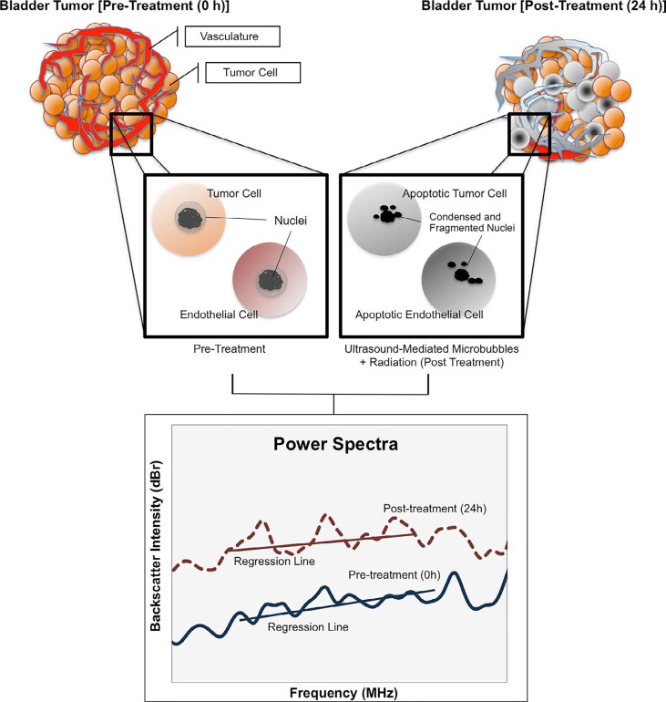
Quantitative ultrasound (QUS) was investigated to monitor bladder cancer treatment response in vivo and to evaluate tumor cell death from combined treatments using ultrasound-stimulated microbubbles and radiation therapy.
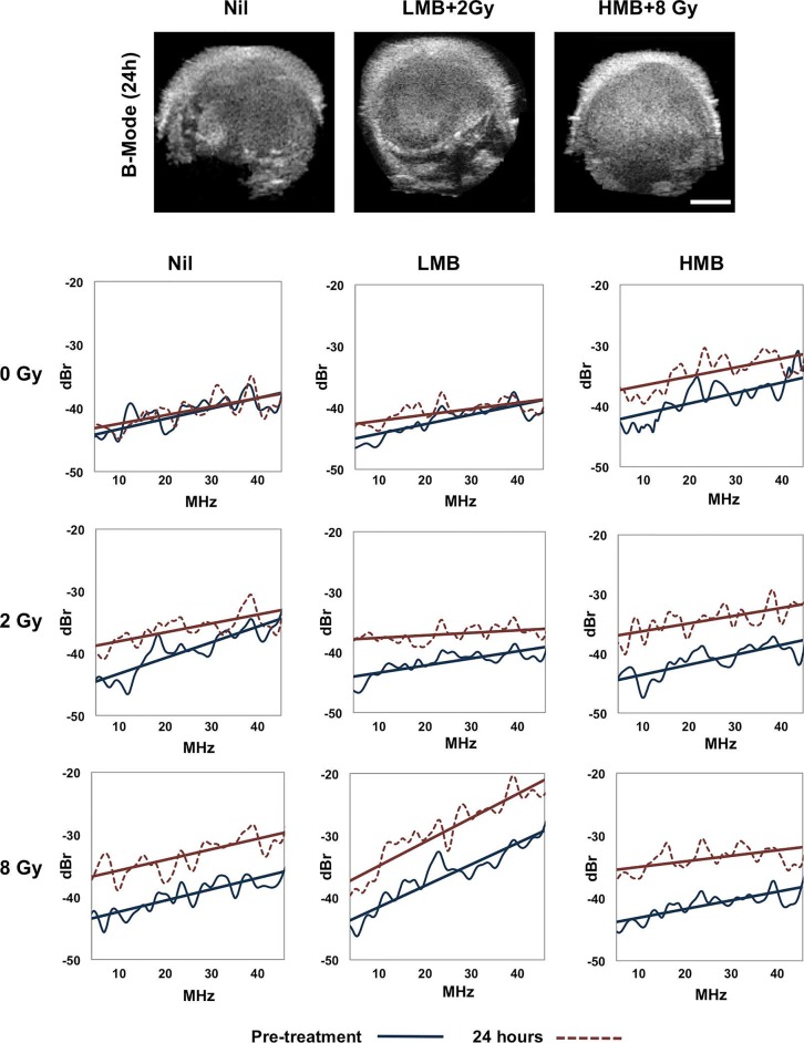
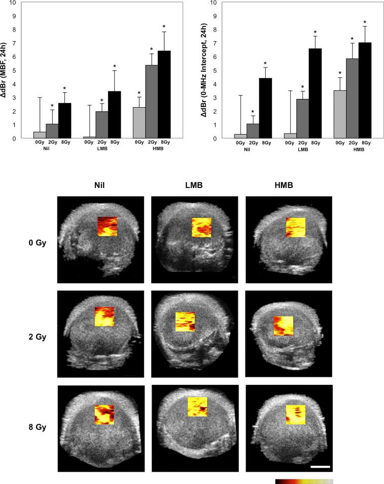
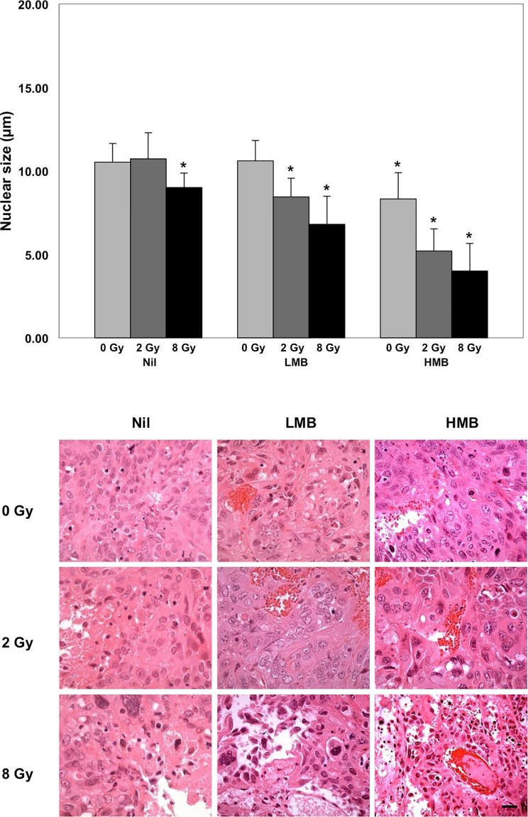
Tumor-bearing mice (n=45), with bladder cancer xenografts (HT- 1376) were exposed to 9 treatment conditions consisting of variable concentrations of ultrasound-stimulated Definity microbubbles [nil, low (1%), high (3%)], combined with single fractionated doses of radiation (0 Gy, 2 Gy, 8 Gy). High frequency (25 MHz) ultrasound was used to collect the raw radiofrequency (RF) data of the backscatter signal from tumors prior to, and 24 hours after treatment in order to obtain QUS parameters. The calculated QUS spectral parameters included the mid-band fit (MBF), and 0-MHz intercept (SI) using a linear regression analysis of the normalized power spectrum.
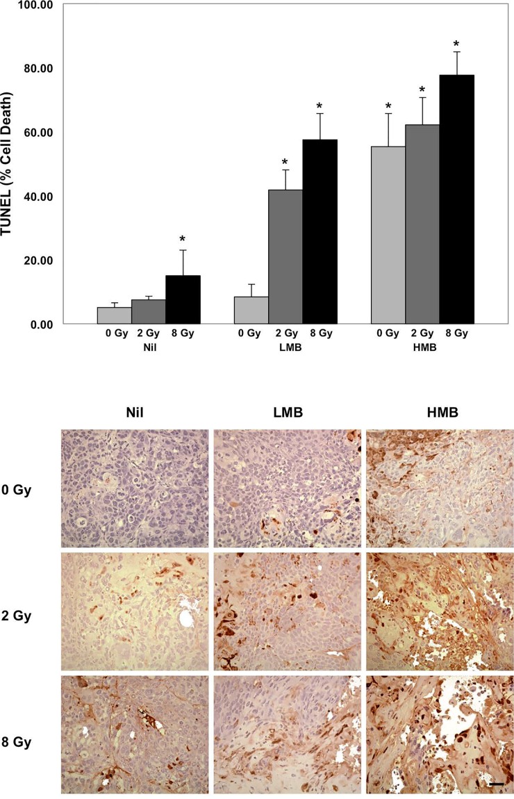
There were maximal increases in QUS parameters following treatments with high concentration microbubbles combined with 8 Gy radiation: (ΔMBF = +6.41 ± 1.40 (±SD) dBr and SI= + 7.01 ± 1.20 (±SD) dBr. Histological data revealed increased cell death, and a reduction in nuclear size with treatments, which was mirrored by changes in quantitative ultrasound parameters. QUS demonstrated markers to detect treatment effects in bladder tumors in vivo.
研究定量超声(QUS)用于在体内监测膀胱癌治疗反应,并评估超声激发微泡与放射治疗联合治疗导致的肿瘤细胞死亡情况。
将45只荷瘤小鼠(移植有膀胱癌异种移植物HT-1376)暴露于9种治疗条件下,这些条件包括不同浓度的超声激发的Definity微泡[无、低(1%)、高(3%)],并与单次分割剂量的放射(0 Gy、2 Gy、8 Gy)联合使用。在治疗前及治疗后24小时,使用高频(25 MHz)超声收集肿瘤后向散射信号的原始射频(RF)数据,以获取QUS参数。计算得到的QUS光谱参数包括中带拟合(MBF)和使用归一化功率谱线性回归分析得到的0-MHz截距(SI)。
高浓度微泡联合8 Gy放射治疗后,QUS参数有最大程度的增加:(ΔMBF = +6.41 ± 1.40(±标准差)dBr,SI = +7.01 ± 1.20(±标准差)dBr)。组织学数据显示,治疗后细胞死亡增加,细胞核大小减小,这与定量超声参数的变化情况相符。QUS证明了在体内检测膀胱肿瘤治疗效果的标志物。