Program in Systems Biology, Department of Biochemistry and Molecular Pharmacology, University of Massachusetts Medical School, Worcester, MA, USA.
Department of Physics, Massachusetts Institute of Technology, Cambridge, MA, USA.
Nat Methods. 2021 Sep;18(9):1046-1055. doi: 10.1038/s41592-021-01248-7. Epub 2021 Sep 3.
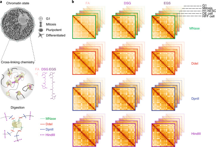
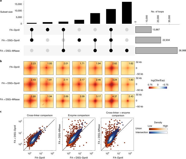
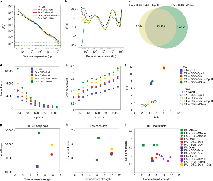
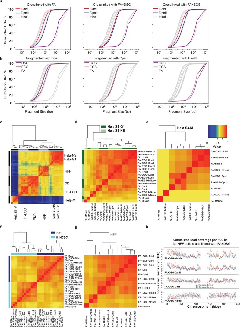
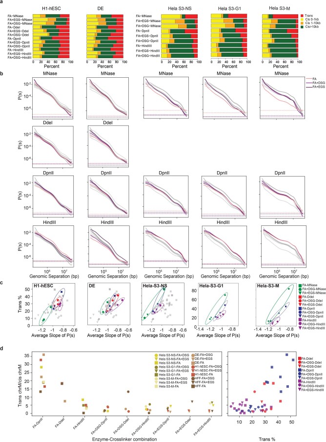
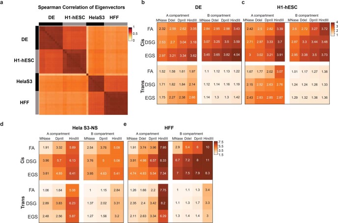
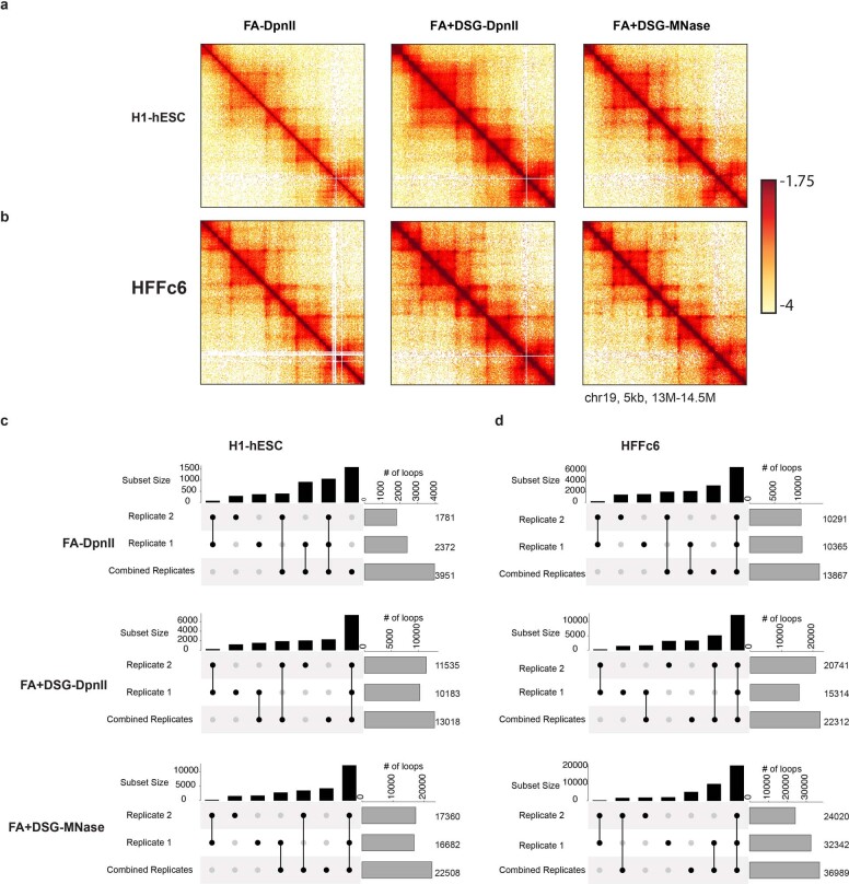
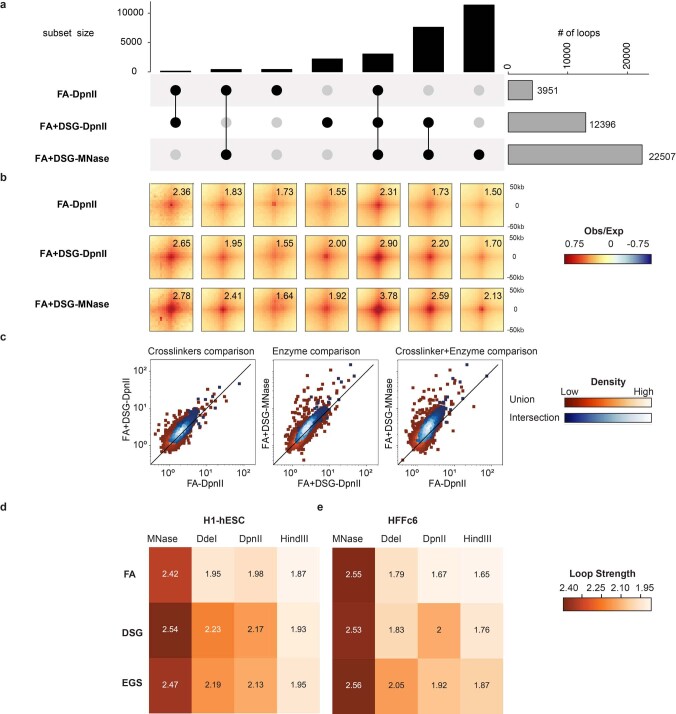
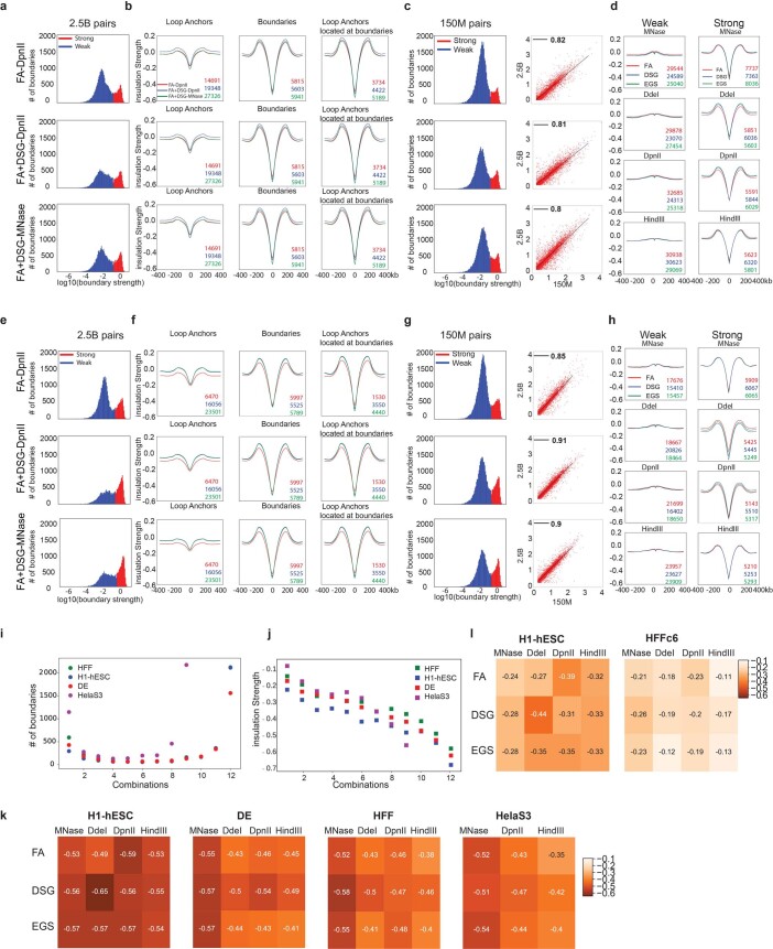
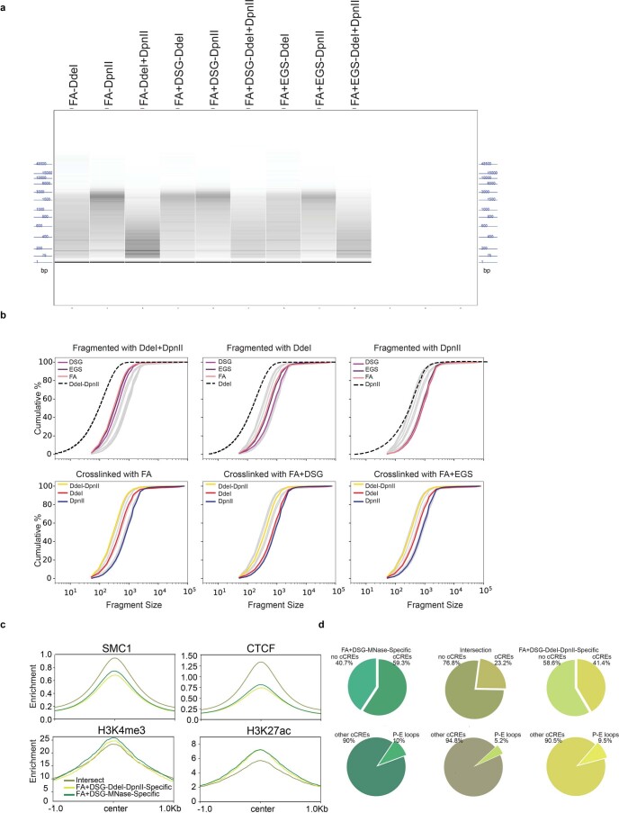
Chromosome conformation capture (3C) assays are used to map chromatin interactions genome-wide. Chromatin interaction maps provide insights into the spatial organization of chromosomes and the mechanisms by which they fold. Hi-C and Micro-C are widely used 3C protocols that differ in key experimental parameters including cross-linking chemistry and chromatin fragmentation strategy. To understand how the choice of experimental protocol determines the ability to detect and quantify aspects of chromosome folding we have performed a systematic evaluation of 3C experimental parameters. We identified optimal protocol variants for either loop or compartment detection, optimizing fragment size and cross-linking chemistry. We used this knowledge to develop a greatly improved Hi-C protocol (Hi-C 3.0) that can detect both loops and compartments relatively effectively. In addition to providing benchmarked protocols, this work produced ultra-deep chromatin interaction maps using Micro-C, conventional Hi-C and Hi-C 3.0 for key cell lines used by the 4D Nucleome project.
染色质构象捕获(3C)实验被用于在全基因组范围内绘制染色质相互作用图谱。染色质相互作用图谱为染色体的空间组织及其折叠机制提供了深入的了解。Hi-C 和 Micro-C 是两种广泛使用的 3C 实验方案,它们在交联化学和染色质片段化策略等关键实验参数上存在差异。为了了解实验方案的选择如何决定检测和量化染色体折叠的能力,我们对 3C 实验参数进行了系统评估。我们确定了用于检测环或隔室的最佳方案变体,优化了片段大小和交联化学。我们利用这些知识开发了一种大大改进的 Hi-C 实验方案(Hi-C 3.0),该方案可以相对有效地检测环和隔室。除了提供基准化的方案外,这项工作还使用 Micro-C、传统 Hi-C 和 Hi-C 3.0 为 4D 核组学项目中使用的关键细胞系生成了超深度染色质相互作用图谱。