Department of Biomedicine, University Hospital Basel, University of Basel, 4056 Basel, Switzerland.
Department of Electronics, Information and Bioengineering, Politecnico di Milano, 20133 Milan, Italy.
Int J Mol Sci. 2021 Sep 3;22(17):9581. doi: 10.3390/ijms22179581.
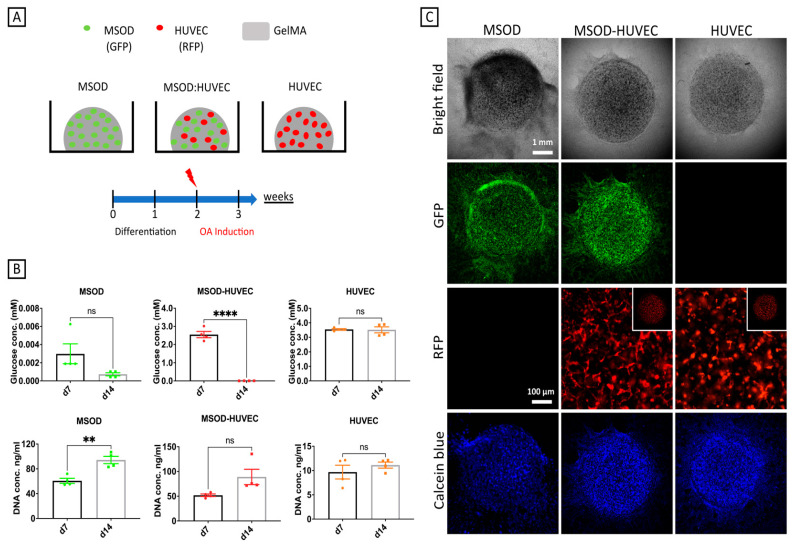
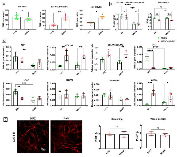
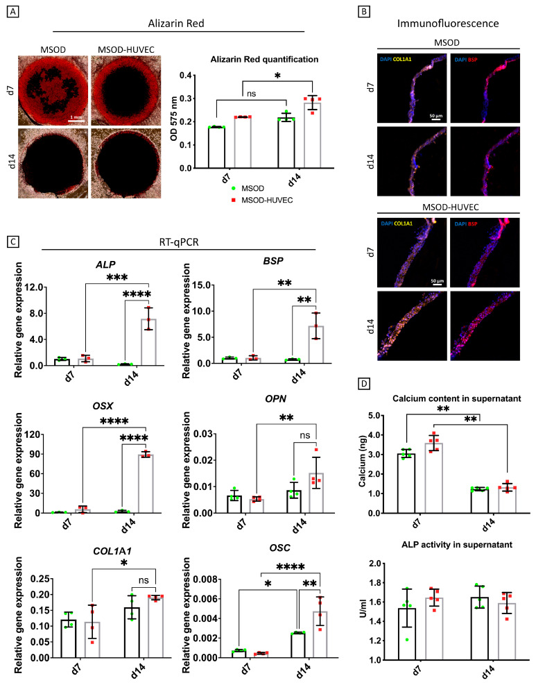
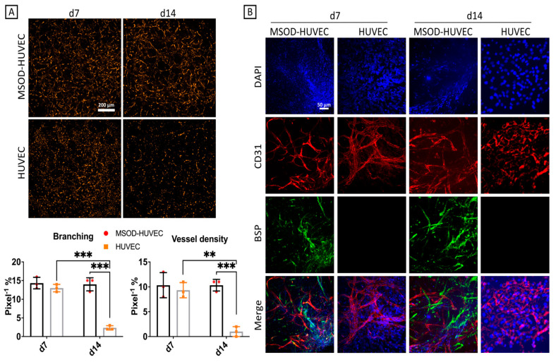
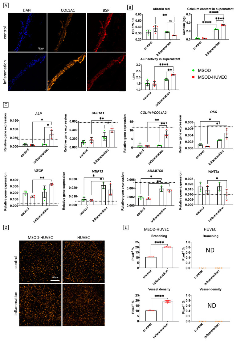
The subchondral bone and its associated vasculature play an important role in the onset of osteoarthritis (OA). Integration of different aspects of the OA environment into multi-cellular and complex human, in vitro models is therefore needed to properly represent the pathology. In this study, we exploited a mesenchymal stromal cell line/endothelial cell co-culture to produce an in vitro human model of vascularized osteogenic tissue. A cocktail of inflammatory cytokines, or conditioned medium from mechanically-induced OA engineered microcartilage, was administered to this vascularized bone model to mimic the inflamed OA environment, hypothesizing that these treatments could induce the onset of specific pathological traits. Exposure to the inflammatory factors led to increased network formation by endothelial cells, reminiscent of the abnormal angiogenesis found in OA subchondral bone, demineralization of the constructs, and increased collagen production, signs of OA related bone sclerosis. Furthermore, inflammation led to augmented expression of osteogenic (alkaline phosphatase (ALP) and osteocalcin (OCN)) and angiogenic (vascular endothelial growth factor (VEGF)) genes. The treatment, with a conditioned medium from the mechanically-induced OA engineered microcartilage, also caused increased demineralization and expression of ALP, OCN, ADAMTS5, and VEGF; however, changes in network formation by endothelial cells were not observed in this second case, suggesting a possible different mechanism of action in inducing OA-like phenotypes. We propose that this vascularized bone model could represent a first step for the in vitro study of bone changes under OA mimicking conditions and possibly serve as a tool in testing anti-OA drugs.
软骨下骨及其相关血管在骨关节炎 (OA) 的发病中起着重要作用。因此,需要将 OA 环境的不同方面整合到多细胞和复杂的人类体外模型中,以正确代表病理学。在这项研究中,我们利用间充质基质细胞系/内皮细胞共培养来产生血管化成骨组织的体外人类模型。将炎症细胞因子鸡尾酒或机械诱导的 OA 工程化微软骨的条件培养基施用于该血管化骨模型,以模拟炎症性 OA 环境,假设这些处理可以诱导特定病理特征的发生。暴露于炎症因子导致内皮细胞形成的网络增加,使人联想到 OA 软骨下骨中发现的异常血管生成、构建体脱矿质和胶原产生增加,这是 OA 相关骨硬化的迹象。此外,炎症导致成骨(碱性磷酸酶 (ALP) 和骨钙素 (OCN))和血管生成(血管内皮生长因子 (VEGF))基因的表达增加。用机械诱导的 OA 工程化微软骨的条件培养基处理也导致脱矿质和 ALP、OCN、ADAMTS5 和 VEGF 的表达增加;然而,在第二种情况下未观察到内皮细胞网络形成的变化,这表明在诱导 OA 样表型方面可能存在不同的作用机制。我们提出,这种血管化骨模型可以代表在模拟 OA 条件下进行骨变化的体外研究的第一步,并可能作为测试抗 OA 药物的工具。