• 文献检索
  • 文档翻译
  • 深度研究
  • 学术资讯
  • Suppr Zotero 插件Zotero 插件
  • 邀请有礼
  • 套餐&价格
  • 历史记录
应用&插件
Suppr Zotero 插件Zotero 插件浏览器插件Mac 客户端Windows 客户端微信小程序
定价
高级版会员购买积分包购买API积分包
服务
文献检索文档翻译深度研究API 文档MCP 服务
关于我们
关于 Suppr公司介绍联系我们用户协议隐私条款
关注我们

Suppr 超能文献

核心技术专利:CN118964589B侵权必究
粤ICP备2023148730 号-1Suppr @ 2026

文献检索

告别复杂PubMed语法,用中文像聊天一样搜索,搜遍4000万医学文献。AI智能推荐,让科研检索更轻松。

立即免费搜索

文件翻译

保留排版,准确专业,支持PDF/Word/PPT等文件格式,支持 12+语言互译。

免费翻译文档

深度研究

AI帮你快速写综述,25分钟生成高质量综述,智能提取关键信息,辅助科研写作。

立即免费体验

使用锥形束计算机断层扫描对具有五条根管的下颌第一前磨牙进行根管治疗:一例报告

Endodontic Management of a Two-rooted Mandibular First Premolar with Five Root Canals with Cone-beam Computed Tomography: A Case Report.

作者信息

Nouroloyouni Ahmad, Lotfi Mehrdad, Milani Amin Salem, Nouroloyouni Sarah

机构信息

Dept. of Endodontics, Faculty of Dentistry, Ardabil University of Medical Sciences, Ardabil, Iran.

Dept. of Endodontics, Faculty of Dentistry, Tabriz University of Medical Sciences, Tabriz, Iran.

出版信息

J Dent (Shiraz). 2021 Sep;22(3):225-228. doi: 10.30476/DENTJODS.2020.83376.1049.

DOI:10.30476/DENTJODS.2020.83376.1049
PMID:34514072
原文链接:https://pmc.ncbi.nlm.nih.gov/articles/PMC8417543/
Abstract

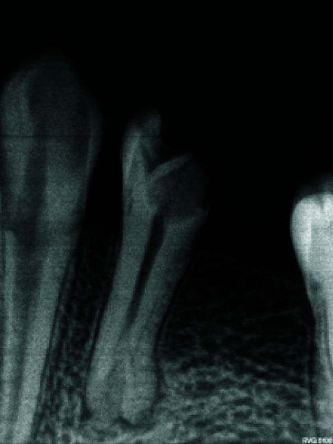
Proper knowledge of the anatomic structure of the root canal system is a vital prerequisite for successful root canal therapy. This report presents the endodontic management a two-rooted lower first premolar with five root canals. A similar case has not been reported to date. The use of cone beam computed tomography (CBCT) in rare and doubtful cases helps establish an accurate diagnosis and render successful endodontic treatment thereafter. This article helps broaden our knowledge about the possible anatomic diversities as to teeth with more roots and root canals than expected normally.

摘要

正确了解根管系统的解剖结构是根管治疗成功的重要前提。本报告介绍了一例有五条根管的下颌第一前磨牙的牙髓治疗情况。迄今为止尚未有类似病例的报道。在罕见和疑难病例中使用锥形束计算机断层扫描(CBCT)有助于准确诊断,并在此后进行成功的牙髓治疗。本文有助于拓宽我们对于牙根和根管数量比正常预期更多的牙齿可能存在的解剖学差异的认识。

https://cdn.ncbi.nlm.nih.gov/pmc/blobs/84b5/8417543/f60ba2dbed90/JDS-22-225-g005.jpg
https://cdn.ncbi.nlm.nih.gov/pmc/blobs/84b5/8417543/7f7bc1aa099e/JDS-22-225-g001.jpg
https://cdn.ncbi.nlm.nih.gov/pmc/blobs/84b5/8417543/4e6e8e0e4978/JDS-22-225-g002.jpg
https://cdn.ncbi.nlm.nih.gov/pmc/blobs/84b5/8417543/d00b2f96decf/JDS-22-225-g003.jpg
https://cdn.ncbi.nlm.nih.gov/pmc/blobs/84b5/8417543/f5d5c147777e/JDS-22-225-g004.jpg
https://cdn.ncbi.nlm.nih.gov/pmc/blobs/84b5/8417543/f60ba2dbed90/JDS-22-225-g005.jpg

相似文献

1
Endodontic Management of a Two-rooted Mandibular First Premolar with Five Root Canals with Cone-beam Computed Tomography: A Case Report.使用锥形束计算机断层扫描对具有五条根管的下颌第一前磨牙进行根管治疗:一例报告
J Dent (Shiraz). 2021 Sep;22(3):225-228. doi: 10.30476/DENTJODS.2020.83376.1049.
2
Endodontic Management of Three-rooted Mandibular First Premolar Using Cone-beam Computed Tomography: A Case Report.使用锥形束计算机断层扫描技术对三根管下颌第一前磨牙进行根管治疗:一例报告
Iran Endod J. 2023;18(2):122-125. doi: 10.22037/iej.v18i2.41169.
3
Cone Beam Computed Tomography Assisted Endodontic Management of a Rare Case of Mandibular First Premolar with Three Roots.锥形束计算机断层扫描辅助下对一例罕见的下颌第一前磨牙三根病例的根管治疗
J Int Oral Health. 2015 Jun;7(6):107-9.
4
Morphological variations of mandibular first premolar on cone-beam computed tomography in a Saudi Arabian sub-population.沙特阿拉伯亚人群下颌第一前磨牙在锥形束计算机断层扫描上的形态变异
Saudi Dent J. 2021 Mar;33(3):150-155. doi: 10.1016/j.sdentj.2019.11.013. Epub 2019 Dec 16.
5
Cone-beam Computed Tomographic Evaluation and Endodontic Management of a Mandibular First Premolar with Type IX Canal Configuration: Case Report.锥形束计算机断层扫描评估及下颌第一前磨牙 9 型根管形态的根管治疗:病例报告。
J Endod. 2017 Jul;43(7):1207-1213. doi: 10.1016/j.joen.2017.01.011. Epub 2017 Apr 25.
6
Cone beam computed tomography evaluation and endodontic management of permanent mandibular second molar with four roots: A rare case report and literature review.锥形束计算机断层扫描评估及四根管下颌第二恒磨牙的根管治疗:一例罕见病例报告及文献综述
J Conserv Dent. 2014 Jul;17(4):385-8. doi: 10.4103/0972-0707.136518.
7
Mandibular first premolar with five root canals: a case report.下颌第一前磨牙 5 根管:1 例报告。
BMC Oral Health. 2020 Sep 10;20(1):253. doi: 10.1186/s12903-020-01241-0.
8
Root canal morphology and variations in mandibular second molar teeth of an Indian population: an in vivo cone-beam computed tomography analysis.印度人群下颌第二磨牙根管形态及变异:体内锥形束 CT 分析。
Clin Oral Investig. 2017 Dec;21(9):2801-2809. doi: 10.1007/s00784-017-2082-6. Epub 2017 Mar 9.
9
Cone-beam computed tomography diagnosis and nonsurgical endodontic management of a taurodontic mandibular first premolar with two roots and four canals: A rare case report.锥形束计算机断层扫描诊断及非手术根管治疗双根四根管的牛牙样下颌第一前磨牙:一例罕见病例报告
J Conserv Dent. 2021 Nov-Dec;24(6):634-639. doi: 10.4103/jcd.jcd_580_21. Epub 2022 Apr 1.
10
Endodontic Management of Mandibular Second Premolar with Type IX Canal Configuration Using Cone-Beam Computed Tomography as a Diagnostic Aid: A Rare Case Report.使用锥形束计算机断层扫描作为诊断辅助手段对具有IX型根管形态的下颌第二前磨牙进行根管治疗:一例罕见病例报告
Contemp Clin Dent. 2018 Jun;9(Suppl 1):S180-S184. doi: 10.4103/ccd.ccd_153_18.

引用本文的文献

1
Mandibular left first premolar with three roots and three canals: A case report.下颌左侧第一前磨牙三根三根管:一例报告。
World J Clin Cases. 2025 May 6;13(13):100822. doi: 10.12998/wjcc.v13.i13.100822.
2
Endodontic treatment of a two-rooted mandibular first premolar with four root canals: a case report.下颌第一前磨牙双根四根管的根管治疗:病例报告
Front Dent Med. 2024 Dec 11;5:1498167. doi: 10.3389/fdmed.2024.1498167. eCollection 2024.
3
Two Palatal Roots in Maxillary First Molar, a Rare Entity: Report of Two Cases.上颌第一磨牙双腭根:一种罕见情况——两例报告

本文引用的文献

1
Anomalous mandibular premolars: a mandibular first premolar with three roots and a mandibular second premolar with a C-shaped canal system.异常下颌前磨牙:一颗有三根的下颌第一前磨牙和一颗具有C形根管系统的下颌第二前磨牙。
Int Endod J. 2008 Nov;41(11):1005-14. doi: 10.1111/j.1365-2591.2008.01451.x.
2
Endodontic management of a mandibular second premolar with four roots and four root canals with the aid of spiral computed tomography: a case report.借助螺旋计算机断层扫描对下颌第二前磨牙四根管四根的根管治疗:一例报告
J Endod. 2008 Jan;34(1):104-7. doi: 10.1016/j.joen.2007.10.004.
3
Use of cone-beam computed tomography to identify root canal systems in vitro.
J Dent (Shiraz). 2024 Jun 1;25(2):178-182. doi: 10.30476/dentjods.2023.98649.2101. eCollection 2024 Jun.
4
Cone-Beam Computed Tomography Assessment of Prevalence of Procedural Errors in Maxillary Posterior Teeth.锥形束计算机断层扫描评估上颌后牙操作失误的发生率。
Biomed Res Int. 2023 Dec 12;2023:4439890. doi: 10.1155/2023/4439890. eCollection 2023.
5
Prevalence and Morphology of C-Shaped Canals in Mandibular First and Second Molars of an Iranian Population: A Cone-Beam Computed Tomography Assessment.伊朗人群下颌第一和第二磨牙 C 形根管的发生率和形态:锥形束 CT 评估。
Scanning. 2023 Oct 3;2023:5628707. doi: 10.1155/2023/5628707. eCollection 2023.
6
Cone-Beam Computed Tomography Assessment of Quality of Endodontic Treatment and Prevalence of Procedural Errors in Mandibular Molars.锥形束计算机断层扫描评估下颌磨牙根管治疗质量和操作失误的发生率。
Int J Clin Pract. 2023 May 19;2023:3558974. doi: 10.1155/2023/3558974. eCollection 2023.
7
Mandibular Second Premolar with Four Canals: A Case Report.下颌第二前磨牙四根管:一例报告
Iran Endod J. 2022;17(4):209-211. doi: 10.22037/iej.v17i4.38604.
使用锥形束计算机断层扫描在体外识别根管系统。
J Endod. 2008 Jan;34(1):87-9. doi: 10.1016/j.joen.2007.10.016.
4
Endodontic applications of cone-beam volumetric tomography.锥形束容积断层扫描在牙髓病学中的应用
J Endod. 2007 Sep;33(9):1121-32. doi: 10.1016/j.joen.2007.06.011. Epub 2007 Jul 19.
5
The root and root canal morphology of the human mandibular first premolar: a literature review.人类下颌第一前磨牙的牙根及根管形态:文献综述
J Endod. 2007 May;33(5):509-16. doi: 10.1016/j.joen.2006.12.004. Epub 2007 Feb 27.
6
Digital and advanced imaging in endodontics: a review.牙髓病学中的数字与先进成像技术:综述
J Endod. 2007 Jan;33(1):1-6. doi: 10.1016/j.joen.2006.08.013.
7
Radiographic evaluation of root canal multiplicity in mandibular first premolars.下颌第一前磨牙根管多样性的影像学评估
J Endod. 2004 Feb;30(2):73-4. doi: 10.1097/00004770-200402000-00002.
8
A standardized endodontic technique utilizing newly designed instruments and filling materials.一种采用新设计器械和充填材料的标准化根管治疗技术。
Oral Surg Oral Med Oral Pathol. 1961 Jan;14:83-91. doi: 10.1016/0030-4220(61)90477-7.
9
Contemporary endodontic retreatments: an analysis based on clinical treatment findings.当代牙髓再治疗:基于临床治疗结果的分析。
J Endod. 2002 Dec;28(12):834-6. doi: 10.1097/00004770-200212000-00010.
10
Analysis of radiologic factors in determining premolar root canal systems.
Oral Surg Oral Med Oral Pathol Oral Radiol Endod. 1999 Dec;88(6):719-22. doi: 10.1016/s1079-2104(99)70016-8.