Division of Nanoscopy, University of Maastricht Multimodal Molecular Imaging Institute, Maastricht, 6229, The Netherlands.
School of Medical Sciences (Discipline of Anatomy and Histology) & Australian Centre for Microscopy & Microanalysis, The University of Sydney, Sydney, NSW, 2006, Australia.
Histochem Cell Biol. 2022 Jan;157(1):27-38. doi: 10.1007/s00418-021-02030-8. Epub 2021 Sep 15.
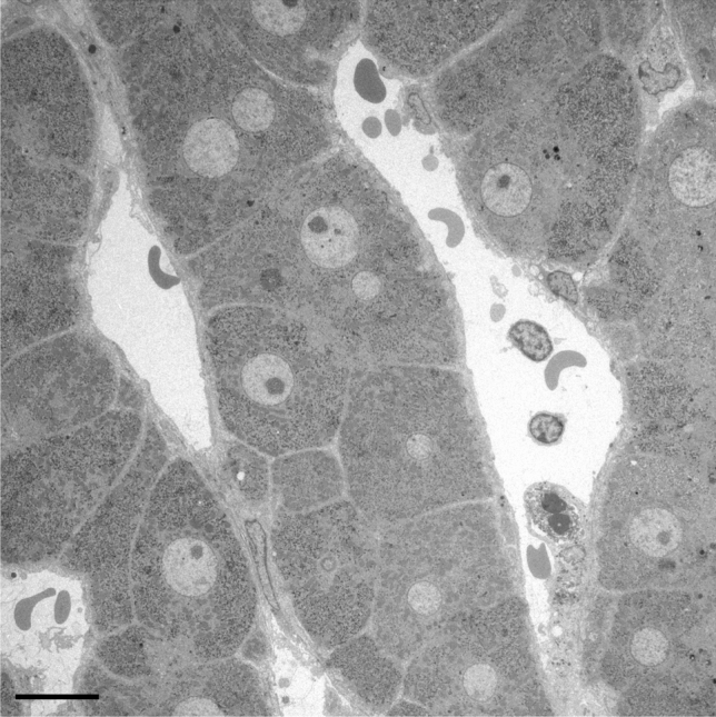
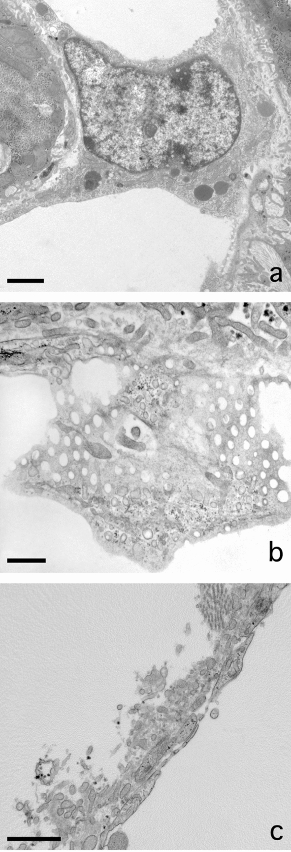
Adapted fixation methods for electron microscopy allowed us to study liver cell fine structure in 217 biopsies of intact human livers over the course of 10 years. The following novel observations and concepts arose: single fat droplets in parenchymal cells can grow to a volume four times larger than the original cell, thereby extremely marginalizing the cytoplasm with all organelles. Necrosis of single parenchymal cells, still containing one huge fat droplet, suggests death by fat in a process of single-cell steatonecrosis. In a later stage of single-cell steatonecrosis, neutrophils and erythrocytes surround the single fat droplet, forming an inflammatory fat follicle indicating the apparent onset of inflammation. Also, fat droplets frequently incorporate masses of filamentous fragments and other material, most probably representing Mallory substance. No other structure or material was found that could possibly represent Mallory bodies. We regularly observe the extrusion of huge fat droplets, traversing the peripheral cytoplasm of parenchymal cells, the Disse space and the endothelium. These fat droplets fill the sinusoid as a sinusoidal lipid embolus. In conclusion, adapted methods of fixation applied to human liver tissue revealed that single, huge fat droplets cause necrosis and inflammation in single parenchymal cells. Fat droplets also collect Mallory substance and give rise to sinusoidal fat emboli. Therefore, degreasing of the liver seems to be an essential therapeutic first step in the self-repairing of non-alcoholic fatty liver disease. This might directly reduce single-cell steatotic necrosis and inflammation as elements in non-alcoholic steatohepatitis progression.
改良的电子显微镜固定方法使我们能够在 10 年的时间里对 217 例完整人类肝脏活检进行肝细胞超微结构研究。由此产生了以下新的观察结果和概念:单个实质细胞中的脂肪滴可以增长到比原始细胞大四倍的体积,从而使所有细胞器都极度边缘化。单个实质细胞发生坏死,仍然含有一个巨大的脂肪滴,提示脂肪在单细胞脂肪坏死过程中导致细胞死亡。在单细胞脂肪坏死的后期阶段,中性粒细胞和红细胞围绕单个脂肪滴,形成一个炎症性脂肪滤泡,表明炎症明显发作。此外,脂肪滴经常包含大量丝状片段和其他物质,很可能代表 Mallory 物质。没有发现其他可能代表 Mallory 小体的结构或物质。我们经常观察到巨大的脂肪滴从实质细胞的周边细胞质、Disse 间隙和内皮细胞中挤出。这些脂肪滴充满窦状隙,形成窦状隙脂质栓子。总之,应用于人类肝组织的改良固定方法表明,单个巨大的脂肪滴会导致单个实质细胞发生坏死和炎症。脂肪滴还会收集 Mallory 物质,并引发窦状隙脂肪栓子。因此,肝脏去脂似乎是治疗非酒精性脂肪性肝病自我修复的必要治疗第一步。这可能直接减少非酒精性脂肪性肝炎进展过程中的单细胞脂肪性坏死和炎症等因素。