Siepker Christopher L, Zimmer Jennifer L, Bedard Kathleen M, Hart Kelsey A, Czerwinski Sarah L, Carmichael K Paige
Iowa State University Veterinary Diagnostic Laboratory, Ames, IA, USA.
Department of Small Animal Medicine & Surgery, College of Veterinary Medicine, University of Georgia, Athens, GA, USA.
Case Rep Vet Med. 2021 Sep 6;2021:2064103. doi: 10.1155/2021/2064103. eCollection 2021.
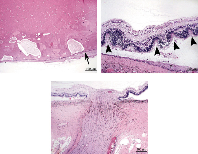
. A two-month-old, female, Aberdeen-Angus calf was presented for congenital cataracts and blindness in both eyes (OU). The dam had a reported history of visual defects (not specified) and had produced other affected calves (per owner history). Ophthalmic examination revealed mature bilateral cataracts, attenuation of the iridic granules, persistent pupillary membranes, and dyscoric pupils. Additionally, the calf had a poor body condition, prognathism, dome-shaped head, excessive nasal drainage, limb contracture, and fever. Histopathology of both eyes revealed lenticular degeneration (congenital cataracts), retinal dysplasia, and optic nerve hypoplasia. BVDV IHC detected antigen within only the left eye (OS), consisting of intrahistiocytic and endothelial immunoreactivity within the ciliary body, iris, and choroid. No BVDV immunoreactivity could be detected in the right eye (OD). This case highlights the unique ocular changes present in in utero BVDV infection of cattle with a different immunohistochemical staining profile than previously described.
一头两个月大的雌性阿伯丁-安格斯犊牛因双眼先天性白内障和失明前来就诊。据报道,其母畜有视觉缺陷病史(未明确说明),并产下过其他患病犊牛(根据畜主病史)。眼科检查发现双侧成熟白内障、虹膜颗粒减少、瞳孔残膜和瞳孔不等大。此外,该犊牛身体状况不佳,有上颌前突、圆顶状头部、鼻腔分泌物过多、肢体挛缩和发热症状。双眼组织病理学检查显示晶状体变性(先天性白内障)、视网膜发育异常和视神经发育不全。牛病毒性腹泻病毒免疫组化检测仅在左眼(OS)检测到抗原,表现为睫状体、虹膜和脉络膜内的组织细胞内和内皮免疫反应性。右眼(OD)未检测到牛病毒性腹泻病毒免疫反应性。该病例突出了牛子宫内感染牛病毒性腹泻病毒时出现的独特眼部变化,其免疫组化染色特征与先前描述的不同。