Department of Mechanical Engineering and Materials Science, Duke University, Durham, NC 27708, USA.
Celldom Inc., San Carlos, CA 94070, USA.
Sci Adv. 2021 Sep 17;7(38):eabf9840. doi: 10.1126/sciadv.abf9840.
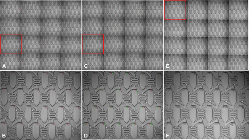
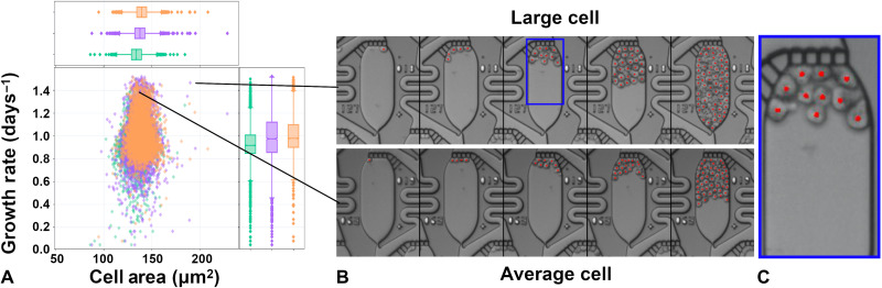
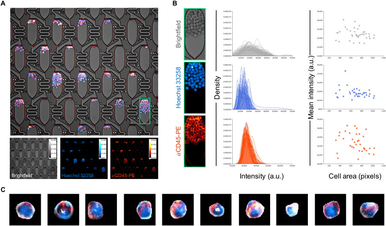
Single-cell analysis tools have made substantial advances in characterizing genomic heterogeneity; however, tools for measuring phenotypic heterogeneity have lagged due to the increased difficulty of handling live biology. Here, we report a single-cell phenotyping tool capable of measuring image-based clonal properties at scales approaching 100,000 clones per experiment. These advances are achieved by exploiting a previously unidentified flow regime in ladder microfluidic networks that, under appropriate conditions, yield a mathematically perfect cell trap. Machine learning and computer vision tools are used to control the imaging hardware and analyze the cellular phenotypic parameters within these images. Using this platform, we quantified the responses of tens of thousands of single cell–derived acute myeloid leukemia (AML) clones to targeted therapy, identifying rare resistance and morphological phenotypes at frequencies down to 0.05%. This approach can be extended to higher-level cellular architectures such as cell pairs and organoids and on-chip live-cell fluorescence assays.
单细胞分析工具在描绘基因组异质性方面取得了重大进展;然而,由于活体生物学处理难度的增加,用于测量表型异质性的工具却一直滞后。在这里,我们报告了一种单细胞表型分析工具,它能够在每个实验中测量基于图像的克隆特性,其规模接近 100000 个克隆。这些进展是通过利用梯式微流控网络中以前未被识别的流型来实现的,在适当的条件下,这种流型产生了数学上完美的细胞陷阱。机器学习和计算机视觉工具被用于控制成像硬件,并分析这些图像中的细胞表型参数。使用该平台,我们定量分析了数万个由急性髓系白血病 (AML) 克隆衍生的单细胞对靶向治疗的反应,以低至 0.05%的频率鉴定出罕见的耐药性和形态表型。这种方法可以扩展到更高层次的细胞结构,如细胞对和类器官,以及芯片上的活细胞荧光检测。