Li Zhiwen, Pu Xiaohong, He Lu, Fu Yao, Li Lin, Xu Yuemei, Guan Wenyan, Fan Xiangshan
Department of Pathology, The Affiliated Drum Tower Hospital, Nanjing University Medical School, Nanjing, China.
Department of Pathology, Nanjing First Hospital, Nanjing Medical University, Nanjing, China.
Front Cardiovasc Med. 2021 Sep 1;8:702215. doi: 10.3389/fcvm.2021.702215. eCollection 2021.
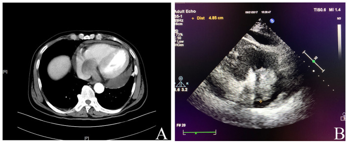
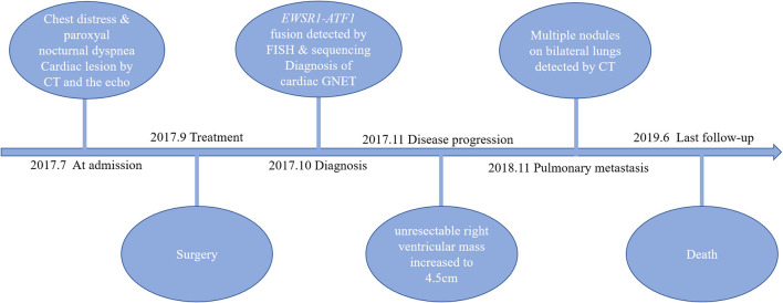
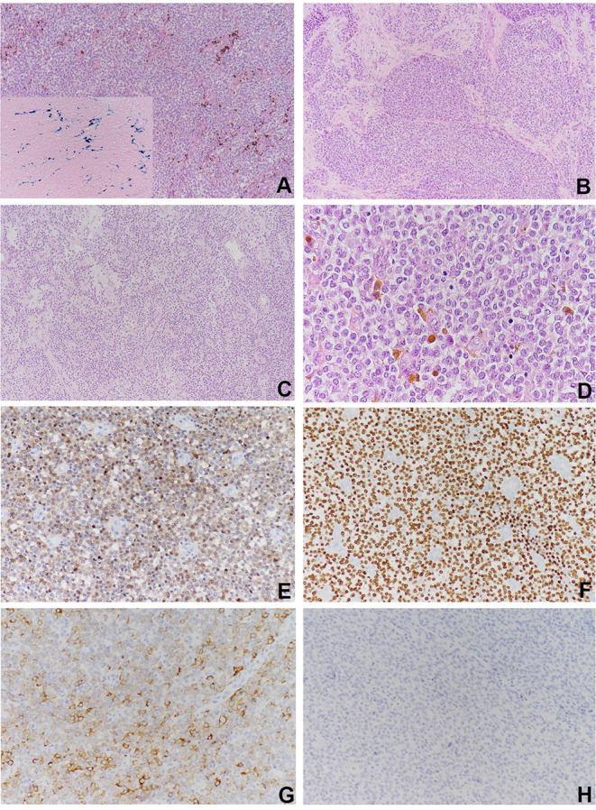
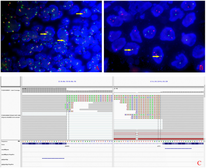
Malignant gastrointestinal neuroectodermal tumor (GNET) is an extremely rare soft tissue sarcoma and has been designated as a new entity recently. At present, GNET virtually exclusively occurs in the gastrointestinal tract. Here we report a case of extra-GNET that arose in the right heart. A 62-year-old male complained of chest distress and breathing difficulty while lying down at night for over 1 month at admission. The radiological findings revealed an occupying lesion involving the right atrium and the right ventricle without any abdominal abnormalities. The patient then underwent a surgical resection. Microscopically, neoplastic cells proliferated in the pattern of nests and sheets with fibrous separation. Focal areas with cellular dyscohesion imparted a vague pseudopapillary pattern. These tumor cells were small to medium in size with fine chromatin and predominantly pale eosinophilic cytoplasm. The nuclei were typically round to oval with somewhat irregular contours and contained small nucleoli. The mitotic figures were easily found. Immunohistochemically, the neoplastic cells were positive for S100 and SOX-10 but negative for HMB-45, A103, and CD99. - rearrangement was detected by fluorescence hybridization and further confirmed by whole-transcriptome sequence analysis. The patient had pulmonary metastasis 8 months later and soon died of the disease. The overall survival of the patient was 20 months. In summary, we reported an extremely rare case of cardiac GNET, indicating that the location of GNET should not be confined to the GI tract as initially defined. Due to the lack of a specific effective treatment and the occurrence of early metastasis, cardiac GNET conferred a poor prognosis. More clinical and experimental studies are warranted to better manage this disease in the future.
恶性胃肠道神经外胚层肿瘤(GNET)是一种极其罕见的软组织肉瘤,最近被认定为一种新的实体瘤。目前,GNET几乎仅发生于胃肠道。在此,我们报告一例发生于右心的胃肠道外GNET病例。一名62岁男性入院前1个多月来夜间平卧时出现胸闷、呼吸困难。影像学检查发现右心房和右心室有占位性病变,腹部未见异常。患者随后接受了手术切除。显微镜下,肿瘤细胞呈巢状和片状增殖,有纤维分隔。局部细胞黏附丧失区域呈现模糊的假乳头样结构。这些肿瘤细胞大小为小到中等,染色质细腻,细胞质主要呈淡嗜酸性。细胞核通常呈圆形至椭圆形,轮廓略不规则,有小核仁。易见核分裂象。免疫组化显示,肿瘤细胞S100和SOX-10阳性,但HMB-45、A103和CD99阴性。通过荧光原位杂交检测到 - 重排,并经全转录组序列分析进一步证实。患者8个月后发生肺转移,很快死于该病。患者总生存期为20个月。总之,我们报告了一例极其罕见的心脏GNET病例,提示GNET的发生部位不应局限于最初定义的胃肠道。由于缺乏特异性有效治疗且早期发生转移,心脏GNET预后较差。未来需要更多的临床和实验研究以更好地诊治该疾病。