Pan Xiu-Wu, Xu Da, Chen Wen-Jin, Chen Jia-Xin, Chen Wei-Jie, Ye Jian-Qing, Gan Si-Shun, Zhou Wang, Song Xu, Shi Lei, Cui Xin-Gang
Department of Urology, Xinhua Hospital, School of Medicine, Shanghai Jiaotong University, 1665 Kongjiang Road, Shanghai, 200092, China.
Depanrtment of Urology, Third Affiliated Hospital of the Second Military Medical University, Shanghai, 201805, China.
Cancer Cell Int. 2021 Sep 20;21(1):486. doi: 10.1186/s12935-021-02161-x.
The benefit of targeted therapy for renal cell carcinoma (RCC) is largely crippled by drug resistance. Rapid disease progression and poor prognosis occur in patients with drug resistance. New treatments demand prompt exploration for clinical therapies. Ubiquitin-specific peptidase 39 (USP39) serves as the pro-tumor factor in several previous studies of other malignant tumors. To investigate the function and mechanism of USP39 in promoting malignant proliferation and angiogenesis of RCC.
We applied ONCOMINE database to analyze the correlation between USP39 expression level and the clinical characteristics of RCC. USP39 knockdown or overexpression plasmids were transfected into 786-O and ACHN cells. The HUVEC received cell supernatants of 786-O and ACHN cells with knockdown or overexpression USP39.The effect of USP39 on RCC was evaluated by MTT assay, cell cycle analysis, colony formation assay and tubule formation assay. The interaction between USP39 and VEGF-A alternative splicing was assessed by affinity purification and mass spectrometry, co-immunoprecipitation and Western blot assays.
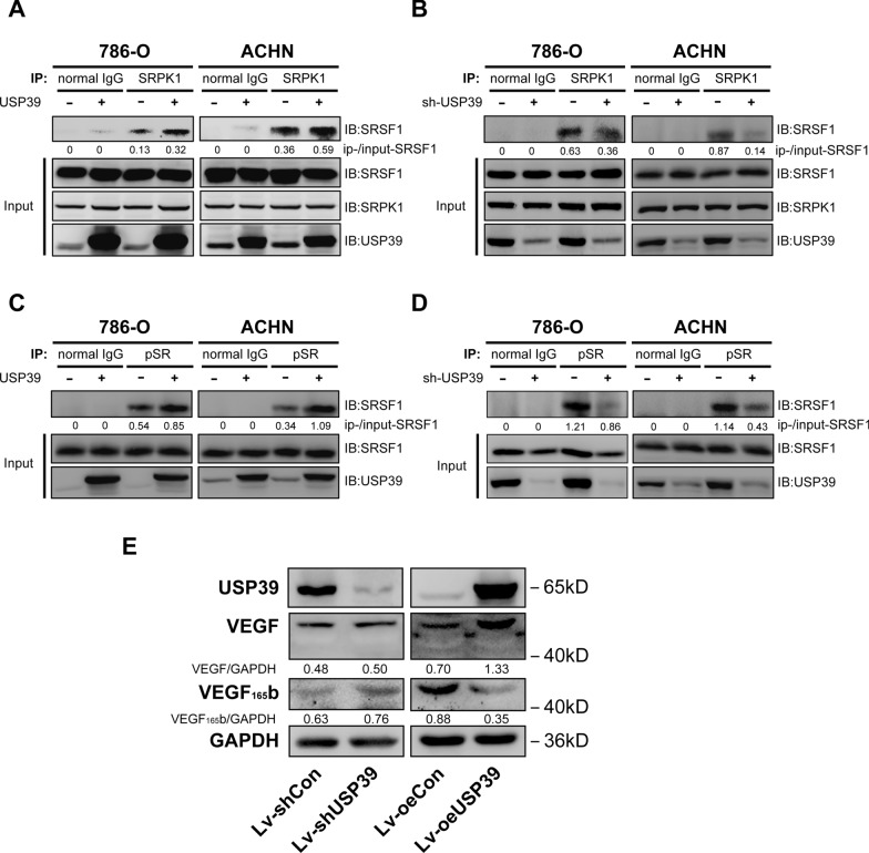
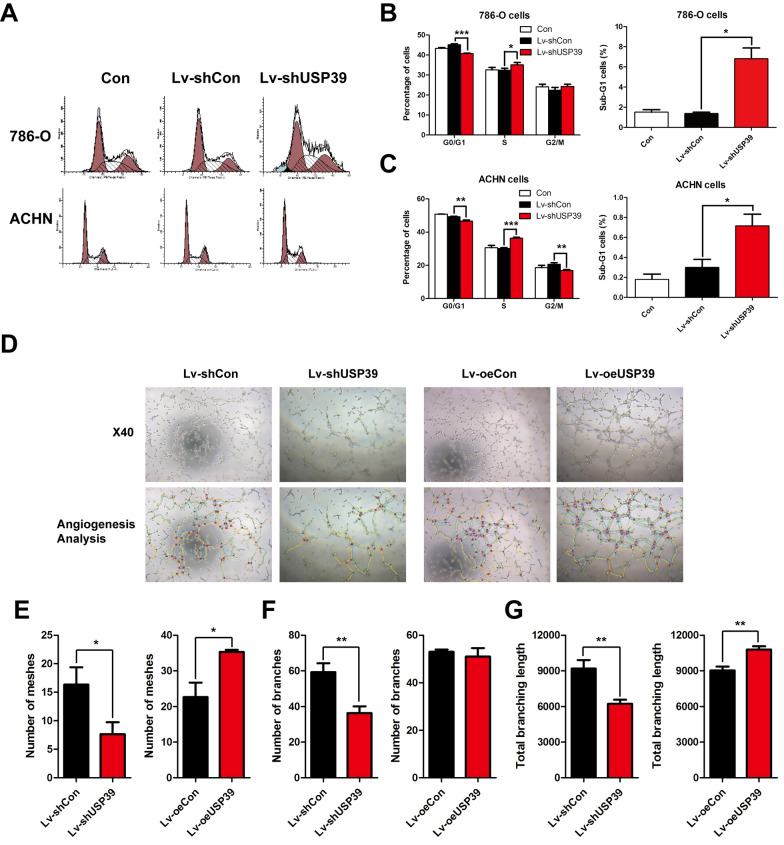
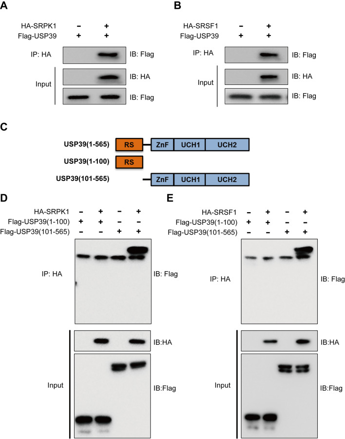
The mRNA expression level of USP39 in RCC was significantly higher than that in normal renal tissue (P < 0.001), and negatively correlated with the survival rate of RCC patients (P < 0.01). Silencing of USP39 in 786-O and ACHN cells inhibited cell proliferation and colony formation, and induced S phase arrest. USP39 overexpression significantly increased the number of tubules (P < 0.05) and branches (P < 0.01) formed by HUVEC cells, and USP39 knockdown produced an opposite effect (P < 0.05). The USP39 fragment directly mediated its binding to SRSF1 and SRPK1, and promoted the phosphorylation of SRSF1 to regulate VEGF-A alternative splicing. USP39 knockdown upregulated the expression of VEGF-A, and USP39 overexpression downregulated the expression of VEGF-A significantly (both P < 0.05).
USP39 acted as a pro-tumor factor by motivating the malignant biological processes of RCC, probably through inhibiting VEGF-A alternative splicing and regulating SRSF1 and SRPK1. USP39 may prove to be a potential therapeutic target for RCC.
靶向治疗对肾细胞癌(RCC)的益处很大程度上因耐药性而受损。耐药患者会出现疾病快速进展和预后不良的情况。新的治疗方法需要迅速探索用于临床治疗。在先前其他恶性肿瘤的多项研究中,泛素特异性肽酶39(USP39)作为促肿瘤因子。旨在研究USP39在促进RCC恶性增殖和血管生成中的作用及机制。
我们应用ONCOMINE数据库分析USP39表达水平与RCC临床特征之间的相关性。将USP39敲低或过表达质粒转染至786 - O和ACHN细胞中。人脐静脉内皮细胞(HUVEC)接受敲低或过表达USP39的786 - O和ACHN细胞的细胞上清液。通过MTT法、细胞周期分析、集落形成试验和小管形成试验评估USP39对RCC的影响。通过亲和纯化和质谱分析、免疫共沉淀和蛋白质印迹试验评估USP39与血管内皮生长因子A(VEGF - A)可变剪接之间的相互作用。
RCC中USP39的mRNA表达水平显著高于正常肾组织(P < 0.001),且与RCC患者的生存率呈负相关(P < 0.01)。在786 - O和ACHN细胞中沉默USP39可抑制细胞增殖和集落形成,并诱导S期阻滞。USP39过表达显著增加了HUVEC细胞形成的小管数量(P < 0.05)和分支数量(P < 0.01),而USP39敲低则产生相反的效果(P < 0.05)。USP39片段直接介导其与丝氨酸/精氨酸丰富剪接因子1(SRSF1)和丝氨酸/精氨酸蛋白激酶1(SRPK1)的结合,并促进SRSF1的磷酸化以调节VEGF - A可变剪接。USP39敲低上调了VEGF - A的表达,而USP39过表达则显著下调了VEGF - A的表达(均P < 0.05)。
USP39可能通过抑制VEGF - A可变剪接并调节SRSF1和SRPK1来推动RCC的恶性生物学过程,从而作为促肿瘤因子发挥作用。USP39可能被证明是RCC的一个潜在治疗靶点。