Koh Siang-Boon, Dontchos Brian N, Bossuyt Veerle, Edmonds Christine, Cristea Simona, Melkonjan Nsan, Mortensen Lindsey, Ma Annie, Beyerlin Kassidy, Denault Elyssa, Niehoff Elizabeth, Hirz Taghreed, Sykes David B, Michor Franziska, Specht Michelle, Lehman Constance, Ellisen Leif W, Spring Laura M
MGH Cancer Center, Massachusetts General Hospital, Boston, MA, USA.
Harvard Medical School, Boston, MA, USA.
NPJ Precis Oncol. 2021 Sep 21;5(1):85. doi: 10.1038/s41698-021-00224-w.
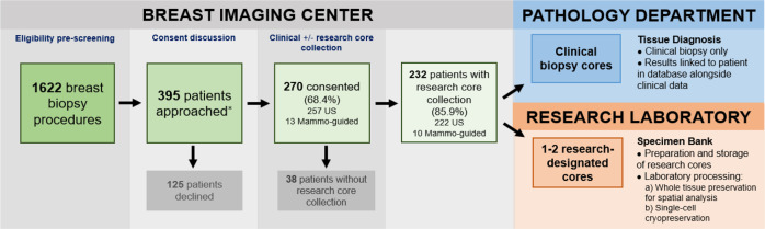
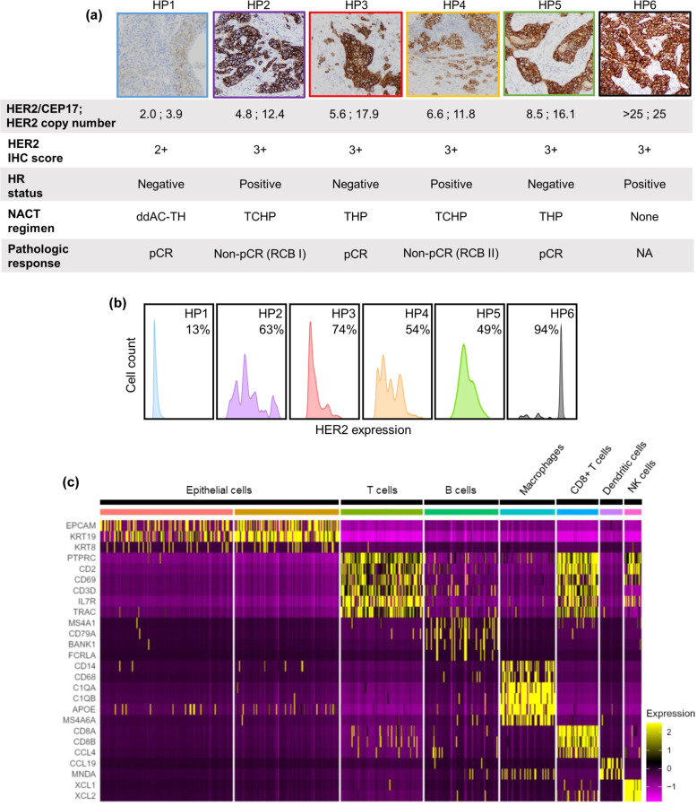
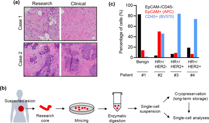
Systematic collection of fresh tissues for research at the time of diagnostic image-guided breast biopsy has the potential to fuel a wide variety of innovative studies. Here we report the initial experience, including safety, feasibility, and laboratory proof-of-principle, with the collection and analysis of research specimens obtained via breast core needle biopsy immediately following routine clinical biopsy at a single institution over a 14-month period. Patients underwent one or two additional core biopsies following collection of all necessary clinical specimens. In total, 395 patients were approached and 270 consented to the research study, yielding a 68.4% consent rate. Among consenting patients, 238 lesions were biopsied for research, resulting in 446 research specimens collected. No immediate complications were observed. Representative research core specimens showed high diagnostic concordance with clinical core biopsies. Flow cytometry demonstrated consistent recovery of hundreds to thousands of viable cells per research core. Among a group of HER2 + tumor research specimens, HER2 assessment by flow cytometry correlated highly with immunohistochemistry (IHC) staining, and in addition revealed extensive inter- and intra-tumoral variation in HER2 levels of potential clinical relevance. Suitability for single-cell transcriptomic analysis was demonstrated for a triple-negative tumor core biopsy, revealing substantial cellular diversity in the tumor immune microenvironment, including a prognostically relevant T cell subpopulation. Thus, collection of fresh tissues for research purposes at the time of diagnostic breast biopsy is safe, feasible and efficient, and may provide a high-yield mechanism to generate a rich tissue repository for a wide variety of cross-disciplinary research.
在诊断性图像引导下进行乳腺活检时系统收集新鲜组织用于研究,有潜力推动开展各种各样的创新性研究。在此,我们报告在一家机构为期14个月的时间里,在常规临床活检后立即通过乳腺粗针活检收集并分析研究标本的初步经验,包括安全性、可行性和实验室原理验证。患者在收集所有必要的临床标本后再接受一到两次额外的粗针活检。总共接触了395名患者,其中270名同意参加该研究,同意率为68.4%。在同意参加研究的患者中,对238个病变进行了活检用于研究,共收集到446份研究标本。未观察到即时并发症。代表性的研究粗针标本与临床粗针活检显示出高度的诊断一致性。流式细胞术表明每个研究粗针能一致地回收数百到数千个活细胞。在一组HER2+肿瘤研究标本中,通过流式细胞术进行的HER2评估与免疫组织化学(IHC)染色高度相关,此外还揭示了HER2水平在肿瘤间和肿瘤内存在广泛的潜在临床相关变异。对三阴性肿瘤粗针活检证明了其适用于单细胞转录组分析,揭示了肿瘤免疫微环境中存在大量细胞多样性,包括一个与预后相关的T细胞亚群。因此,在诊断性乳腺活检时收集新鲜组织用于研究目的是安全、可行且高效的,并且可能提供一种高产机制,以生成一个丰富的组织库用于各种各样的跨学科研究。