Department of Clinical Medicine, University of Bergen, 5021, Bergen, Norway.
Mohn Medical Imaging and Visualization Centre, Department of Radiology, Haukeland University Hospital, 5021, Bergen, Norway.
J Transl Med. 2021 Sep 26;19(1):406. doi: 10.1186/s12967-021-03086-9.
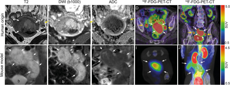
Pelvic magnetic resonance imaging (MRI) and whole-body positron emission tomography-computed tomography (PET-CT) play an important role at primary diagnostic work-up and in detecting recurrent disease in endometrial cancer (EC) patients, however the preclinical use of these imaging methods is currently limited. We demonstrate the feasibility and utility of MRI and dynamic F-fluorodeoxyglucose (FDG)-PET imaging for monitoring tumor progression and assessing chemotherapy response in an orthotopic organoid-based patient-derived xenograft (O-PDX) mouse model of EC.
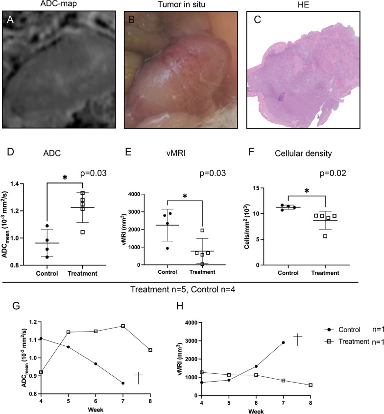
18 O-PDX mice (grade 3 endometrioid EC, stage IIIC1), selectively underwent weekly T2-weighted MRI (total scans = 32), diffusion-weighted MRI (DWI) (total scans = 9) and dynamic F-FDG-PET (total scans = 26) during tumor progression. MRI tumor volumes (vMRI), tumor apparent diffusion coefficient values (ADC) and metabolic tumor parameters from F-FDG-PET including maximum and mean standard uptake values (SUV/SUV), metabolic tumor volume (MTV), total lesion glycolysis (TLG) and metabolic rate of F-FDG (MR) were calculated. Further, nine mice were included in a chemotherapy treatment study (treatment; n = 5, controls; n = 4) and tumor ADC-values were compared to changes in vMRI and cellular density from histology at endpoint. A Mann-Whitney test was used to evaluate differences between groups.
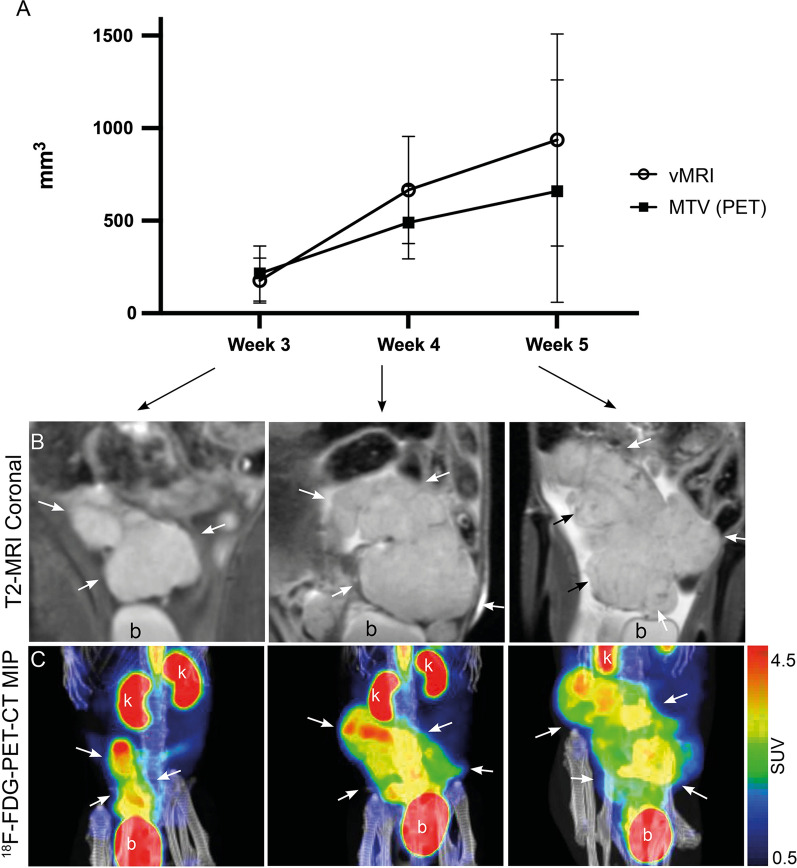
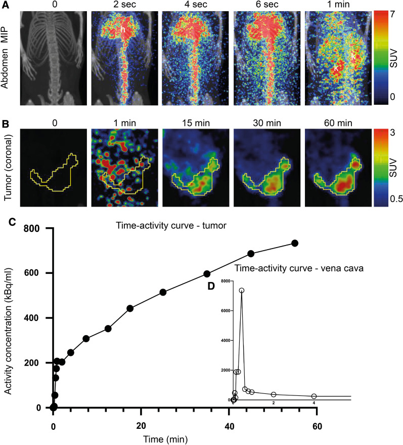
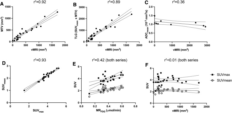
Tumors with large tumor volumes (vMRI) had higher metabolic activity (MTV and TLG) in a clear linear relationship (r = 0.92 and 0.89, respectively). Non-invasive calculation of MR from dynamic F-FDG-PET (mean MR = 0.39 μmol/min) was feasible using an image-derived input function. Treated mice had higher tumor ADC (p = 0.03), lower vMRI (p = 0.03) and tumor cellular density (p = 0.02) than non-treated mice, all indicating treatment response.
Preclinical imaging mirroring clinical imaging methods in EC is highly feasible for monitoring tumor progression and treatment response in the present orthotopic organoid mouse model.
盆腔磁共振成像(MRI)和全身正电子发射断层扫描-计算机断层扫描(PET-CT)在子宫内膜癌(EC)患者的初始诊断和检测复发性疾病方面发挥着重要作用,然而,这些影像学方法的临床前应用目前受到限制。我们展示了 MRI 和动态 F-氟脱氧葡萄糖(FDG)-PET 成像在监测肿瘤进展和评估 EC 患者的基于同源器官的患者衍生异种移植(O-PDX)小鼠模型中化疗反应的可行性和实用性。
18 只 O-PDX 小鼠(3 级子宫内膜样 EC,IIIIC1 期),在肿瘤进展过程中选择性地每周进行 T2 加权 MRI(总扫描数=32)、扩散加权 MRI(DWI)(总扫描数=9)和动态 F-FDG-PET(总扫描数=26)。计算 MRI 肿瘤体积(vMRI)、肿瘤表观扩散系数值(ADC)和 F-FDG-PET 的代谢肿瘤参数,包括最大和平均标准摄取值(SUV/SUV)、代谢肿瘤体积(MTV)、总病变糖酵解(TLG)和 F-FDG 代谢率(MR)。进一步,9 只小鼠被纳入化疗治疗研究(治疗组;n=5,对照组;n=4),并比较终点时肿瘤 ADC 值与组织学上 vMRI 和细胞密度的变化。使用 Mann-Whitney 检验评估组间差异。
具有较大肿瘤体积(vMRI)的肿瘤具有更高的代谢活性(MTV 和 TLG),呈明显的线性关系(r=0.92 和 0.89)。使用图像衍生的输入函数,可以从动态 F-FDG-PET 无创计算 MR(平均 MR=0.39 μmol/min)。与未治疗的小鼠相比,治疗的小鼠的肿瘤 ADC 更高(p=0.03)、vMRI 更低(p=0.03)和肿瘤细胞密度更低(p=0.02),所有这些都表明治疗反应。
在目前的同源器官小鼠模型中,EC 临床成像方法的临床前成像高度可行,可用于监测肿瘤进展和治疗反应。