Sibert Bryan S, Kim Joseph Y, Yang Jie E, Wright Elizabeth R
Department of Biochemistry, University of Wisconsin, Madison; Cryo-Electron Microscopy Research Center, Department of Biochemistry, University of Wisconsin, Madison; Midwest Center for Cryo-Electron Tomography, Department of Biochemistry, University of Wisconsin, Madison.
Department of Biochemistry, University of Wisconsin, Madison; Department of Chemistry, University of Wisconsin, Madison.
J Vis Exp. 2021 Sep 13(175). doi: 10.3791/62992.
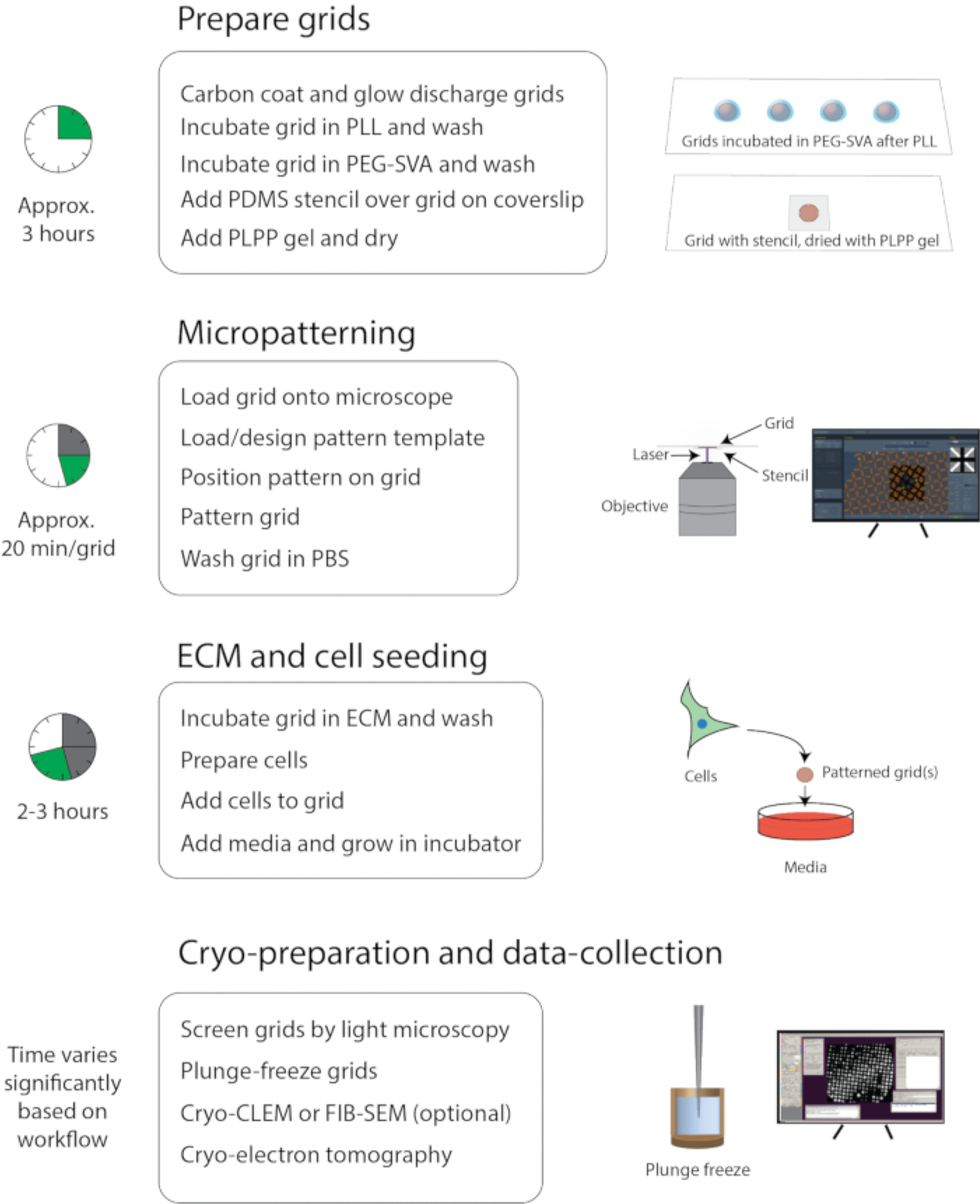
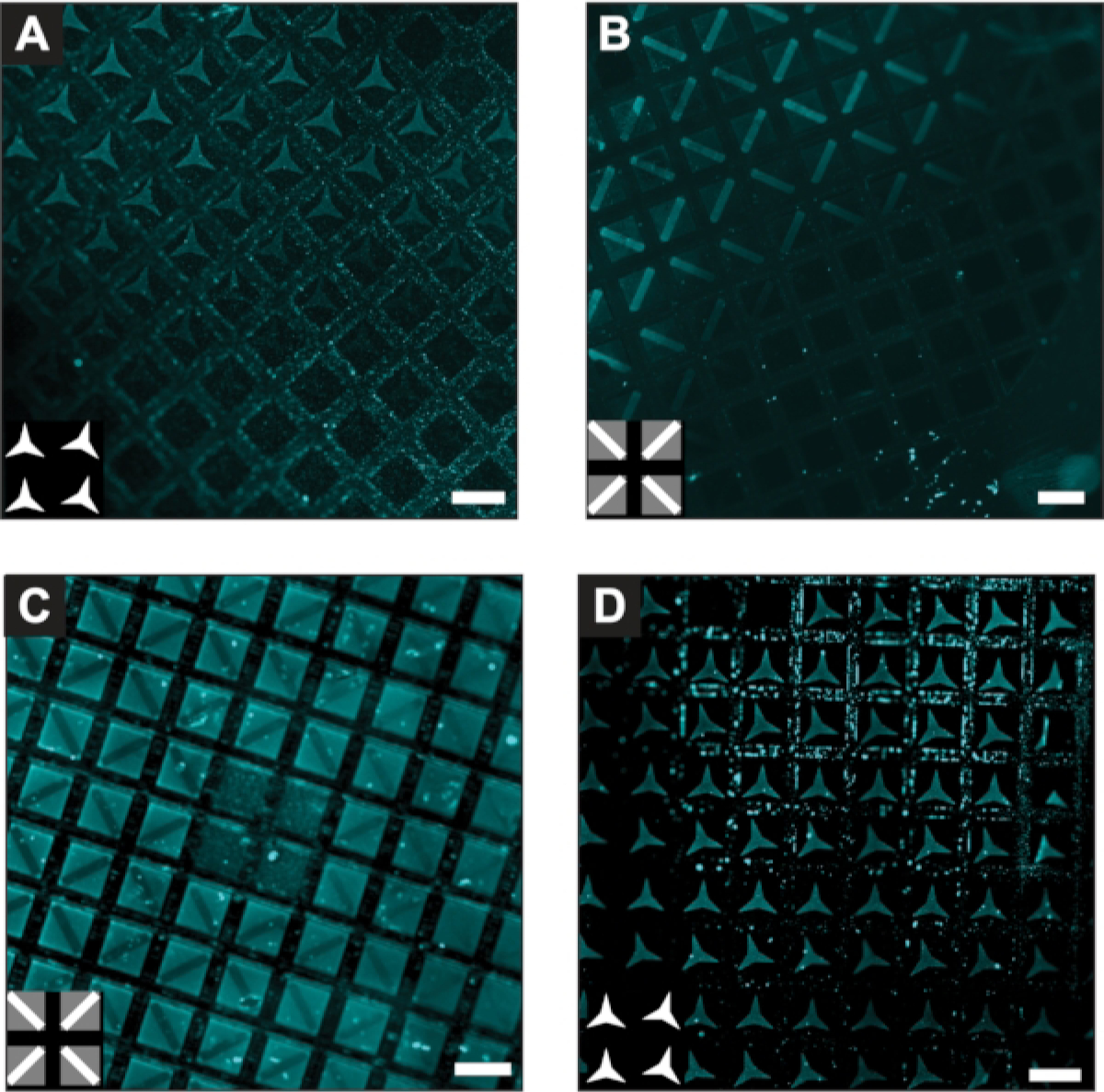
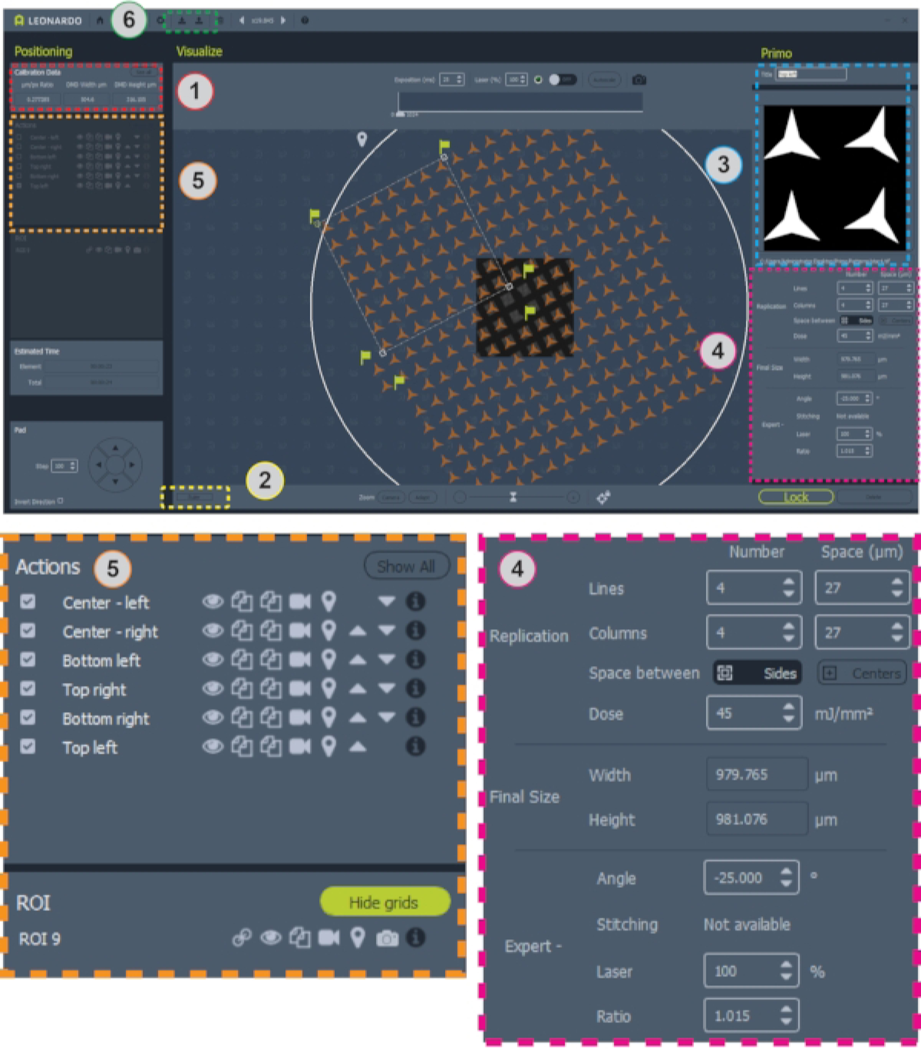
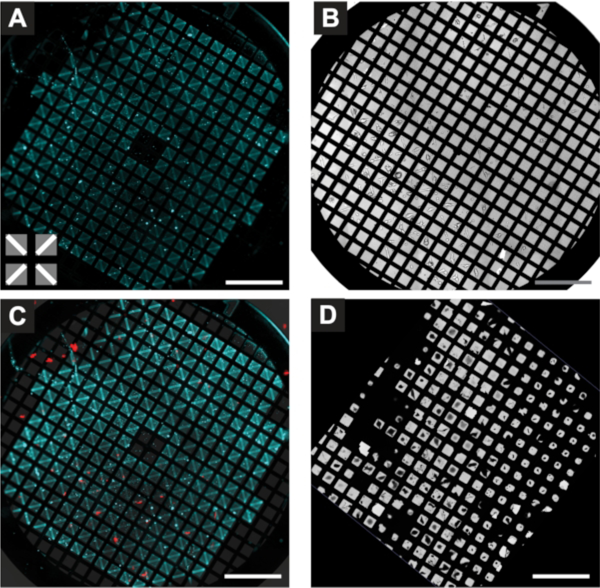
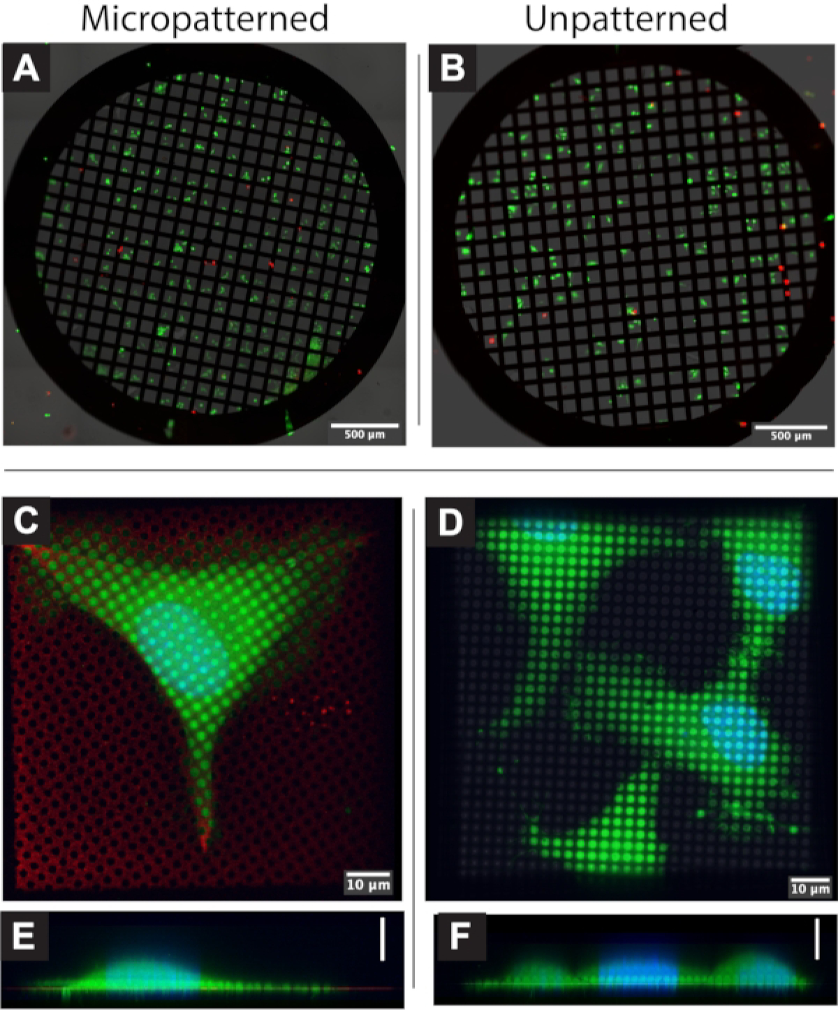
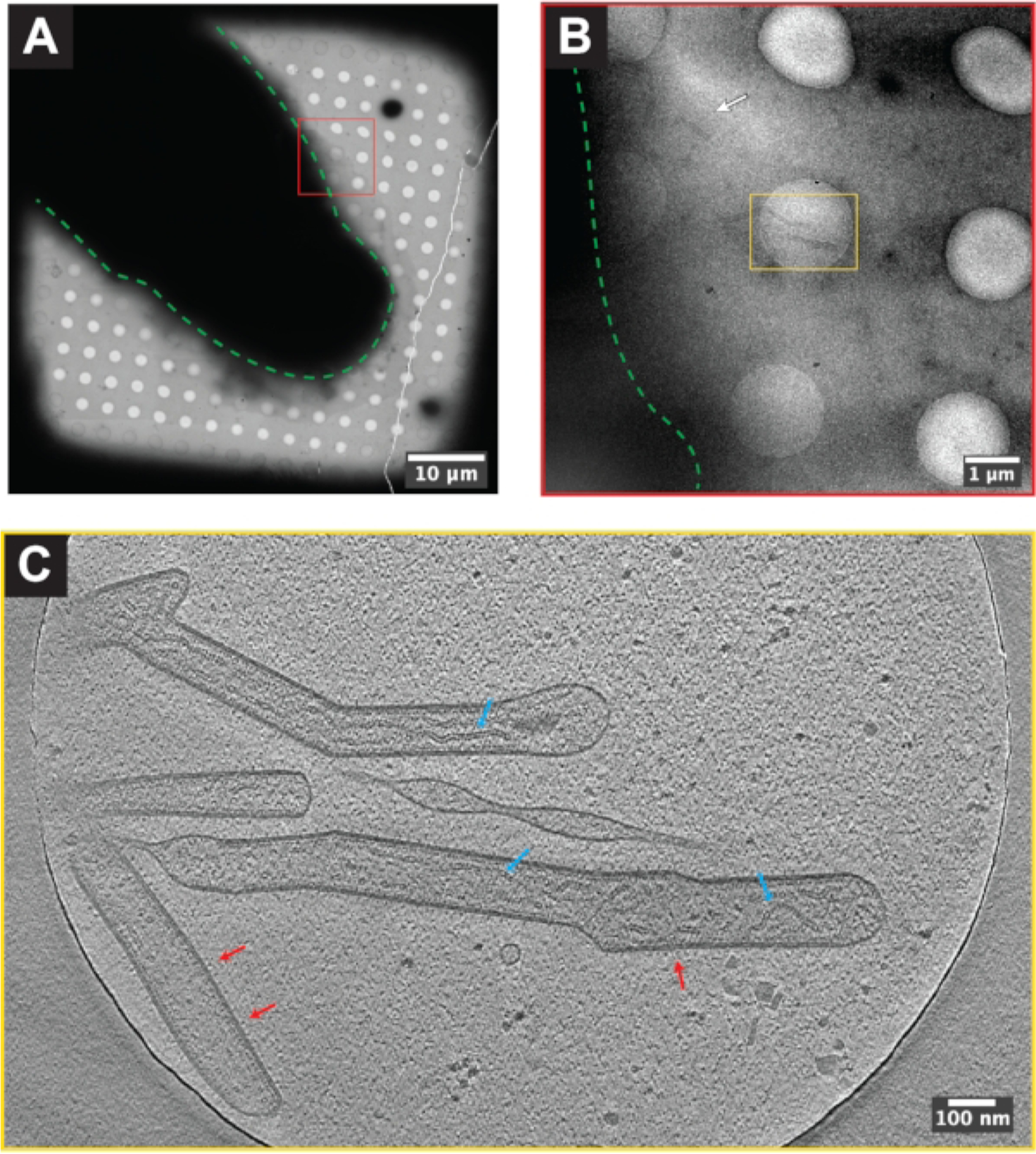
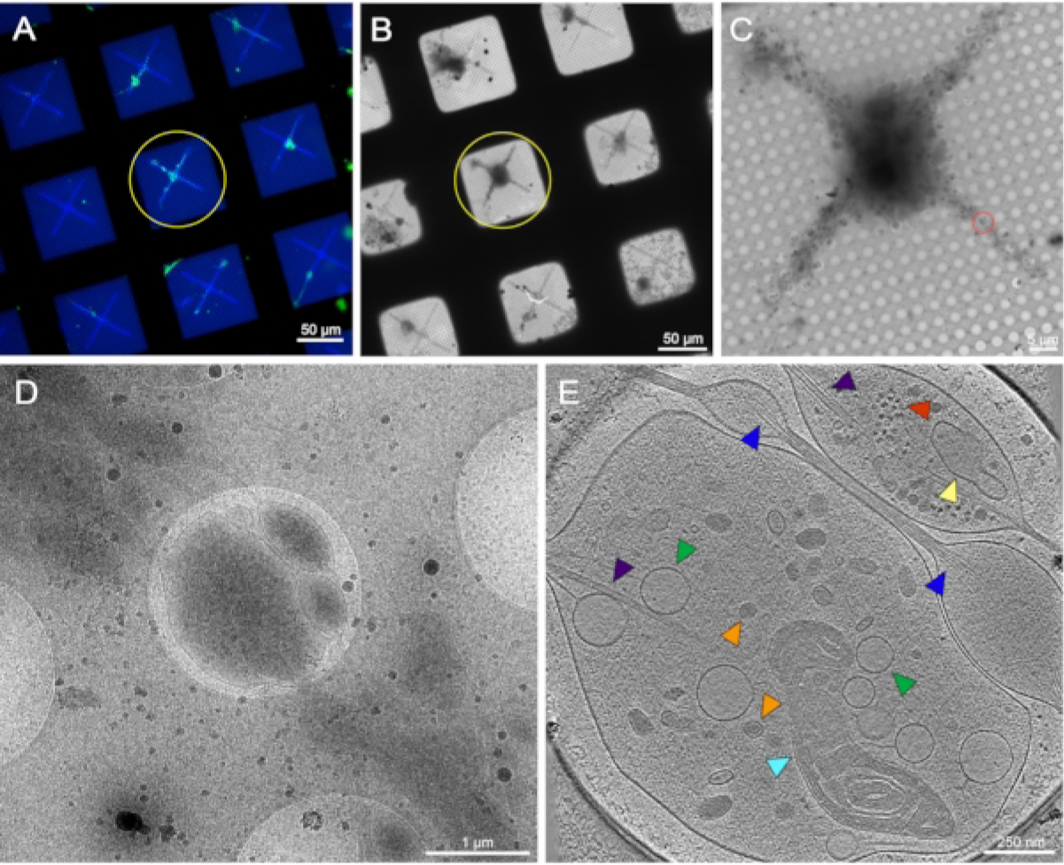
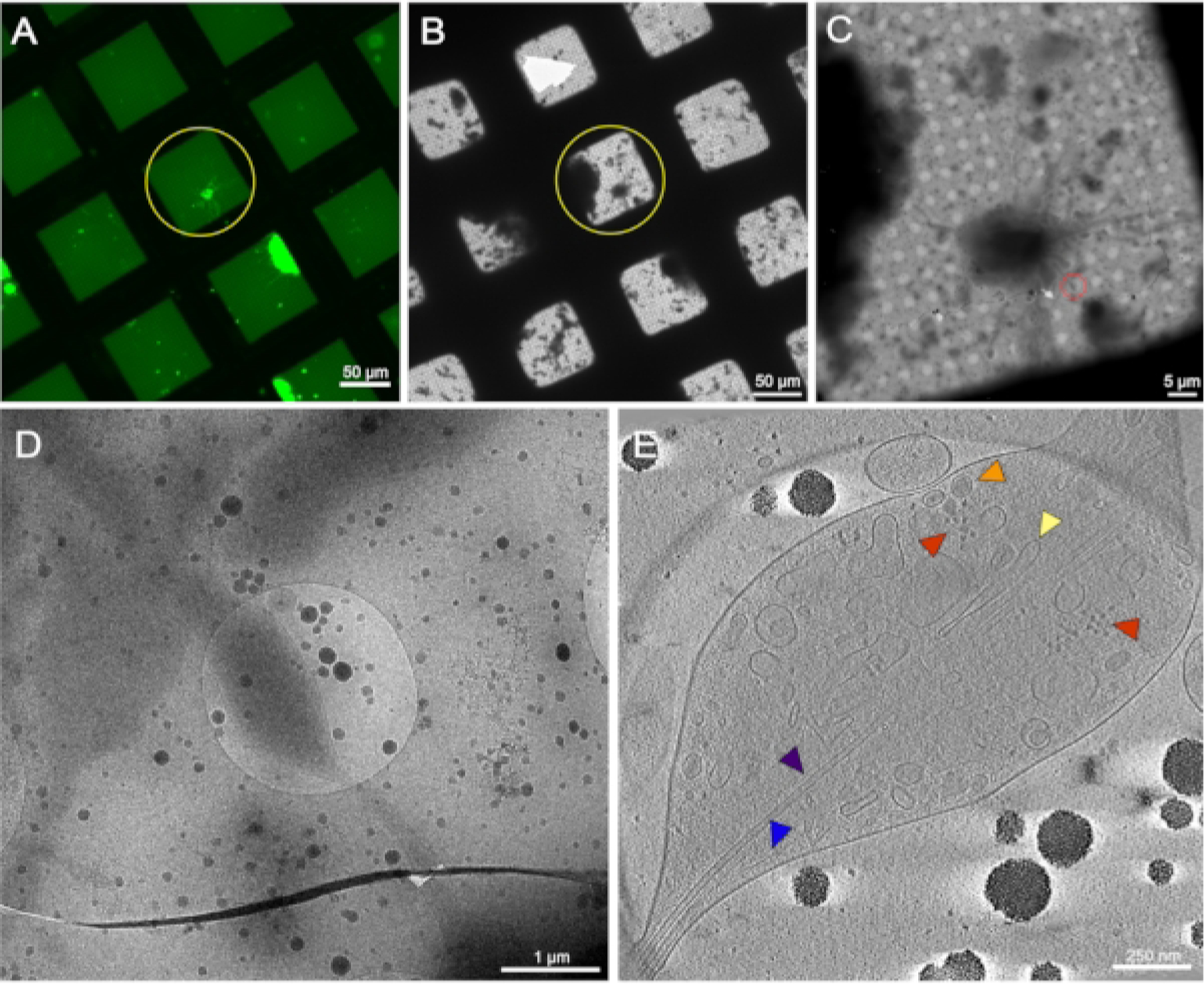
Whole-cell cryo-electron tomography (cryo-ET) is a powerful technology that is used to produce nanometer-level resolution structures of macromolecules present in the cellular context and preserved in a near-native frozen-hydrated state. However, there are challenges associated with culturing and/or adhering cells onto TEM grids in a manner that is suitable for tomography while retaining the cells in their physiological state. Here, a detailed step-by-step protocol is presented on the use of micropatterning to direct and promote eukaryotic cell growth on TEM grids. During micropatterning, cell growth is directed by depositing extra-cellular matrix (ECM) proteins within specified patterns and positions on the foil of the TEM grid while the other areas remain coated with an anti-fouling layer. Flexibility in the choice of surface coating and pattern design makes micropatterning broadly applicable for a wide range of cell types. Micropatterning is useful for studies of structures within individual cells as well as more complex experimental systems such as host-pathogen interactions or differentiated multi-cellular communities. Micropatterning may also be integrated into many downstream whole-cell cryo-ET workflows, including correlative light and electron microscopy (cryo-CLEM) and focused-ion beam milling (cryo-FIB).
全细胞冷冻电子断层扫描(cryo-ET)是一项强大的技术,用于生成细胞环境中存在的、处于近天然冷冻水合状态的大分子的纳米级分辨率结构。然而,以适合断层扫描的方式培养细胞和/或将细胞附着到透射电子显微镜(TEM)网格上,并使细胞保持其生理状态,存在一些挑战。在此,我们介绍了一种详细的分步方案,用于利用微图案化技术引导和促进真核细胞在TEM网格上生长。在微图案化过程中,通过在TEM网格箔片上特定的图案和位置沉积细胞外基质(ECM)蛋白来引导细胞生长,而其他区域则保持涂有防污层。表面涂层和图案设计选择的灵活性使得微图案化广泛适用于多种细胞类型。微图案化对于研究单个细胞内的结构以及更复杂的实验系统(如宿主-病原体相互作用或分化的多细胞群落)很有用。微图案化还可以集成到许多下游全细胞冷冻电子断层扫描工作流程中,包括相关光电子显微镜(cryo-CLEM)和聚焦离子束铣削(cryo-FIB)。