Laboratory of Neuroinflammation, Hospital Nacional de Paraplejicos, 45071 Toledo, Spain.
School of Biological & Chemical Sciences, Queen Mary University of London, London E1 4NS, UK.
Cells. 2021 Aug 28;10(9):2235. doi: 10.3390/cells10092235.
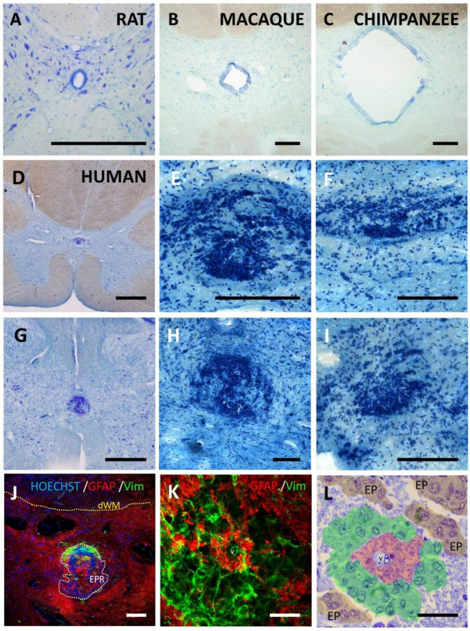
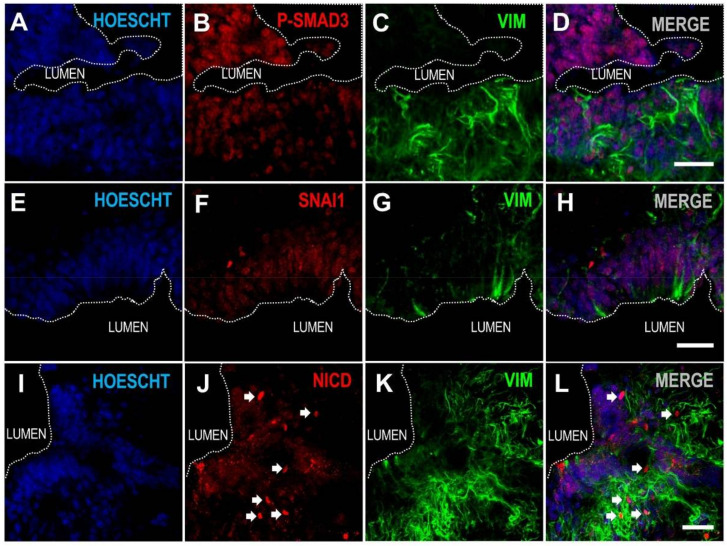
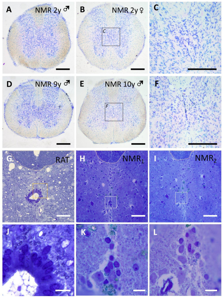
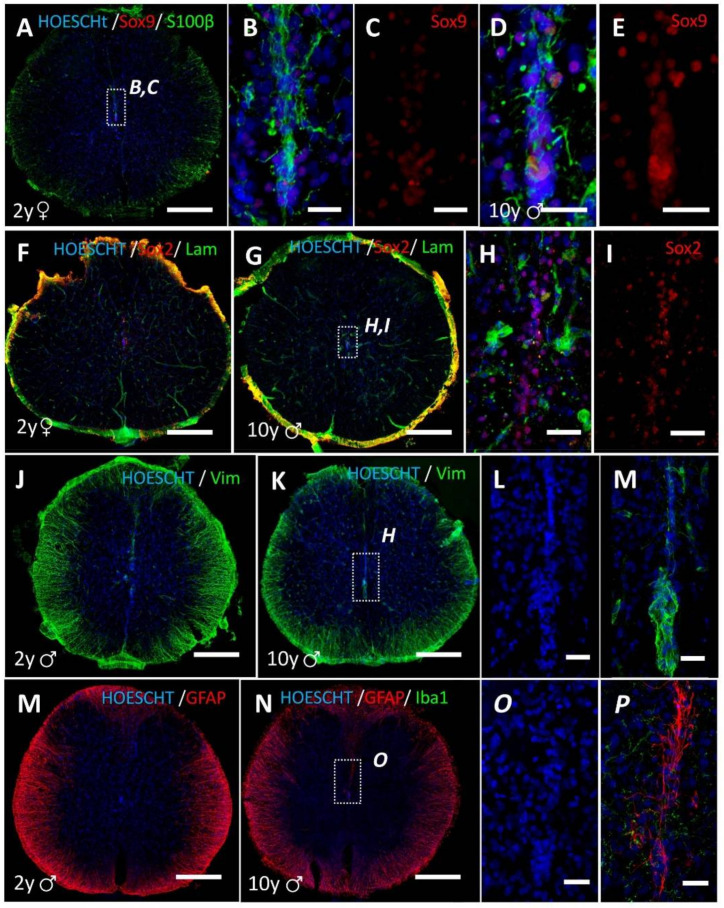
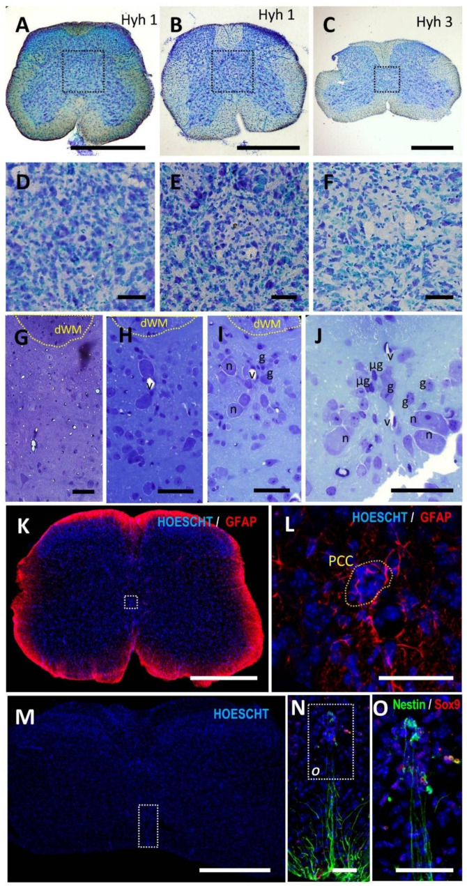
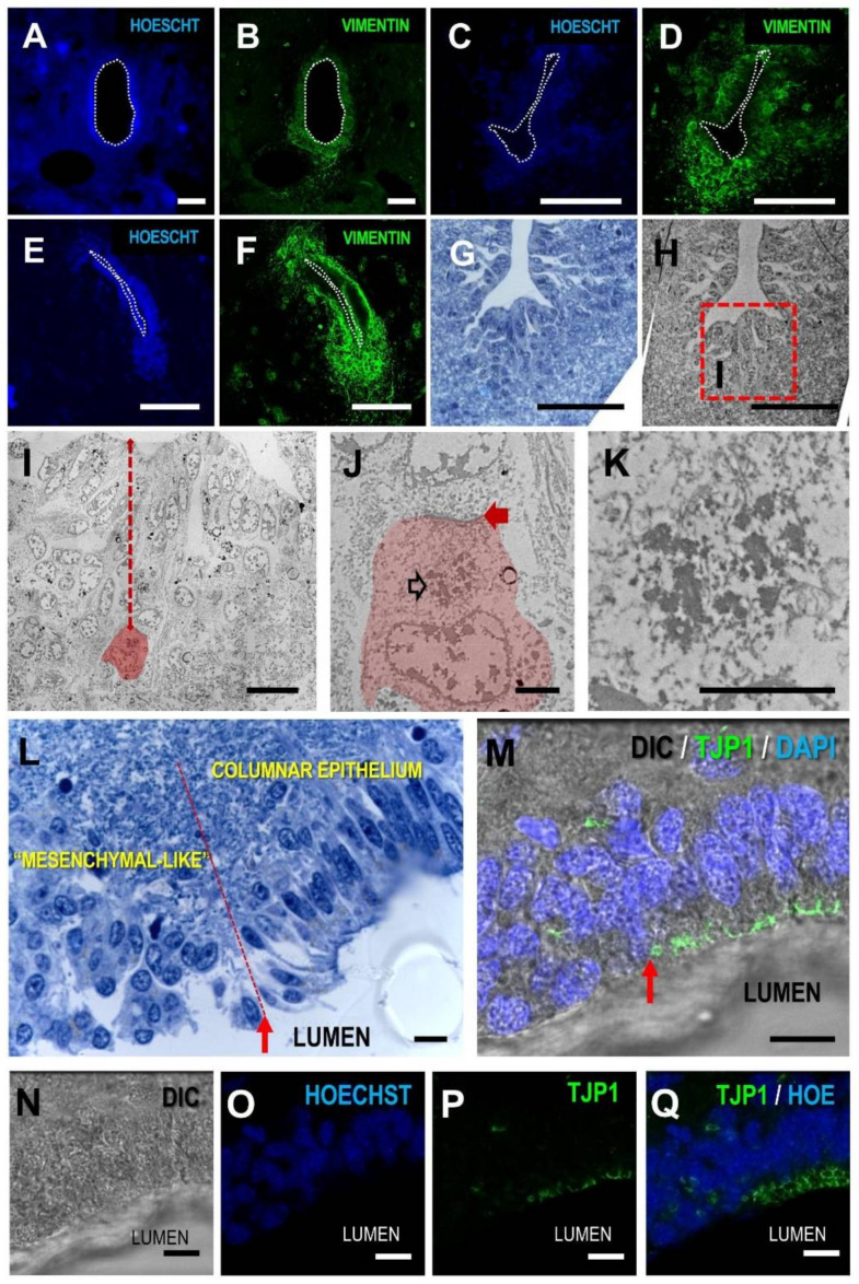
In species that regenerate the injured spinal cord, the ependymal region is a source of new cells and a prominent coordinator of regeneration. In mammals, cells at the ependymal region proliferate in normal conditions and react after injury, but in humans, the central canal is lost in the majority of individuals from early childhood. It is replaced by a structure that does not proliferate after damage and is formed by large accumulations of ependymal cells, strong astrogliosis and perivascular pseudo-rosettes. We inform here of two additional mammals that lose the central canal during their lifetime: the Naked Mole-Rat (NMR, ) and the mutant hyh () mice. The morphological study of their spinal cords shows that the tissue substituting the central canal is not similar to that found in humans. In both NMR and hyh mice, the central canal is replaced by tissue reminiscent of normal lamina X and may include small groups of ependymal cells in the midline, partially resembling specific domains of the former canal. However, no features of the adult human ependymal remnant are found, suggesting that this structure is a specific human trait. In order to shed some more light on the mechanism of human central canal closure, we provide new data suggesting that canal patency is lost by delamination of the ependymal epithelium, in a process that includes apical polarity loss and the expression of signaling mediators involved in epithelial to mesenchymal transitions.
在能够再生受损脊髓的物种中,室管膜区域是新细胞的来源,也是再生的主要协调者。在哺乳动物中,室管膜区域的细胞在正常情况下会增殖,并在受伤后发生反应,但在人类中,中央管在大多数个体从幼年早期开始就会丢失。它被一种不会在损伤后增殖的结构所取代,这种结构由大量的室管膜细胞、强烈的星形胶质增生和血管周围假玫瑰花结组成。我们在这里报告另外两种在其生命周期中失去中央管的哺乳动物:裸鼹鼠(Naked Mole-Rat,NMR)和突变型 hyh()小鼠。对它们脊髓的形态学研究表明,替代中央管的组织与在人类中发现的组织不同。在 NMR 和 hyh 小鼠中,中央管被类似于正常 X 层的组织所取代,其中可能包括中线的小群室管膜细胞,部分类似于前管的特定区域。然而,没有发现成人人类室管膜残余物的特征,这表明这种结构是人类特有的特征。为了进一步阐明人类中央管闭合的机制,我们提供了新的数据,表明中央管的通畅性是通过室管膜上皮的分层丧失而丧失的,这一过程包括顶端极性丧失和参与上皮-间质转化的信号介质的表达。