• 文献检索
  • 文档翻译
  • 深度研究
  • 学术资讯
  • Suppr Zotero 插件Zotero 插件
  • 邀请有礼
  • 套餐&价格
  • 历史记录
应用&插件
Suppr Zotero 插件Zotero 插件浏览器插件Mac 客户端Windows 客户端微信小程序
定价
高级版会员购买积分包购买API积分包
服务
文献检索文档翻译深度研究API 文档MCP 服务
关于我们
关于 Suppr公司介绍联系我们用户协议隐私条款
关注我们

Suppr 超能文献

核心技术专利:CN118964589B侵权必究
粤ICP备2023148730 号-1Suppr @ 2026

文献检索

告别复杂PubMed语法,用中文像聊天一样搜索,搜遍4000万医学文献。AI智能推荐,让科研检索更轻松。

立即免费搜索

文件翻译

保留排版,准确专业,支持PDF/Word/PPT等文件格式,支持 12+语言互译。

免费翻译文档

深度研究

AI帮你快速写综述,25分钟生成高质量综述,智能提取关键信息,辅助科研写作。

立即免费体验

成纤维细胞激活和心肌纤维化的机制:从 FB 特异性条件性小鼠模型中得到的经验教训。

Mechanisms of Fibroblast Activation and Myocardial Fibrosis: Lessons Learned from FB-Specific Conditional Mouse Models.

机构信息

Division of Cardiovascular Disease, The University of Alabama at Birmingham, Birmingham, AL 35294, USA.

School of Health Professions, The University of Alabama at Birmingham, Birmingham, AL 35294, USA.

出版信息

Cells. 2021 Sep 14;10(9):2412. doi: 10.3390/cells10092412.

DOI:10.3390/cells10092412
PMID:34572061
原文链接:https://pmc.ncbi.nlm.nih.gov/articles/PMC8471002/
Abstract

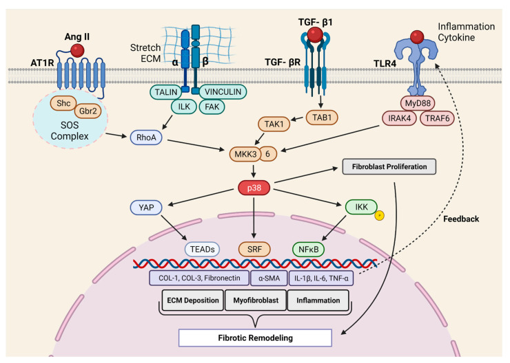
Heart failure (HF) is a leading cause of morbidity and mortality across the world. Cardiac fibrosis is associated with HF progression. Fibrosis is characterized by the excessive accumulation of extracellular matrix components. This is a physiological response to tissue injury. However, uncontrolled fibrosis leads to adverse cardiac remodeling and contributes significantly to cardiac dysfunction. Fibroblasts (FBs) are the primary drivers of myocardial fibrosis. However, until recently, FBs were thought to play a secondary role in cardiac pathophysiology. This review article will present the evolving story of fibroblast biology and fibrosis in cardiac diseases, emphasizing their recent shift from a supporting to a leading role in our understanding of the pathogenesis of cardiac diseases. Indeed, this story only became possible because of the emergence of FB-specific mouse models. This study includes an update on the advancements in the generation of FB-specific mouse models. Regarding the underlying mechanisms of myocardial fibrosis, we will focus on the pathways that have been validated using FB-specific, in vivo mouse models. These pathways include the TGF-β/SMAD3, p38 MAPK, Wnt/β-Catenin, G-protein-coupled receptor kinase (GRK), and Hippo signaling. A better understanding of the mechanisms underlying fibroblast activation and fibrosis may provide a novel therapeutic target for the management of adverse fibrotic remodeling in the diseased heart.

摘要

心力衰竭(HF)是全球发病率和死亡率的主要原因。心脏纤维化与 HF 进展有关。纤维化的特征是细胞外基质成分的过度积累。这是对组织损伤的生理反应。然而,不受控制的纤维化会导致不良的心脏重构,并对心脏功能障碍有重大贡献。成纤维细胞(FBs)是心肌纤维化的主要驱动因素。然而,直到最近,FBs 才被认为在心脏病理生理学中起次要作用。这篇综述文章将介绍成纤维细胞生物学和纤维化在心脏疾病中的发展历程,强调它们在我们对心脏疾病发病机制的理解中从辅助作用转变为主要作用。事实上,这个故事之所以成为可能,仅仅是因为 FB 特异性小鼠模型的出现。本研究包括对 FB 特异性小鼠模型生成进展的更新。关于心肌纤维化的潜在机制,我们将重点介绍使用 FB 特异性、体内小鼠模型验证的途径。这些途径包括 TGF-β/SMAD3、p38 MAPK、Wnt/β-Catenin、G 蛋白偶联受体激酶(GRK)和 Hippo 信号通路。更好地了解成纤维细胞激活和纤维化的机制可能为心脏疾病中不良纤维性重塑的管理提供新的治疗靶点。

https://cdn.ncbi.nlm.nih.gov/pmc/blobs/5907/8471002/5670f6cd5bc2/cells-10-02412-g004.jpg
https://cdn.ncbi.nlm.nih.gov/pmc/blobs/5907/8471002/620fb0f88532/cells-10-02412-g001.jpg
https://cdn.ncbi.nlm.nih.gov/pmc/blobs/5907/8471002/53d208c86d6c/cells-10-02412-g002.jpg
https://cdn.ncbi.nlm.nih.gov/pmc/blobs/5907/8471002/55b25f9a7a02/cells-10-02412-g003.jpg
https://cdn.ncbi.nlm.nih.gov/pmc/blobs/5907/8471002/5670f6cd5bc2/cells-10-02412-g004.jpg

相似文献

1
Mechanisms of Fibroblast Activation and Myocardial Fibrosis: Lessons Learned from FB-Specific Conditional Mouse Models.成纤维细胞激活和心肌纤维化的机制:从 FB 特异性条件性小鼠模型中得到的经验教训。
Cells. 2021 Sep 14;10(9):2412. doi: 10.3390/cells10092412.
2
Cardiac Fibrosis: The Fibroblast Awakens.心脏纤维化:成纤维细胞觉醒
Circ Res. 2016 Mar 18;118(6):1021-40. doi: 10.1161/CIRCRESAHA.115.306565.
3
SKI activates the Hippo pathway via LIMD1 to inhibit cardiac fibroblast activation.SKI 通过 LIMD1 激活 Hippo 通路抑制心肌成纤维细胞的激活。
Basic Res Cardiol. 2021 Apr 13;116(1):25. doi: 10.1007/s00395-021-00865-9.
4
Modeling Cardiac Fibrosis in Mice: (Myo)Fibroblast Phenotype After Ischemia.小鼠心脏纤维化建模:缺血后的(肌)成纤维细胞表型
Methods Mol Biol. 2017;1627:123-137. doi: 10.1007/978-1-4939-7113-8_9.
5
Pharmacological and Activated Fibroblast Targeting of Gβγ-GRK2 After Myocardial Ischemia Attenuates Heart Failure Progression.心肌缺血后对Gβγ-GRK2进行药理学和靶向活化成纤维细胞治疗可减轻心力衰竭进展。
J Am Coll Cardiol. 2017 Aug 22;70(8):958-971. doi: 10.1016/j.jacc.2017.06.049.
6
Nuclear Receptor Nur77 Controls Cardiac Fibrosis through Distinct Actions on Fibroblasts and Cardiomyocytes.核受体 Nur77 通过对成纤维细胞和心肌细胞的不同作用控制心脏纤维化。
Int J Mol Sci. 2021 Feb 5;22(4):1600. doi: 10.3390/ijms22041600.
7
Entanglement of GSK-3β, β-catenin and TGF-β1 signaling network to regulate myocardial fibrosis.GSK-3β、β-连环蛋白和TGF-β1信号网络的相互作用调控心肌纤维化
J Mol Cell Cardiol. 2017 Sep;110:109-120. doi: 10.1016/j.yjmcc.2017.07.011. Epub 2017 Jul 27.
8
Fibroblast-specific TGF-β-Smad2/3 signaling underlies cardiac fibrosis.成纤维细胞特异性转化生长因子-β- Smad2/3信号传导是心脏纤维化的基础。
J Clin Invest. 2017 Oct 2;127(10):3770-3783. doi: 10.1172/JCI94753. Epub 2017 Sep 11.
9
Featured Article: TGF-β1 dominates extracellular matrix rigidity for inducing differentiation of human cardiac fibroblasts to myofibroblasts.特色文章:TGF-β1 通过控制细胞外基质硬度诱导人心肌成纤维细胞分化为肌成纤维细胞。
Exp Biol Med (Maywood). 2018 Apr;243(7):601-612. doi: 10.1177/1535370218761628. Epub 2018 Mar 4.
10
Inhibition of autophagy inhibits the conversion of cardiac fibroblasts to cardiac myofibroblasts.自噬的抑制会抑制心脏成纤维细胞向心肌成纤维细胞的转化。
Oncotarget. 2016 Nov 29;7(48):78516-78531. doi: 10.18632/oncotarget.12392.

引用本文的文献

1
Amphiregulin in Fibrotic Diseases and Cancer.双调蛋白在纤维化疾病和癌症中的作用
Int J Mol Sci. 2025 Jul 19;26(14):6945. doi: 10.3390/ijms26146945.
2
Morphometric and Molecular Interplay in Hypertension-Induced Cardiac Remodeling with an Emphasis on the Potential Therapeutic Implications.高血压诱导的心脏重塑中的形态计量学与分子相互作用:着重探讨潜在治疗意义
Int J Mol Sci. 2025 Apr 24;26(9):4022. doi: 10.3390/ijms26094022.
3
Low-density lipoprotein receptor-related protein 6 ameliorates cardiac hypertrophy by regulating CTSD/HSP90α signaling during pressure overload.

本文引用的文献

1
Repurposing Nintedanib for pathological cardiac remodeling and dysfunction.尼达尼布在病理性心脏重构和功能障碍中的再利用。
Pharmacol Res. 2021 Jul;169:105605. doi: 10.1016/j.phrs.2021.105605. Epub 2021 May 13.
2
The double face of miR-320: cardiomyocytes-derived miR-320 deteriorated while fibroblasts-derived miR-320 protected against heart failure induced by transverse aortic constriction.miR-320 的双重面孔:心肌细胞来源的 miR-320 恶化,而成纤维细胞来源的 miR-320 可预防由主动脉缩窄引起的心力衰竭。
Signal Transduct Target Ther. 2021 Feb 18;6(1):69. doi: 10.1038/s41392-020-00445-8.
3
AMPKα1 deletion in myofibroblasts exacerbates post-myocardial infarction fibrosis by a connexin 43 mechanism.
低密度脂蛋白受体相关蛋白6在压力过载期间通过调节CTSD/HSP90α信号通路改善心脏肥大。
Acta Pharmacol Sin. 2025 Mar;46(3):606-617. doi: 10.1038/s41401-024-01415-4. Epub 2025 Jan 8.
4
Wnt Signaling Inhibitors as Therapeutic Approach in Ischemic Heart Disease.Wnt信号通路抑制剂作为缺血性心脏病的治疗方法
Molecules. 2024 Dec 17;29(24):5958. doi: 10.3390/molecules29245958.
5
Free-breathing non-contrast T1ρ dispersion magnetic resonance imaging of myocardial interstitial fibrosis in comparison with extracellular volume fraction.自由呼吸非对比T1ρ弥散磁共振成像评估心肌间质纤维化与细胞外容积分数的比较
J Cardiovasc Magn Reson. 2024;26(2):101093. doi: 10.1016/j.jocmr.2024.101093. Epub 2024 Sep 6.
6
Pharmacological mechanisms by which baicalin ameliorates cardiovascular disease.黄芩苷改善心血管疾病的药理机制。
Front Pharmacol. 2024 Aug 9;15:1415971. doi: 10.3389/fphar.2024.1415971. eCollection 2024.
7
Evaluation of the Canonical Wnt Signaling Pathway in the Hearts of Hypertensive Rats of Various Etiologies.评价各种病因高血压大鼠心脏中的经典 Wnt 信号通路。
Int J Mol Sci. 2024 Jun 11;25(12):6428. doi: 10.3390/ijms25126428.
8
Fibroblasts orchestrate cellular crosstalk in the heart through the ECM.成纤维细胞通过细胞外基质协调心脏中的细胞间相互作用。
Nat Cardiovasc Res. 2022 Apr;1(4):312-321. doi: 10.1038/s44161-022-00043-7. Epub 2022 Apr 13.
9
In Vitro Assessment of Cardiac Fibroblast Activation at Physiologic Stiffness.在生理弹性下评估心脏成纤维细胞的激活
Curr Protoc. 2024 Apr;4(4):e1025. doi: 10.1002/cpz1.1025.
10
Cardioprotective Effects of the GRK2 Inhibitor Paroxetine on Isoproterenol-Induced Cardiac Remodeling by Modulating NF-κB Mediated Prohypertrophic and Profibrotic Gene Expression.帕罗西汀通过调节 NF-κB 介导的心肌肥厚和纤维化相关基因表达对异丙肾上腺素诱导的心脏重构的心脏保护作用。
Int J Mol Sci. 2023 Dec 8;24(24):17270. doi: 10.3390/ijms242417270.
肌成纤维细胞中 AMPKα1 的缺失通过缝隙连接蛋白 43 机制加剧心肌梗死后的纤维化。
Basic Res Cardiol. 2021 Feb 9;116(1):10. doi: 10.1007/s00395-021-00846-y.
4
GRK5 is a regulator of fibroblast activation and cardiac fibrosis.GRK5 是成纤维细胞活化和心脏纤维化的调节因子。
Proc Natl Acad Sci U S A. 2021 Feb 2;118(5). doi: 10.1073/pnas.2012854118.
5
Targeting 5-HT Receptor Signaling Prevents Border Zone Expansion and Improves Microstructural Remodeling After Myocardial Infarction.靶向 5-羟色胺受体信号可防止心肌梗死后边缘区扩张并改善结构重塑。
Circulation. 2021 Mar 30;143(13):1317-1330. doi: 10.1161/CIRCULATIONAHA.120.051517. Epub 2021 Jan 21.
6
β2-Adrenergic Receptors Increase Cardiac Fibroblast Proliferation Through the Gαs/ERK1/2-Dependent Secretion of Interleukin-6.β2-肾上腺素能受体通过 Gαs/ERK1/2 依赖性分泌白细胞介素-6 增加心肌成纤维细胞增殖。
Int J Mol Sci. 2020 Nov 12;21(22):8507. doi: 10.3390/ijms21228507.
7
Blockade of Fibroblast YAP Attenuates Cardiac Fibrosis and Dysfunction Through MRTF-A Inhibition.成纤维细胞YAP的阻断通过抑制MRTF-A减轻心脏纤维化和功能障碍。
JACC Basic Transl Sci. 2020 Sep 28;5(9):931-945. doi: 10.1016/j.jacbts.2020.07.009. eCollection 2020 Sep.
8
Transforming growth factor-β in tissue fibrosis.组织纤维化中的转化生长因子-β。
J Exp Med. 2020 Feb 13;217(3):e20190103. doi: 10.1084/jem.20190103. Print 2020 Mar 2.
9
Infarct Collagen Topography Regulates Fibroblast Fate via p38-Yes-Associated Protein Transcriptional Enhanced Associate Domain Signals.梗死胶原形貌通过 p38-Yes 相关蛋白转录增强相关结构域信号调节成纤维细胞命运。
Circ Res. 2020 Oct 23;127(10):1306-1322. doi: 10.1161/CIRCRESAHA.119.316162. Epub 2020 Sep 4.
10
Cardiac Fibroblast Diversity.心肌成纤维细胞多样性。
Annu Rev Physiol. 2020 Feb 10;82:63-78. doi: 10.1146/annurev-physiol-021119-034527.