Jeican Ionuț Isaia, Gheban Dan, Barbu-Tudoran Lucian, Inișca Patricia, Albu Camelia, Ilieș Maria, Albu Silviu, Vică Mihaela Laura, Matei Horea Vladi, Tripon Septimiu, Lazăr Mihaela, Aluaș Maria, Siserman Costel Vasile, Muntean Monica, Trombitas Veronica, Iuga Cristina Adela, Opincariu Iulian, Junie Lia Monica
Department of Head and Neck Surgery and Otorhinolaryngology, University Clinical Hospital of Railway Company, Iuliu Hatieganu University of Medicine and Pharmacy, 400015 Cluj-Napoca, Romania.
Department of Anatomy and Embryology, Iuliu Hatieganu University of Medicine and Pharmacy, 400006 Cluj-Napoca, Romania.
J Clin Med. 2021 Sep 12;10(18):4110. doi: 10.3390/jcm10184110.
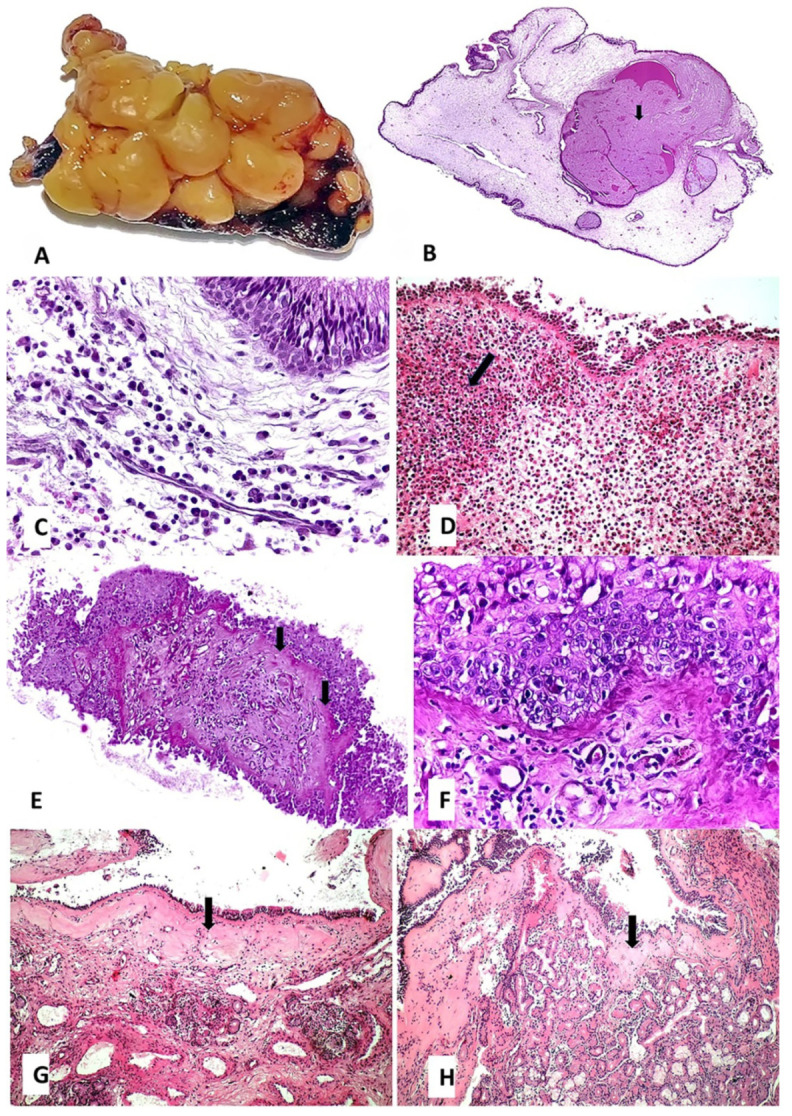
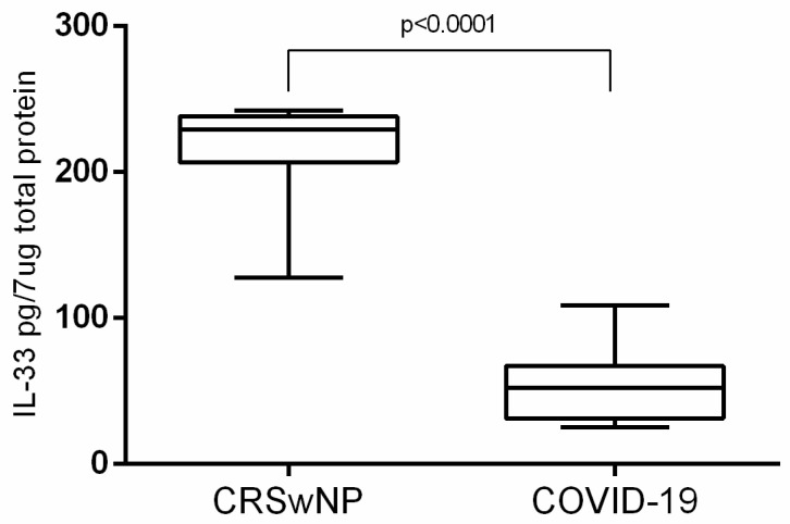
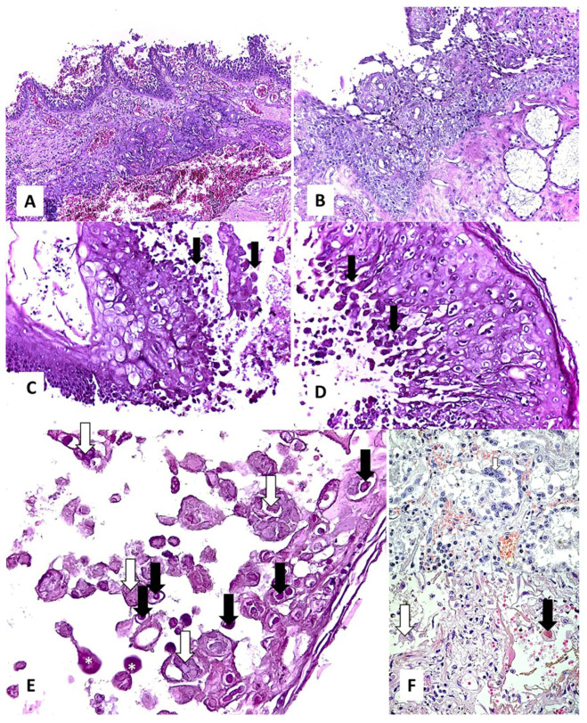
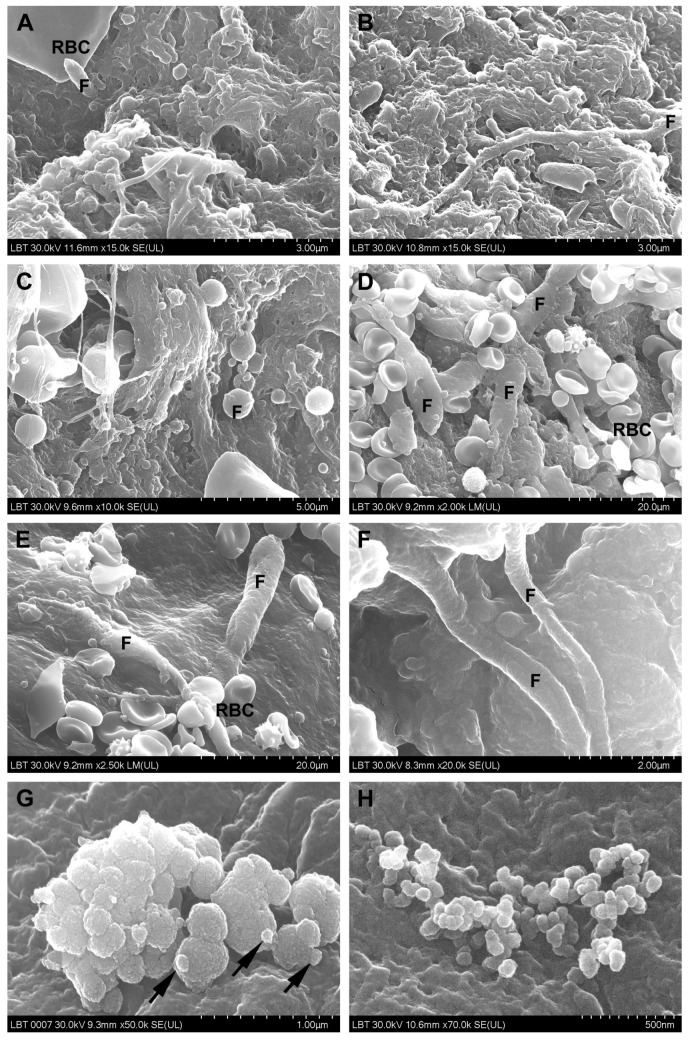
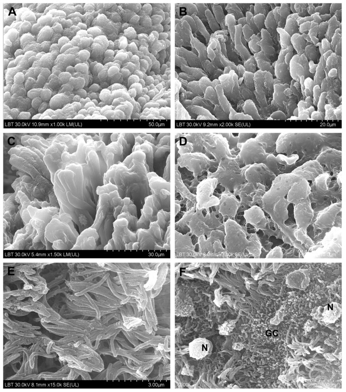
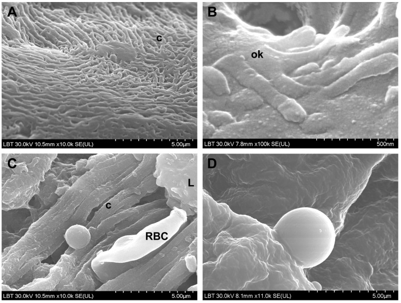
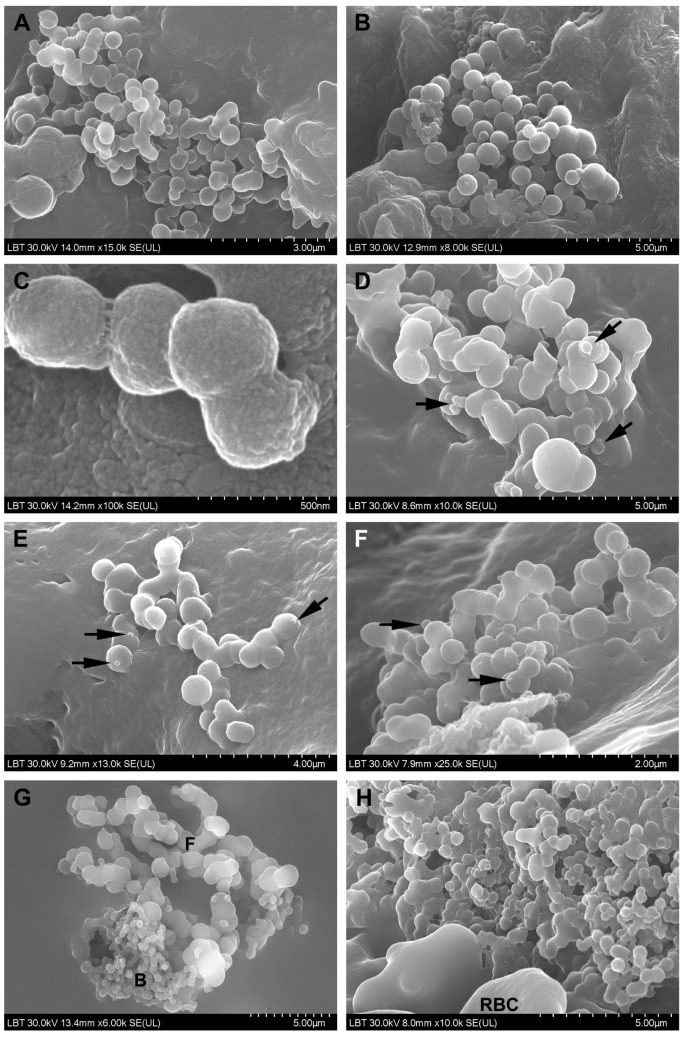
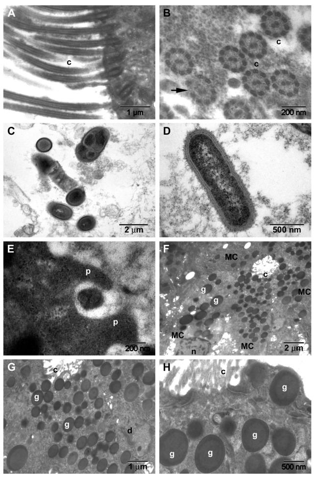
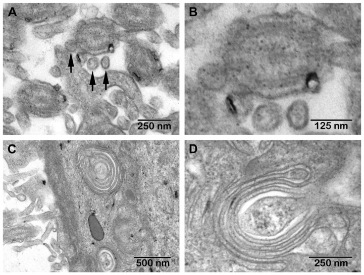
(1) Background: Chronic rhinosinusitis with nasal polyps (CRSwNP) is one of the most studied rhinological disorders. Modifications of the respiratory nasal mucosa in COVID-19 patients are so far unknown. This paper presents a comparative morphological characterization of the respiratory nasal mucosa in CRSwNP versus COVID-19 and tissue interleukin (IL)-33 concentration. (2) Methods: We analyzed CRSwNP and COVID-19 samples through histopathology, scanning and transmission electron microscopy and performed proteomic determination of IL-33. (3) Results: Histopathologically, stromal edema ( < 0.0001) and basal membrane thickening ( = 0.0768) were found more frequently in CRSwNP than in COVID-19. Inflammatory infiltrate was mainly eosinophil-dominant in CRSwNP and lymphocyte-dominant in COVID-19 ( = 0.3666). A viral cytopathic effect was identified in COVID-19. Scanning electron microscopy detected biofilms only in CRSwNP, while most COVID-19 samples showed microbial aggregates ( = 0.0148) and immune cells ( = 0.1452). Transmission electron microscopy of CRSwNP samples identified biofilms, mucous cell hyperplasia ( = 0.0011), eosinophils, fibrocytes, mastocytes, and collagen fibers. Extracellular suggestive structures for SARS-CoV-2 and multiple Golgi apparatus in epithelial cells were detected in COVID-19 samples. The tissue IL-33 concentration in CRSwNP (210.0 pg/7 μg total protein) was higher than in COVID-19 (52.77 pg/7 μg total protein) ( < 0.0001), also suggesting a different inflammatory pattern. (4) Conclusions: The inflammatory pattern is different in each of these disorders. Results suggested the presence of nasal dysbiosis in both conditions, which could be a determining factor in CRSwNP and a secondary factor in COVID-19.
(1) 背景:伴鼻息肉的慢性鼻-鼻窦炎(CRSwNP)是研究最多的鼻科疾病之一。目前尚不清楚新冠病毒病(COVID-19)患者鼻呼吸黏膜的改变情况。本文呈现了CRSwNP与COVID-19患者鼻呼吸黏膜的比较形态学特征以及组织白细胞介素(IL)-33浓度。(2) 方法:我们通过组织病理学、扫描和透射电子显微镜分析了CRSwNP和COVID-19样本,并对IL-33进行了蛋白质组学测定。(3) 结果:组织病理学上,CRSwNP中基质水肿(<0.0001)和基底膜增厚(=0.0768)的发生率高于COVID-19。CRSwNP中的炎性浸润以嗜酸性粒细胞为主,而COVID-19中以淋巴细胞为主(=0.3666)。在COVID-19中发现了病毒细胞病变效应。扫描电子显微镜仅在CRSwNP中检测到生物膜,而大多数COVID-19样本显示有微生物聚集体(=0.0148)和免疫细胞(=0.1452)。CRSwNP样本的透射电子显微镜检查发现了生物膜、黏液细胞增生(=0.0011)、嗜酸性粒细胞、纤维细胞、肥大细胞和胶原纤维。在COVID-19样本中检测到上皮细胞内提示为严重急性呼吸综合征冠状病毒2(SARS-CoV-2)的结构和多个高尔基体。CRSwNP中的组织IL-33浓度(210.0 pg/7μg总蛋白)高于COVID-19(52.77 pg/7μg总蛋白)(<0.0001),这也提示了不同的炎症模式。(4) 结论:这些疾病中的每一种炎症模式都不同。结果表明这两种情况均存在鼻生态失调,这可能是CRSwNP的决定因素,而在COVID-19中是次要因素。