Lee Chung-Fei, Hsu Yung-Heng, Lin Yu-Chien, Nguyen Thu-Trang, Chen Hsiang-Wen, Nabilla Sasza Chyntara, Hou Shao-Yi, Chang Feng-Cheng, Chung Ren-Jei
Department of Chemical Engineering and Biotechnology, National Taipei University of Technology (Taipei Tech.), Taipei 10608, Taiwan.
Bone and Joint Research Center, Chang Gung Memorial Hospital, Linko 33305, Taiwan.
Polymers (Basel). 2021 Sep 16;13(18):3123. doi: 10.3390/polym13183123.
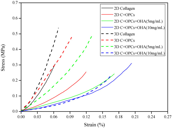
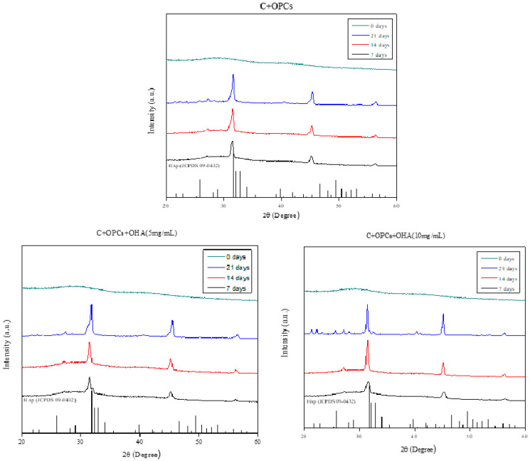
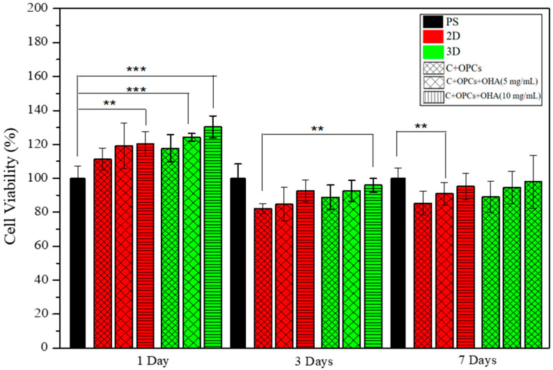
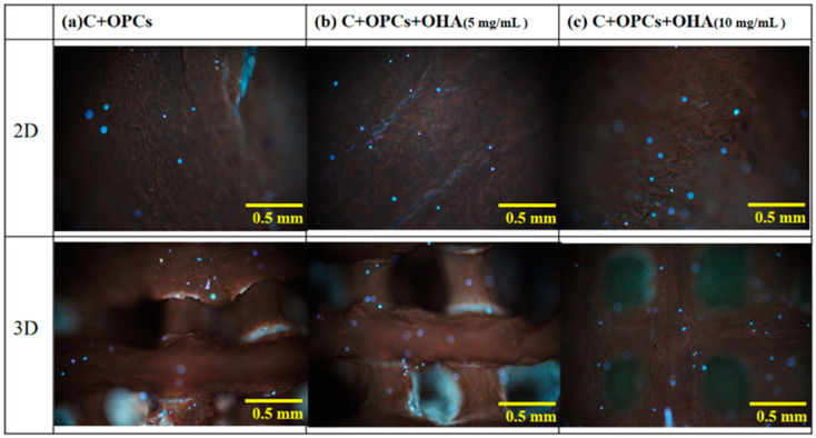
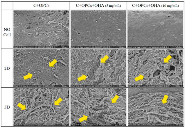
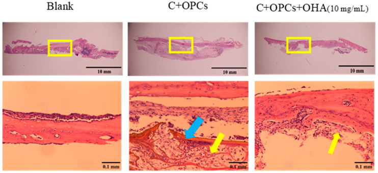
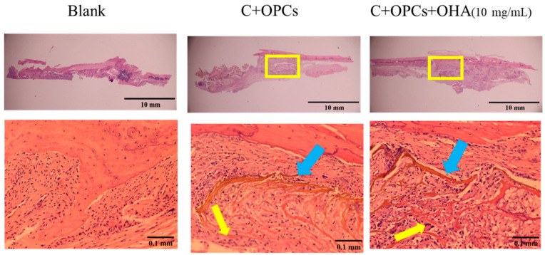
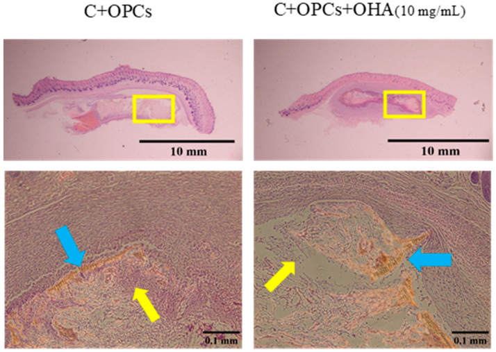
Articular cartilage defects affect millions of people worldwide, including children, adolescents, and adults. Progressive wear and tear of articular cartilage can lead to progressive tissue loss, further exposing the bony ends and leaving them unprotected, which may ultimately cause osteoarthritis (degenerative joint disease). Unlike other self-repairing tissues, cartilage has a low regenerative capacity; once injured, the cartilage is much more difficult to heal. Consequently, developing methods to repair this defect remains a challenge in clinical practice. In recent years, tissue engineering applications have employed the use of three-dimensional (3D) porous scaffolds for growing cells to regenerate damaged cartilage. However, these scaffolds are mainly chemically synthesized polymers or are crosslinked using organic solvents. Utilizing 3D printing technologies to prepare biodegradable natural composite scaffolds could replace chemically synthesized polymers with more natural polymers or low-toxicity crosslinkers. In this study, collagen/oligomeric proanthocyanidin/oxidized hyaluronic acid composite scaffolds showing high biocompatibility and excellent mechanical properties were prepared. The compressive strengths of the scaffolds were between 0.25-0.55 MPa. Cell viability of the 3D scaffolds reached up to 90%, which indicates that they are favorable surfaces for the deposition of apatite. An in vivo test was performed using the Sprague Dawley (SD) rat skull model. Histological images revealed signs of angiogenesis and new bone formation. Therefore, 3D collagen-based scaffolds can be used as potential candidates for articular cartilage repair.
关节软骨缺损影响着全球数百万人,包括儿童、青少年和成年人。关节软骨的渐进性磨损会导致组织逐渐流失,进一步暴露骨端且使其失去保护,这最终可能导致骨关节炎(退行性关节疾病)。与其他自我修复组织不同,软骨的再生能力较低;一旦受损,软骨愈合起来要困难得多。因此,开发修复这种缺损的方法在临床实践中仍然是一项挑战。近年来,组织工程应用采用三维(3D)多孔支架来培养细胞以再生受损软骨。然而,这些支架主要是化学合成聚合物或使用有机溶剂交联而成。利用3D打印技术制备可生物降解的天然复合支架可以用更天然的聚合物或低毒性交联剂取代化学合成聚合物。在本研究中,制备了具有高生物相容性和优异力学性能的胶原蛋白/低聚原花青素/氧化透明质酸复合支架。这些支架的抗压强度在0.25 - 0.55兆帕之间。3D支架的细胞活力高达90%,这表明它们是磷灰石沉积的良好表面。使用斯普拉格·道利(SD)大鼠颅骨模型进行了体内试验。组织学图像显示有血管生成和新骨形成的迹象。因此,基于3D胶原蛋白的支架可作为关节软骨修复的潜在候选材料。