• 文献检索
  • 文档翻译
  • 深度研究
  • 学术资讯
  • Suppr Zotero 插件Zotero 插件
  • 邀请有礼
  • 套餐&价格
  • 历史记录
应用&插件
Suppr Zotero 插件Zotero 插件浏览器插件Mac 客户端Windows 客户端微信小程序
定价
高级版会员购买积分包购买API积分包
服务
文献检索文档翻译深度研究API 文档MCP 服务
关于我们
关于 Suppr公司介绍联系我们用户协议隐私条款
关注我们

Suppr 超能文献

核心技术专利:CN118964589B侵权必究
粤ICP备2023148730 号-1Suppr @ 2026

文献检索

告别复杂PubMed语法,用中文像聊天一样搜索,搜遍4000万医学文献。AI智能推荐,让科研检索更轻松。

立即免费搜索

文件翻译

保留排版,准确专业,支持PDF/Word/PPT等文件格式,支持 12+语言互译。

免费翻译文档

深度研究

AI帮你快速写综述,25分钟生成高质量综述,智能提取关键信息,辅助科研写作。

立即免费体验

KDM6A 通过非经典激活素途径调节细胞可塑性和胰腺癌进展。

KDM6A Regulates Cell Plasticity and Pancreatic Cancer Progression by Noncanonical Activin Pathway.

机构信息

Department of Pathology & Clinical Labs, University of Michigan, Ann Arbor, Michigan; Xiangya Hospital, Central South University, Changsha, Hunan, People's Republic of China.

Proteios Technology, Seattle, Washington.

出版信息

Cell Mol Gastroenterol Hepatol. 2022;13(2):643-667. doi: 10.1016/j.jcmgh.2021.09.014. Epub 2021 Sep 25.

DOI:10.1016/j.jcmgh.2021.09.014
PMID:34583087
原文链接:https://pmc.ncbi.nlm.nih.gov/articles/PMC8715196/
Abstract

BACKGROUND & AIMS: Inactivating mutations of KDM6A, a histone demethylase, were frequently found in pancreatic ductal adenocarcinoma (PDAC). We investigated the role of KDM6A (lysine demethylase 6A) in PDAC development.

METHODS

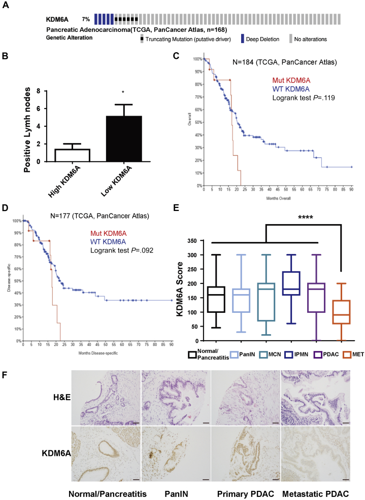
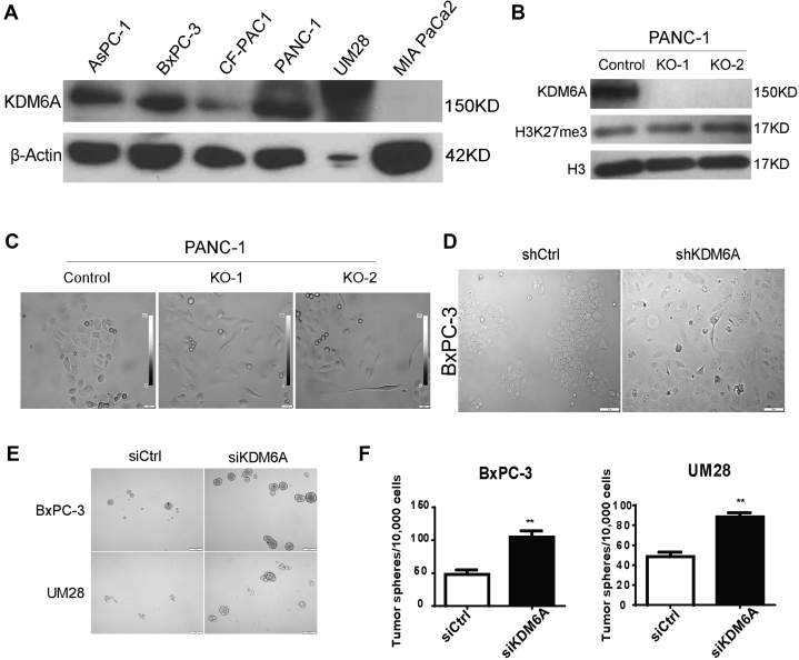
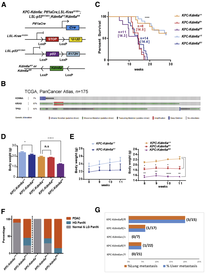
We performed a pancreatic tissue microarray analysis of KDM6A protein levels. We used human PDAC cell lines for KDM6A knockout and knockdown experiments. We performed bromouridine sequencing analysis to elucidate the effects of KDM6A loss on global transcription. We performed studies with Ptf1a; LSL-Kras; Trp53; Kdm6a, Ptf1a; Kdm6a, and orthotopic xenograft mice to investigate the impacts of Kdm6a deficiency on pancreatic tumorigenesis and pancreatitis.

RESULTS

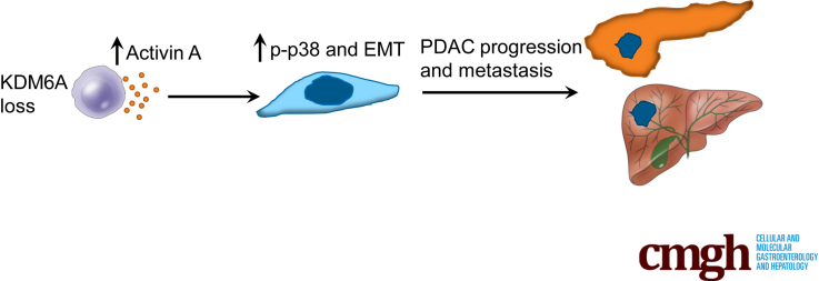
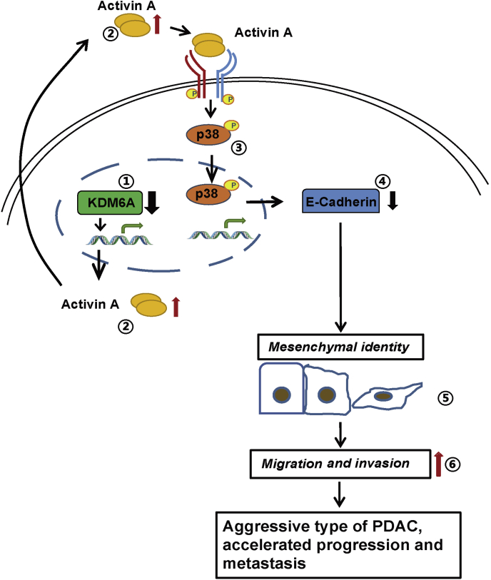
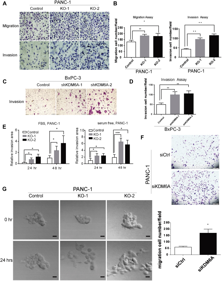
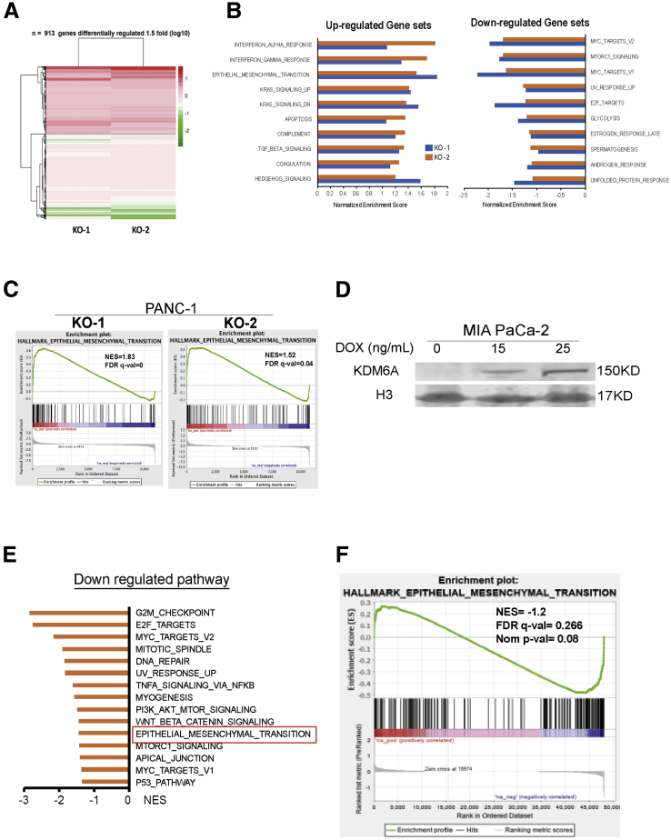
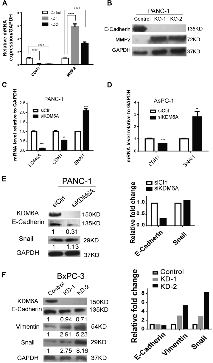
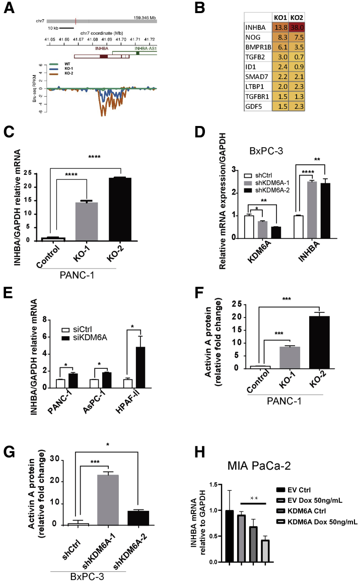
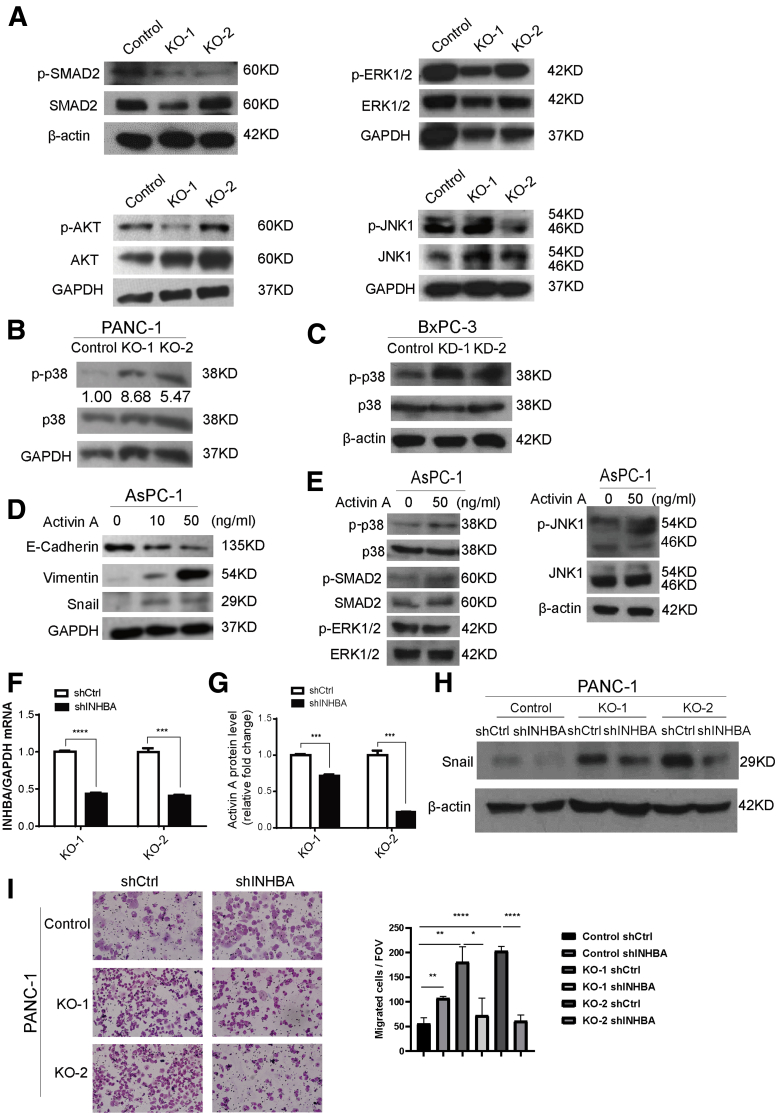
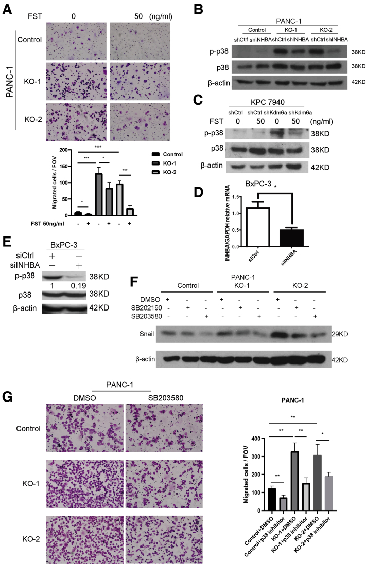
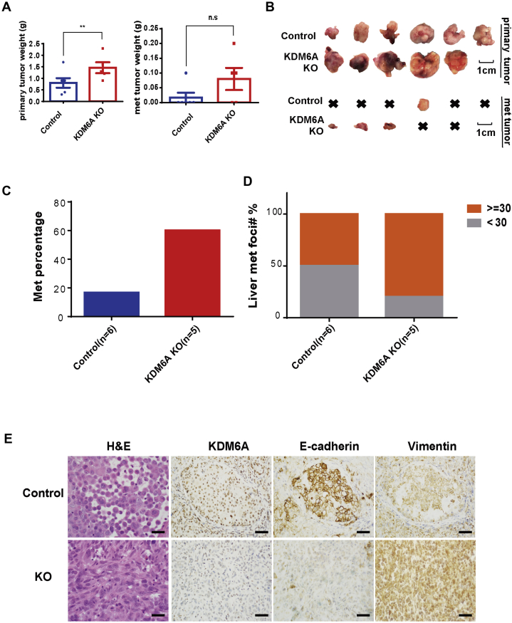
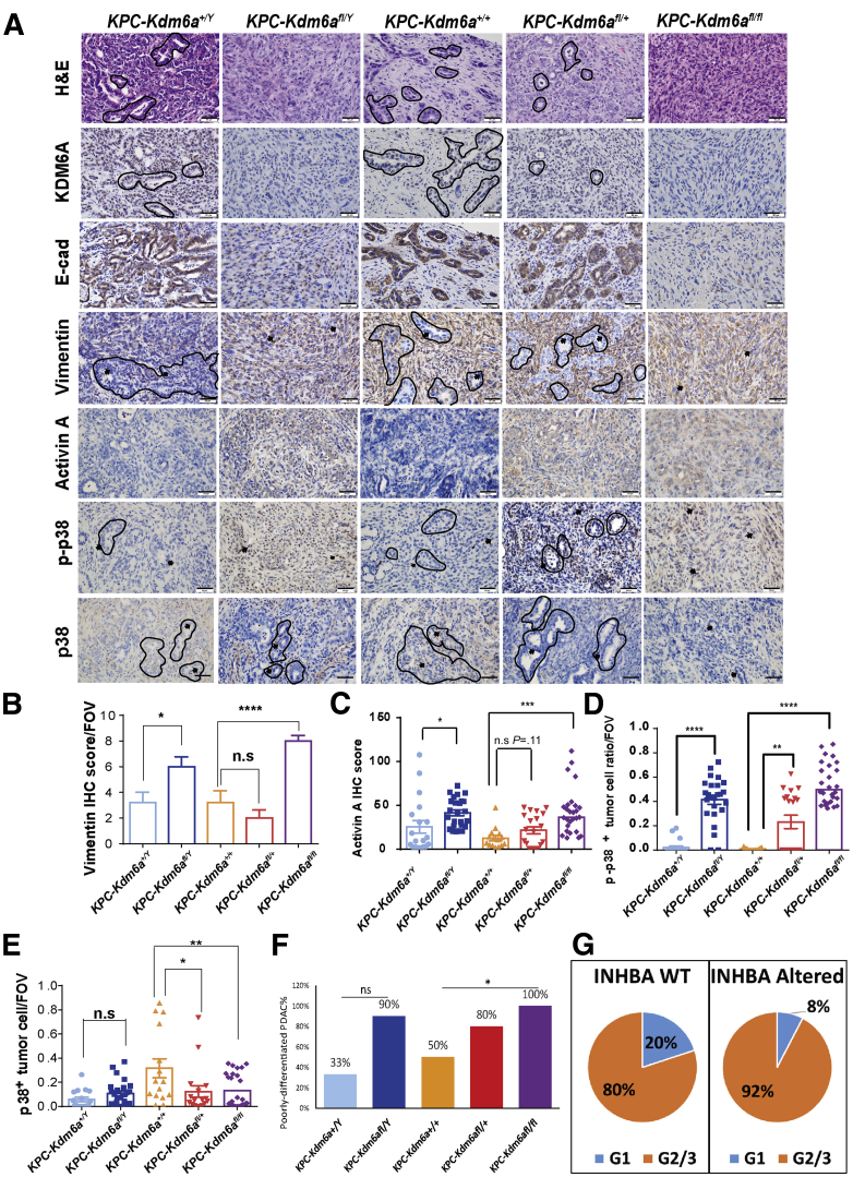
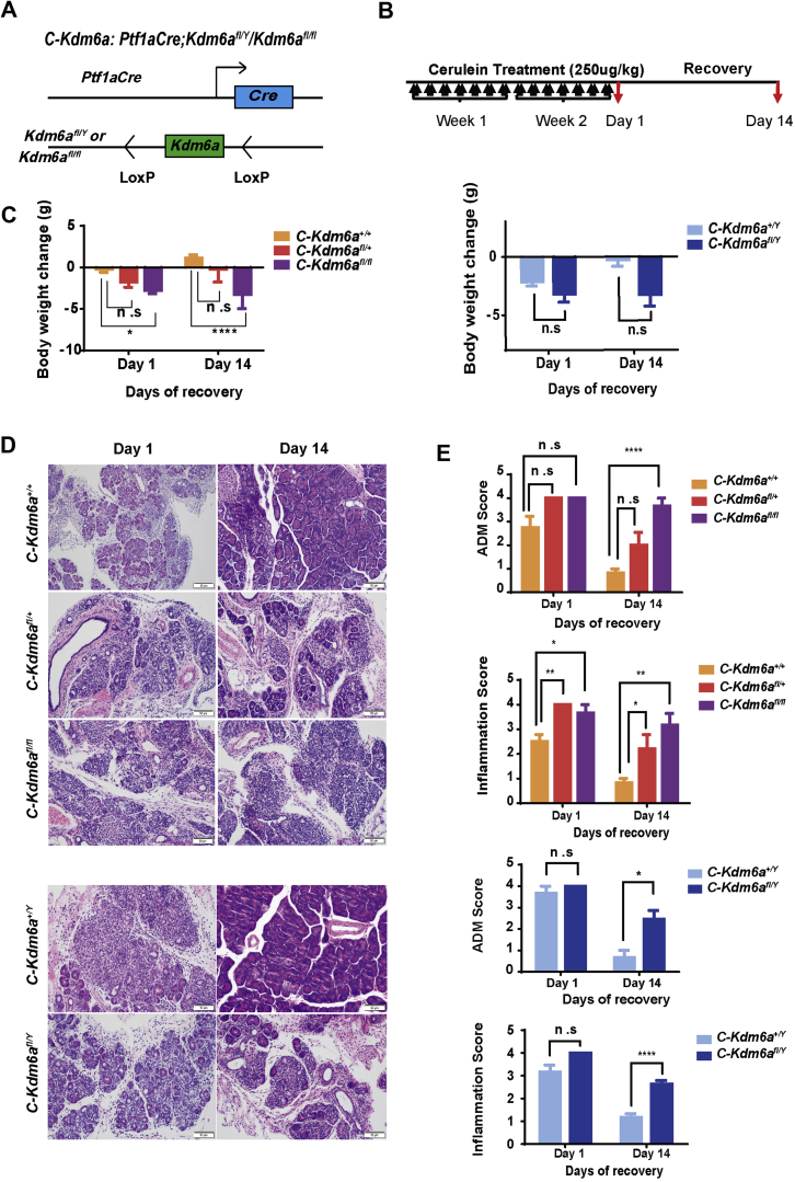
Loss of KDM6A was associated with metastasis in PDAC patients. Bromouridine sequencing analysis showed up-regulation of the epithelial-mesenchymal transition pathway in PDAC cells deficient in KDM6A. Loss of KDM6A promoted mesenchymal morphology, migration, and invasion in PDAC cells in vitro. Mechanistically, activin A and subsequent p38 activation likely mediated the role of KDM6A loss. Inhibiting either activin A or p38 reversed the effect. Pancreas-specific Kdm6a-knockout mice pancreata showed accelerated PDAC progression, developed a more aggressive undifferentiated type of PDAC, and increased metastases in the background of Kras and p53 mutations. Kdm6a-deficient pancreata in a pancreatitis model had a delayed recovery with increased PDAC precursor lesions compared with wild-type pancreata.

CONCLUSIONS

Loss of KDM6A accelerates PDAC progression and metastasis, most likely by a noncanonical p38-dependent activin A pathway. KDM6A also promotes pancreatic tissue recovery from pancreatitis. Activin A might be used as a therapeutic target for KDM6A-deficient PDACs.

摘要

背景与目的

组蛋白去甲基化酶 KDM6A 的失活突变在胰腺导管腺癌(PDAC)中经常发生。我们研究了 KDM6A(赖氨酸去甲基酶 6A)在 PDAC 发展中的作用。

方法

我们对 KDM6A 蛋白水平进行了胰腺组织微阵列分析。我们使用人 PDAC 细胞系进行 KDM6A 敲除和敲低实验。我们进行了溴尿嘧啶测序分析,以阐明 KDM6A 缺失对全局转录的影响。我们使用 Ptf1a; LSL-Kras; Trp53; Kdm6a; Ptf1a; Kdm6a 和原位异种移植小鼠进行研究,以研究 Kdm6a 缺失对胰腺肿瘤发生和胰腺炎的影响。

结果

KDM6A 的缺失与 PDAC 患者的转移有关。溴尿嘧啶测序分析显示,在缺乏 KDM6A 的 PDAC 细胞中,上皮-间充质转化途径上调。KDM6A 的缺失促进了 PDAC 细胞在体外的间充质形态、迁移和侵袭。在机制上,激活素 A 和随后的 p38 激活可能介导了 KDM6A 缺失的作用。抑制激活素 A 或 p38 均可逆转这种作用。胰腺特异性 Kdm6a 敲除小鼠的胰腺显示出 PDAC 进展加速,发展出更具侵袭性的未分化型 PDAC,并且在 Kras 和 p53 突变的背景下转移增加。与野生型胰腺相比,胰腺炎模型中 Kdm6a 缺失的胰腺具有延迟的恢复,并且 PDAC 前体病变增加。

结论

KDM6A 的缺失加速了 PDAC 的进展和转移,这很可能是通过非典型的 p38 依赖性激活素 A 途径。KDM6A 还促进了胰腺炎后胰腺组织的恢复。激活素 A 可能被用作 KDM6A 缺陷型 PDAC 的治疗靶点。

https://cdn.ncbi.nlm.nih.gov/pmc/blobs/60d9/8715196/c5eee7bcab7e/gr13.jpg
https://cdn.ncbi.nlm.nih.gov/pmc/blobs/60d9/8715196/aeac494162cd/fx1.jpg
https://cdn.ncbi.nlm.nih.gov/pmc/blobs/60d9/8715196/b63b5b27068b/gr1.jpg
https://cdn.ncbi.nlm.nih.gov/pmc/blobs/60d9/8715196/4222ad25090f/gr2.jpg
https://cdn.ncbi.nlm.nih.gov/pmc/blobs/60d9/8715196/c591cbf711e5/gr3.jpg
https://cdn.ncbi.nlm.nih.gov/pmc/blobs/60d9/8715196/a2f350de323c/gr4.jpg
https://cdn.ncbi.nlm.nih.gov/pmc/blobs/60d9/8715196/b4db2189d402/gr5.jpg
https://cdn.ncbi.nlm.nih.gov/pmc/blobs/60d9/8715196/351c173b9839/gr6.jpg
https://cdn.ncbi.nlm.nih.gov/pmc/blobs/60d9/8715196/641d972efa16/gr7.jpg
https://cdn.ncbi.nlm.nih.gov/pmc/blobs/60d9/8715196/1aee003de6cf/gr8.jpg
https://cdn.ncbi.nlm.nih.gov/pmc/blobs/60d9/8715196/5f026c35f067/gr9.jpg
https://cdn.ncbi.nlm.nih.gov/pmc/blobs/60d9/8715196/3456711eea2c/gr10.jpg
https://cdn.ncbi.nlm.nih.gov/pmc/blobs/60d9/8715196/f9f5d233c425/gr11.jpg
https://cdn.ncbi.nlm.nih.gov/pmc/blobs/60d9/8715196/81877bf2d28c/gr12.jpg
https://cdn.ncbi.nlm.nih.gov/pmc/blobs/60d9/8715196/c5eee7bcab7e/gr13.jpg

相似文献

1
KDM6A Regulates Cell Plasticity and Pancreatic Cancer Progression by Noncanonical Activin Pathway.KDM6A 通过非经典激活素途径调节细胞可塑性和胰腺癌进展。
Cell Mol Gastroenterol Hepatol. 2022;13(2):643-667. doi: 10.1016/j.jcmgh.2021.09.014. Epub 2021 Sep 25.
2
SETDB1 Inhibits p53-Mediated Apoptosis and Is Required for Formation of Pancreatic Ductal Adenocarcinomas in Mice.SETDB1抑制p53介导的细胞凋亡,是小鼠胰腺导管腺癌形成所必需的。
Gastroenterology. 2020 Aug;159(2):682-696.e13. doi: 10.1053/j.gastro.2020.04.047. Epub 2020 Apr 28.
3
KDM6A Loss Recruits Tumor-Associated Neutrophils and Promotes Neutrophil Extracellular Trap Formation in Pancreatic Cancer.KDM6A 缺失招募肿瘤相关中性粒细胞并促进胰腺癌中中性粒细胞胞外诱捕网的形成。
Cancer Res. 2022 Nov 15;82(22):4247-4260. doi: 10.1158/0008-5472.CAN-22-0968.
4
Interleukin 22 Signaling Regulates Acinar Cell Plasticity to Promote Pancreatic Tumor Development in Mice.白细胞介素 22 信号调节腺泡细胞可塑性,促进小鼠胰腺肿瘤发展。
Gastroenterology. 2020 Apr;158(5):1417-1432.e11. doi: 10.1053/j.gastro.2019.12.010. Epub 2019 Dec 14.
5
HNF1A recruits KDM6A to activate differentiated acinar cell programs that suppress pancreatic cancer.HNF1A 招募 KDM6A 以激活分化的腺泡细胞程序,从而抑制胰腺癌。
EMBO J. 2020 May 4;39(9):e102808. doi: 10.15252/embj.2019102808. Epub 2020 Mar 10.
6
Pancreatic Premalignant Lesions Secrete Tissue Inhibitor of Metalloproteinases-1, Which Activates Hepatic Stellate Cells Via CD63 Signaling to Create a Premetastatic Niche in the Liver.胰腺前恶性病变分泌组织金属蛋白酶抑制剂-1,通过 CD63 信号激活肝星状细胞,在肝脏中形成一个前转移龛。
Gastroenterology. 2016 Nov;151(5):1011-1024.e7. doi: 10.1053/j.gastro.2016.07.043. Epub 2016 Aug 6.
7
Loss of KDM6A characterizes a poor prognostic subtype of human pancreatic cancer and potentiates HDAC inhibitor lethality.KDM6A 的缺失是人类胰腺癌预后不良的亚型特征,并增强了 HDAC 抑制剂的致死性。
Int J Cancer. 2019 Jul 1;145(1):192-205. doi: 10.1002/ijc.32072. Epub 2018 Dec 30.
8
Loss of Activin Receptor Type 1B Accelerates Development of Intraductal Papillary Mucinous Neoplasms in Mice With Activated KRAS.激活素受体1B型缺失加速KRAS激活小鼠导管内乳头状黏液性肿瘤的发展。
Gastroenterology. 2016 Jan;150(1):218-228.e12. doi: 10.1053/j.gastro.2015.09.013. Epub 2015 Sep 25.
9
ETS-Transcription Factor ETV1 Regulates Stromal Expansion and Metastasis in Pancreatic Cancer.ETS转录因子ETV1调控胰腺癌的基质扩张和转移。
Gastroenterology. 2016 Sep;151(3):540-553.e14. doi: 10.1053/j.gastro.2016.06.005. Epub 2016 Jun 16.
10
Sex specificity of pancreatic cancer cachexia phenotypes, mechanisms, and treatment in mice and humans: role of Activin.胰腺癌恶病质表型、机制和治疗在小鼠和人类中的性别特异性:激活素的作用。
J Cachexia Sarcopenia Muscle. 2022 Aug;13(4):2146-2161. doi: 10.1002/jcsm.12998. Epub 2022 May 5.

引用本文的文献

1
Knockdown of eIF3a alleviates pulmonary arterial hypertension by inhibiting endothelial-to-mesenchymal transition via TGFβ1/SMAD pathway.敲低真核生物翻译起始因子3a(eIF3a)可通过转化生长因子β1(TGFβ1)/SMAD信号通路抑制内皮-间充质转化,从而减轻肺动脉高压。
J Transl Med. 2025 May 9;23(1):524. doi: 10.1186/s12967-025-06505-3.
2
Insights into the mechanisms driven by H3K4 KMTs in pancreatic cancer.揭示组蛋白 H3K4 甲基转移酶驱动的胰腺癌发生机制。
Biochem J. 2024 Aug 7;481(15):983-997. doi: 10.1042/BCJ20230374.
3
Unveiling the Molecular Landscape of Pancreatic Ductal Adenocarcinoma: Insights into the Role of the COMPASS-like Complex.
揭示胰腺导管腺癌的分子图谱:COMPASS 样复合物的作用解析。
Int J Mol Sci. 2024 May 7;25(10):5069. doi: 10.3390/ijms25105069.
4
Clinical Evaluation of the Pancreatic Cancer Microenvironment: Opportunities and Challenges.胰腺癌微环境的临床评估:机遇与挑战
Cancers (Basel). 2024 Feb 15;16(4):794. doi: 10.3390/cancers16040794.
5
TP53 missense mutation reveals gain-of-function properties in small-sized KRAS transformed pancreatic ductal adenocarcinoma.TP53 错义突变揭示了小尺寸 KRAS 转化的胰腺导管腺癌中的功能获得特性。
J Transl Med. 2023 Dec 1;21(1):872. doi: 10.1186/s12967-023-04742-y.
6
Epigenetic control of pancreatic cancer metastasis.表观遗传调控胰腺癌转移。
Cancer Metastasis Rev. 2023 Dec;42(4):1113-1131. doi: 10.1007/s10555-023-10132-z. Epub 2023 Sep 2.
7
Molecular profile of metastasis, cell plasticity and EMT in pancreatic cancer: a pre-clinical connection to aggressiveness and drug resistance.胰腺癌转移、细胞可塑性和 EMT 的分子特征:临床前侵袭性和耐药性的联系。
Cancer Metastasis Rev. 2024 Mar;43(1):29-53. doi: 10.1007/s10555-023-10125-y. Epub 2023 Jul 15.
8
Sex-based differences in effector cells of the adaptive immune system during Alzheimer's disease and related dementias.适应性免疫系统效应细胞在阿尔茨海默病及相关痴呆中的性别差异。
Neurobiol Dis. 2023 Aug;184:106202. doi: 10.1016/j.nbd.2023.106202. Epub 2023 Jun 15.
9
Uridine-derived ribose fuels glucose-restricted pancreatic cancer.尿苷衍生的核糖为葡萄糖限制的胰腺癌供能。
Nature. 2023 Jun;618(7963):151-158. doi: 10.1038/s41586-023-06073-w. Epub 2023 May 17.
10
Pancreatic cancer: Advances and challenges.胰腺癌:进展与挑战。
Cell. 2023 Apr 13;186(8):1729-1754. doi: 10.1016/j.cell.2023.02.014.