Zentout Siham, Smith Rebecca, Jacquier Marine, Huet Sébastien
Univ Rennes, CNRS, IGDR (Institut de Génétique et Développement de Rennes)-UMR 6290, BIOSIT-UMS 3480, Rennes, France.
Institut Universitaire de France, Paris, France.
Front Cell Dev Biol. 2021 Sep 13;9:730998. doi: 10.3389/fcell.2021.730998. eCollection 2021.
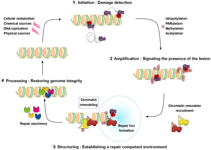
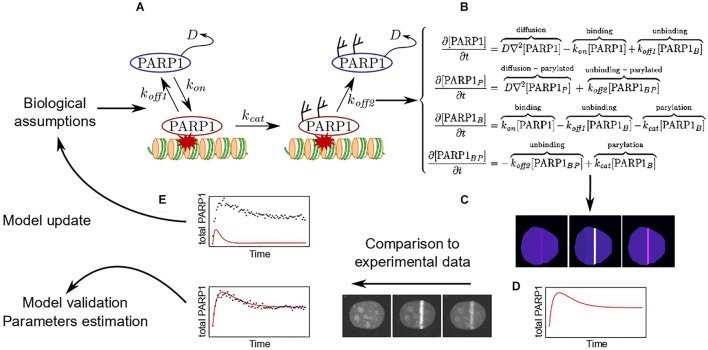
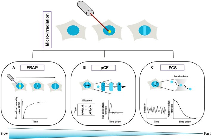
DNA repair requires a coordinated effort from an array of factors that play different roles in the DNA damage response from recognizing and signaling the presence of a break, creating a repair competent environment, and physically repairing the lesion. Due to the rapid nature of many of these events, live-cell microscopy has become an invaluable method to study this process. In this review we outline commonly used tools to induce DNA damage under the microscope and discuss spatio-temporal analysis tools that can bring added information regarding protein dynamics at sites of damage. In particular, we show how to go beyond the classical analysis of protein recruitment curves to be able to assess the dynamic association of the repair factors with the DNA lesions as well as the target-search strategies used to efficiently find these lesions. Finally, we discuss how the use of mathematical models, combined with experimental evidence, can be used to better interpret the complex dynamics of repair proteins at DNA lesions.
DNA修复需要一系列因素的协同作用,这些因素在DNA损伤反应中发挥着不同的作用,包括识别和标记断裂的存在、营造有利于修复的环境以及对损伤进行物理修复。由于这些事件大多具有快速性,活细胞显微镜技术已成为研究这一过程的重要方法。在本综述中,我们概述了在显微镜下诱导DNA损伤常用的工具,并讨论了时空分析工具,这些工具可以提供有关损伤部位蛋白质动态的额外信息。特别是,我们展示了如何超越对蛋白质募集曲线的经典分析,以便能够评估修复因子与DNA损伤的动态关联以及用于有效找到这些损伤的靶标搜索策略。最后,我们讨论了如何结合数学模型和实验证据,更好地解释DNA损伤处修复蛋白的复杂动态。