HIV and TB Research Directorate, Ethiopian Public Health Institute (EPHI), Addis Ababa, Ethiopia.
Mycobacterial Disease Research Directorate, Armauer Hansen Research Institute (AHRI), Addis Ababa, Ethiopia.
PLoS One. 2021 Oct 1;16(10):e0258122. doi: 10.1371/journal.pone.0258122. eCollection 2021.
PDL1 and its interaction with PD1 is implicated in immune dysfunction in TB and HIV. The expression of PDL1 on multiple subsets of monocytes as well as their associations with cytokines and microbial products have not been well studied.
HIV (TB-HIV+), TB (TB+HIV-) and TB/HIV co-infected (TB+HIV+) patients as well as apparently healthy controls (TB-HIV-) were recruited. TB and HIV patients were treatment naïve while TB/HIV patients were both ART naïve and experienced but not yet started TB therapy. Monocyte subsets were evaluated for PDL1 expression by flow cytometry; plasma TNFα, IL6, IP10, IFNγ and IL10 were measured by Luminex; and cytokine mRNA from purified monocytes quantitated by qPCR. The association of PDL1 with cytokines, clinical and microbial indices, including HIV viral load, TB smear microscopy and TB urinary lipoarabinomannan (LAM) were assessed.
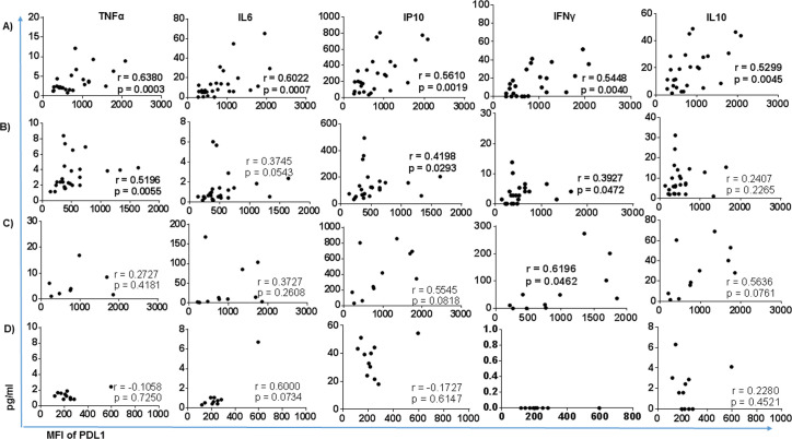
Monocyte expression of PDL1 was significantly higher in TB, HIV and TB/HIV co-infected patients compared with healthy controls (p = 0.0001), with the highest levels in TB/HIV co-infected patients. The highest expression of PDL1 was on intermediate (CD14+CD16+) monocytes in all participant groups. PDL1 strongly correlated with HIV viral load in TB/HIV while weakly correlated in HIV. PDL1 levels moderately correlated with plasma TNFα, IL6, IP10, IFNγ and IL10 level in TB subjects whereas weakly correlated with TNFα and IP10 in HIV patients. However, cytokine mRNA from purified monocytes showed no association with either plasma cytokines or monocyte PDL1 expression, implying that if cytokines modulate PDL1, they are likely not originating from circulating monocytes themselves. These results underscore the importance of further characterization of multiple monocyte subsets and their phenotypic and functional differences in different disease states.
PDL1 及其与 PD1 的相互作用与 TB 和 HIV 中的免疫功能障碍有关。PDL1 在单核细胞多个亚群上的表达及其与细胞因子和微生物产物的关联尚未得到很好的研究。
招募了 HIV(TB-HIV+)、TB(TB+HIV-)和 TB/HIV 合并感染(TB+HIV+)患者以及明显健康的对照者(TB-HIV-)。TB 和 HIV 患者均为治疗初治,而 TB/HIV 患者均为 ART 初治且已接受但尚未开始 TB 治疗。通过流式细胞术评估单核细胞亚群的 PDL1 表达;通过 Luminex 测量血浆 TNFα、IL6、IP10、IFNγ 和 IL10;通过 qPCR 定量纯化单核细胞中的细胞因子 mRNA。评估了 PDL1 与细胞因子、临床和微生物指标(包括 HIV 病毒载量、TB 涂片显微镜检查和 TB 尿液脂阿拉伯甘露聚糖(LAM))的关联。
与健康对照组相比,TB、HIV 和 TB/HIV 合并感染患者的单核细胞 PDL1 表达显著升高(p = 0.0001),TB/HIV 合并感染患者的表达最高。在所有患者组中,PDL1 的表达最高的是中间(CD14+CD16+)单核细胞。在 TB/HIV 中,PDL1 与 HIV 病毒载量呈强相关,而在 HIV 中呈弱相关。在 TB 患者中,PDL1 水平与血浆 TNFα、IL6、IP10、IFNγ 和 IL10 水平中度相关,而在 HIV 患者中与 TNFα 和 IP10 弱相关。然而,从纯化的单核细胞中提取的细胞因子 mRNA 与血浆细胞因子或单核细胞 PDL1 表达均无相关性,这意味着如果细胞因子调节 PDL1,它们可能不是来自循环单核细胞本身。这些结果强调了进一步表征不同疾病状态下的多个单核细胞亚群及其表型和功能差异的重要性。