Chatzinikolaou Eva, Keklikoglou Kleoniki
Hellenic Centre for Marine Research (HCMR), Institute of Marine Biology, Biotechnology and Aquaculture (IMBBC), Heraklion, Crete, Greece Hellenic Centre for Marine Research (HCMR), Institute of Marine Biology, Biotechnology and Aquaculture (IMBBC) Heraklion, Crete Greece.
Biology Department, University of Crete, Heraklion, Crete, Greece Biology Department, University of Crete Heraklion, Crete Greece.
Biodivers Data J. 2021 Sep 15;9:e71542. doi: 10.3897/BDJ.9.e71542. eCollection 2021.
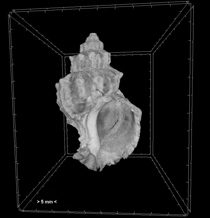
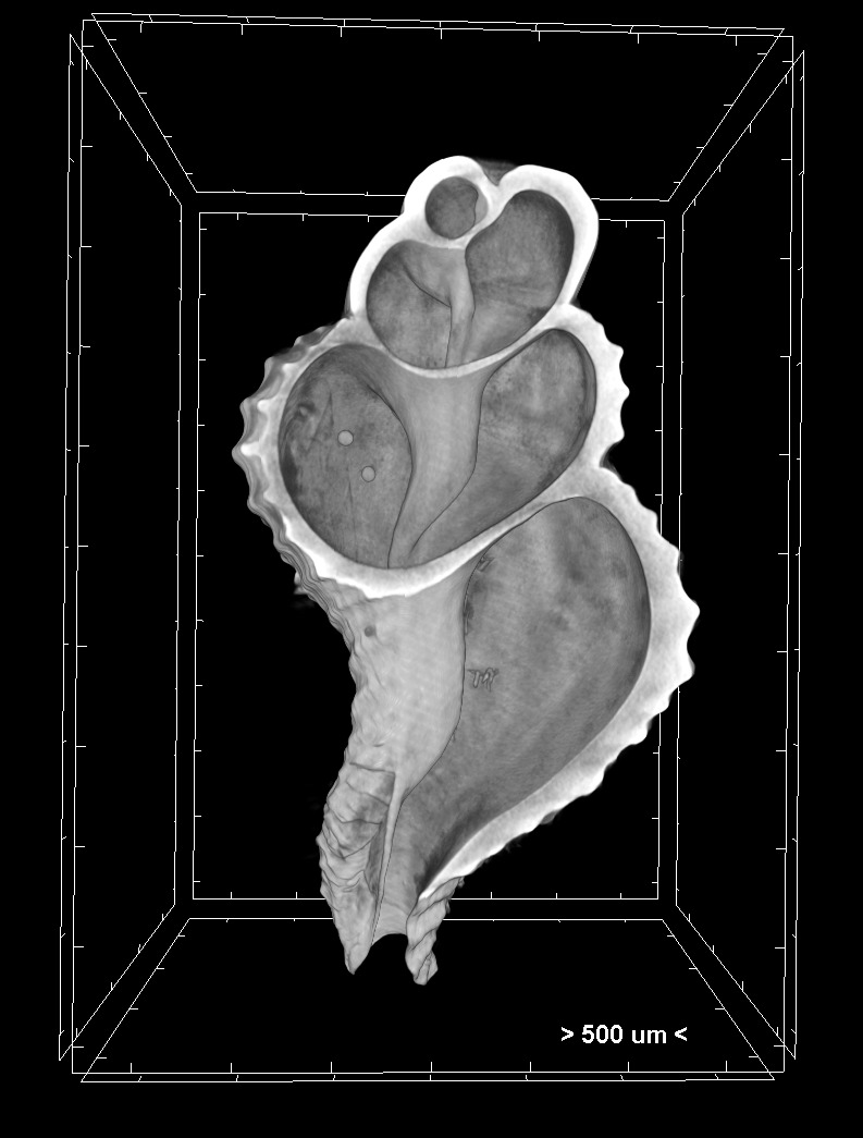
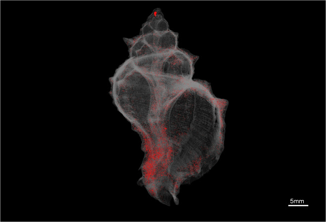
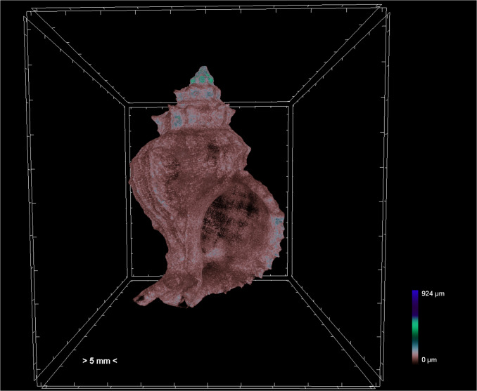
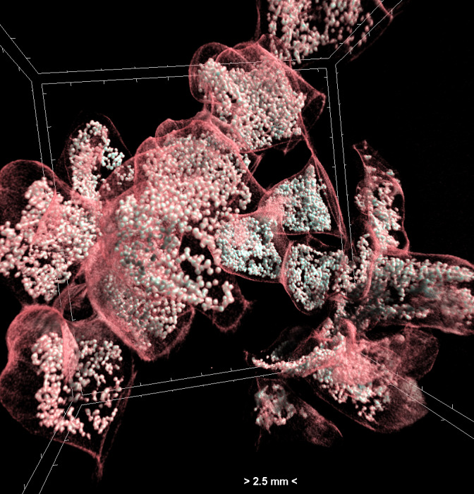
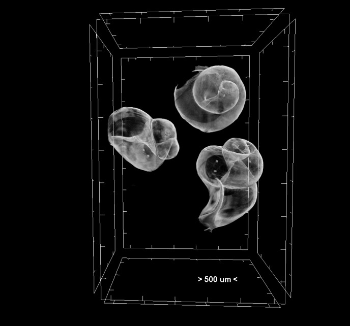
Micro-computed tomography (micro-CT) is a high-resolution 3D-imaging technique which is now increasingly applied in biological studies focusing on taxonomy and functional morphology. The creation of virtual representations of specimens can increase availability of otherwise underexploited and inaccessible samples. The 3D model dataset can be also further processed through volume rendering and morphometric analysis. The success of micro-CT as a visualisation technique depends on several methodological manipulations, including the use of contrast enhancing staining agents, filters, scanning mediums, containers, exposure time and frame averaging. The aim of this study was to standardise a series of micro-CT scanning and 3D analysis protocols for a marine gastropod species, . The analytical protocols have followed all the developmental stages of this gastropod, from egg capsules and embryos to juveniles and adults.
微计算机断层扫描(micro-CT)是一种高分辨率三维成像技术,目前越来越多地应用于专注于分类学和功能形态学的生物学研究中。创建标本的虚拟模型可以提高那些原本未得到充分利用且难以获取的样本的可用性。三维模型数据集还可以通过体绘制和形态计量分析进行进一步处理。微计算机断层扫描作为一种可视化技术的成功取决于多种方法操作,包括使用造影增强染色剂、滤镜、扫描介质、容器、曝光时间和帧平均。本研究的目的是为一种海洋腹足类物种标准化一系列微计算机断层扫描和三维分析方案。分析方案涵盖了这种腹足类动物从卵囊和胚胎到幼体和成体的所有发育阶段。