Moon Jieun, Kim Pilhan
Graduate School of Nanoscience and Technology, Korea Advanced Institute of Science and Technology (KAIST), Daejeon, Korea.
KI for Health Science and Technology (KIHST), Korea Advanced Institute of Science and Technology (KAIST), Daejeon, Korea.
J Lipid Atheroscler. 2021 Sep;10(3):313-321. doi: 10.12997/jla.2021.10.3.313. Epub 2021 Jun 28.
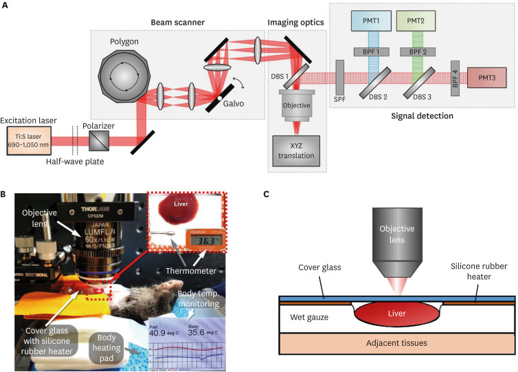
The liver plays a central role in lipid metabolism. During fasting and feeding, the fatty acid trafficking between adipose tissue and liver induces accumulation and dissociation of dynamic hepatic lipid droplets (LDs). Herein, we established an intravital 2-photon imaging technique to longitudinally visualize the dynamic alteration of hepatic LD deposition during fasting and refeeding in the liver of live mouse.
Intravital 2-photon imaging of liver was performed to observe hepatic LD alteration induced by fasting for different periods of time, 12, 24, and 48 hours followed by refeeding. Hepatic LDs were fluorescently labelled by intravenous injection of Seoul-Flour 44 and visualized by custom-built intravital 2-photon microscope.
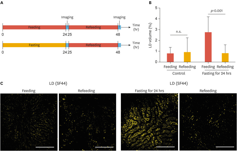
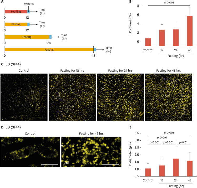
Significant increases of the number and size of hepatic LDs were observed by intravital 2-photon imaging of the liver after 12 hours of fasting. The degree of hepatic LD accumulation continuously increased with fasting up to 48 hours. Remarkably, with refeeding for 24 hours, the hepatic LDs accumulated by fasting were fully dissociated and the LD occupancy in the liver was recovered to the normal state.
Utilizing intravital 2-photon microscope with systemic fluorescent labeling of LD in live mice, dynamic alterations of hepatic LDs such as accumulation and dissociation by fasting and refeeding were successfully visualized at a subcellular level . The established method enabling the visualization of LDs will be a useful tool to investigate the pathophysiology of various diseases associated with dysregulated lipid metabolism.
肝脏在脂质代谢中起核心作用。在禁食和进食期间,脂肪组织与肝脏之间的脂肪酸转运诱导了动态肝脂滴(LDs)的积累和解离。在此,我们建立了一种活体双光子成像技术,以纵向观察活体小鼠肝脏在禁食和再进食期间肝脂滴沉积的动态变化。
对肝脏进行活体双光子成像,观察禁食不同时间段(12、24和48小时)后再进食所诱导的肝脂滴变化。通过静脉注射首尔荧光44对肝脂滴进行荧光标记,并使用定制的活体双光子显微镜进行观察。
禁食12小时后,通过肝脏活体双光子成像观察到肝脂滴的数量和大小显著增加。随着禁食时间延长至48小时,肝脂滴积累程度持续增加。值得注意的是,再进食24小时后,禁食所积累的肝脂滴完全解离,肝脏中的脂滴占有率恢复到正常状态。
利用活体双光子显微镜对活体小鼠的脂滴进行全身荧光标记,成功在亚细胞水平可视化了肝脂滴的动态变化,如禁食和再进食引起的积累和解离。所建立的能够可视化脂滴的方法将成为研究与脂质代谢失调相关的各种疾病病理生理学的有用工具。