Ruple Bradley A, Godwin Joshua S, Mesquita Paulo H C, Osburn Shelby C, Sexton Casey L, Smith Morgan A, Ogletree Jeremy C, Goodlett Michael D, Edison Joseph L, Ferrando Arny A, Fruge Andrew D, Kavazis Andreas N, Young Kaelin C, Roberts Michael D
School of Kinesiology, Auburn University, Auburn, AL, United States.
Athletics Department, Auburn University, Auburn, AL, United States.
Front Physiol. 2021 Sep 24;12:728683. doi: 10.3389/fphys.2021.728683. eCollection 2021.
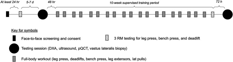
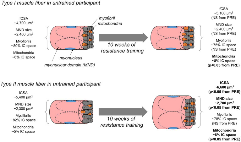
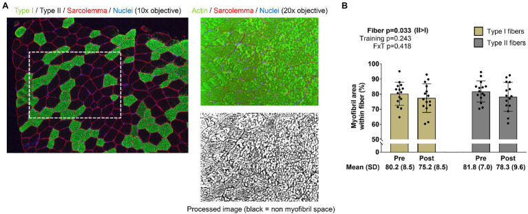
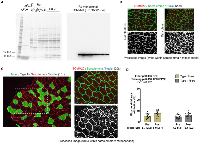
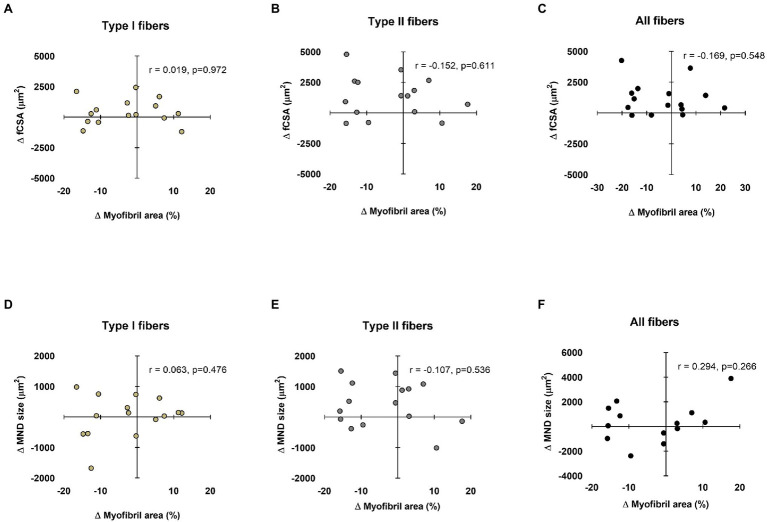
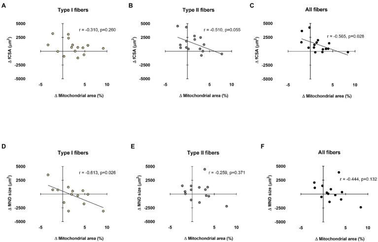
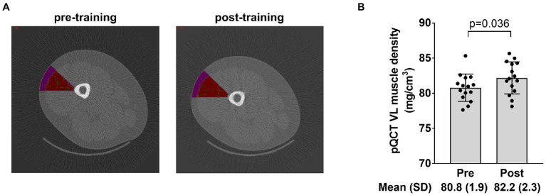
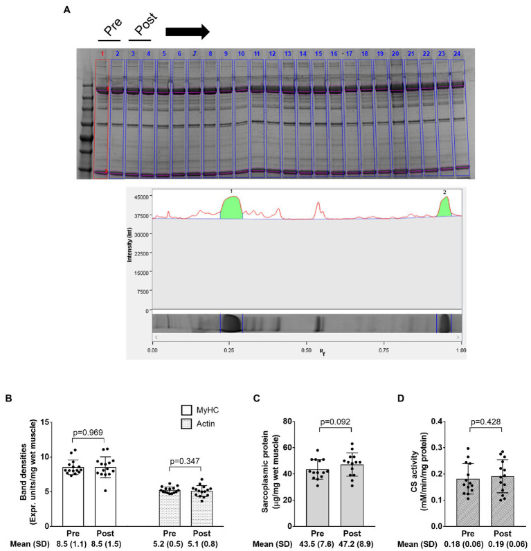
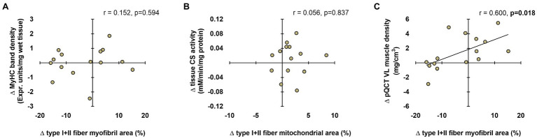
Resistance training increases muscle fiber hypertrophy, but the morphological adaptations that occur within muscle fibers remain largely unresolved. Fifteen males with minimal training experience (24±4years, 23.9±3.1kg/m body mass index) performed 10weeks of conventional, full-body resistance training (2× weekly). Body composition, the radiological density of the vastus lateralis muscle using peripheral quantitative computed tomography (pQCT), and vastus lateralis muscle biopsies were obtained 1week prior to and 72h following the last training bout. Quantification of myofibril and mitochondrial areas in type I (positive for MyHC I) and II (positive for MyHC IIa/IIx) fibers was performed using immunohistochemistry (IHC) techniques. Relative myosin heavy chain and actin protein abundances per wet muscle weight as well as citrate synthase (CS) activity assays were also obtained on tissue lysates. Training increased whole-body lean mass, mid-thigh muscle cross-sectional area, mean and type II fiber cross-sectional areas (fCSA), and maximal strength values for leg press, bench press, and deadlift (<0.05). The intracellular area occupied by myofibrils in type I or II fibers was not altered with training, suggesting a proportional expansion of myofibrils with fCSA increases. However, our histological analysis was unable to differentiate whether increases in myofibril number or girth occurred. Relative myosin heavy chain and actin protein abundances also did not change with training. IHC indicated training increased mitochondrial areas in both fiber types (=0.018), albeit CS activity levels remained unaltered with training suggesting a discordance between these assays. Interestingly, although pQCT-derived muscle density increased with training (=0.036), suggestive of myofibril packing, a positive association existed between training-induced changes in this metric and changes in mean fiber myofibril area (=0.600, =0.018). To summarize, our data imply that shorter-term resistance training promotes a proportional expansion of the area occupied by myofibrils and a disproportional expansion of the area occupied by mitochondria in type I and II fibers. Additionally, IHC and biochemical techniques should be viewed independently from one another given the lack of agreement between the variables assessed herein. Finally, the pQCT may be a viable tool to non-invasively track morphological changes (specifically myofibril density) in muscle tissue.
抗阻训练可增加肌纤维肥大,但肌纤维内发生的形态学适应在很大程度上仍未得到解决。15名训练经验最少的男性(年龄24±4岁,体重指数23.9±3.1kg/m)进行了为期10周的常规全身抗阻训练(每周2次)。在最后一次训练前1周和训练后72小时采集身体成分、使用外周定量计算机断层扫描(pQCT)测量的股外侧肌放射密度以及股外侧肌活检样本。使用免疫组织化学(IHC)技术对I型(肌球蛋白重链I阳性)和II型(肌球蛋白重链IIa/IIx阳性)纤维中的肌原纤维和线粒体面积进行定量分析。还对组织裂解物进行了每湿肌肉重量的相对肌球蛋白重链和肌动蛋白蛋白丰度以及柠檬酸合酶(CS)活性测定。训练增加了全身瘦体重、大腿中部肌肉横截面积、平均和II型纤维横截面积(fCSA)以及腿举、卧推和硬拉的最大力量值(<0.05)。训练后I型或II型纤维中肌原纤维所占的细胞内面积没有改变,这表明随着fCSA增加,肌原纤维呈比例扩张。然而,我们的组织学分析无法区分肌原纤维数量或周长的增加情况。相对肌球蛋白重链和肌动蛋白蛋白丰度也不会随训练而改变。免疫组织化学显示训练增加了两种纤维类型的线粒体面积(=0.018),尽管CS活性水平并未随训练而改变,这表明这些检测结果之间存在不一致。有趣的是,尽管pQCT得出的肌肉密度随训练而增加(=0.036),提示肌原纤维堆积,但该指标训练诱导的变化与平均纤维肌原纤维面积的变化之间存在正相关(=0.600,=0.018)。总之,我们的数据表明,短期抗阻训练促进了I型和II型纤维中肌原纤维所占面积的比例扩张以及线粒体所占面积的非比例扩张。此外,鉴于本文评估的变量之间缺乏一致性,免疫组织化学和生化技术应相互独立看待。最后,pQCT可能是一种可行的工具,用于无创追踪肌肉组织中的形态学变化(特别是肌原纤维密度)。