Huang Jiling, Gong Zhigang, Kong Yingnan, Huang Yanwen, Wang Hui, Kang Yingjie, Zhan Songhua
Department of Radiology, Shuguang Hospital Affiliated to Shanghai University of Traditional Chinese Medicine, Shanghai 201203, China.
Evid Based Complement Alternat Med. 2021 Oct 1;2021:7992688. doi: 10.1155/2021/7992688. eCollection 2021.
To investigate the effect of electroacupuncture (EA) on cognitive dysfunction in rats with hepatic encephalopathy and its underlying mechanism.
Fifty Wistar rats were randomly divided into a normal group ( = 10) and model group ( = 40). Rat models of hepatic encephalopathy were established by administration of carbon tetrachloride and thioacetamide for a total of 12 weeks. At the 9th week after modeling, rats with cognitive impairment in the model group were identified by conducting the Morris water maze test, which were then randomly divided into a control group (CCl) and treatment groups including EA group (CCl + EA), lactulose group (CCl + Lac), and EA plus lactulose group (CCl + CM), with 9 rats in each group. At the end of the 9th week, rats in CCl + Lac and CCl + CM groups had lactulose gavage at a dose of 10 mL/kg body weight, while normal control and CCl groups had gavage with the same volume of normal saline once a day for 21 days until the end of the experiment. Rats in CCl + EA and CCl + CM groups underwent acupuncture at Baihui (GV[DU]20), Shenting (GV[DU]24), and Zusanli (ST36) acupoints, among which EA at Baihui and Shenting acupoints were given once daily for 30 min lasting for 21 consecutive days. The effect of the treatment was measured by the Morris water maze test for learning and memory ability and magnetic resonance spectroscopy (MRS) for neuronal metabolism in the hippocampus of rats with hepatic encephalopathy. Pathological change in the rat hippocampus was observed by HE staining, while serum ammonia and liver function markers were detected. Western blot and real-time fluorescent quantitative PCR were used to detect the expressions of specific genes and proteins in the brain tissue.
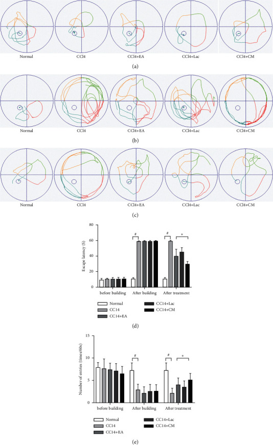
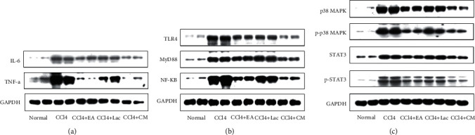
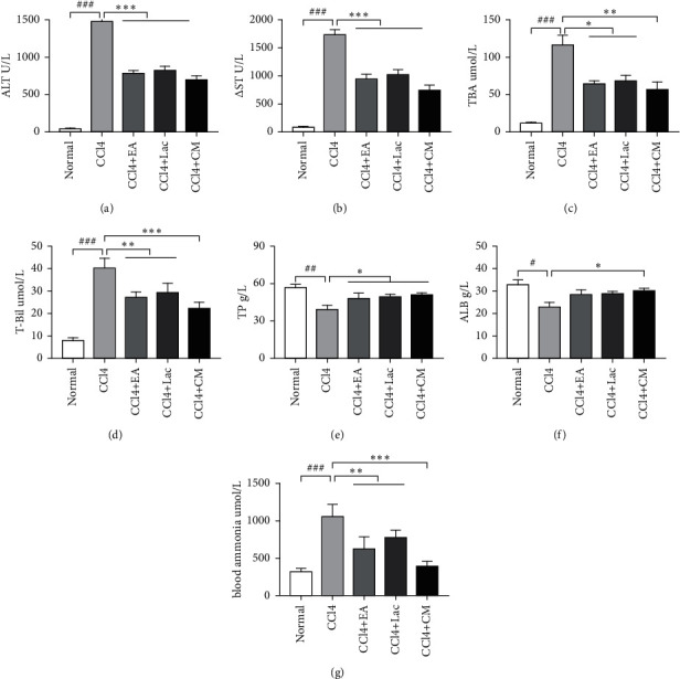
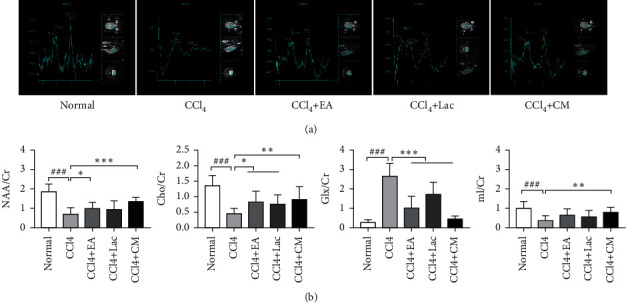
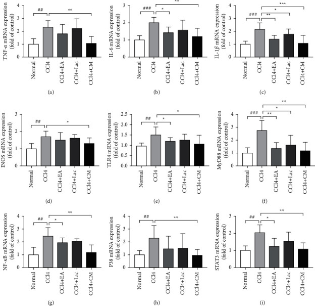
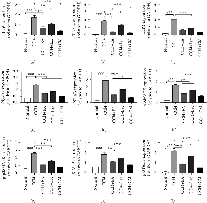
Compared with those in the control group, rats undergoing EA had significantly shortened escape latency and increased number of platform crossing. H&E staining confirmed that EA improved brain tissue necrosis and ameliorated nuclear pyknosis in rats with hepatic encephalopathy. Significantly decreased levels of serum ammonia, alanine aminotransferase (ALT), aspartate transaminase (AST), total bilirubin (TBil), and total bile acid (TBA) were observed in rats undergoing EA, as well as improved levels of total protein (TP) and albumin (ALB). In addition, EA inhibited the brain expressions of TNF-, IL-1, IL-6, iNOS, TLR4, MyD88, NF-B, p38MAPK, phosphorylated (p)-p38MAPK, STAT3, and p-STAT3 genes, as well as protein expressions of TNF-, IL-6, TLR4, MyD88, NF-B, p38MAPK, p-p38MAPK, STAT3, and p-STAT3. MRS showed increased Glx/Cr and decreased NAA/Cr, Cho/Cr and mI/Cr in the control group, and EA significantly reversed such changes in Glx/Cr and mI/Cr values.
EA ameliorated the production of excessive proinflammatory cytokines in the hippocampus of rats with cognitive dysfunction secondary to hepatic encephalopathy, which also gave rise to subsequent changes such as reduced blood ammonia level, brain-protective activated astrocytes, and lower degree of brain tissue injury. The p38MAPK/STAT3 and TLR4/MyD88/NF-B signaling pathways may be involved. EA can also improve the metabolism of NAA and Cho in the rat hippocampus and thereby improve learning and memory abilities.
探讨电针(EA)对肝性脑病大鼠认知功能障碍的影响及其潜在机制。
将50只Wistar大鼠随机分为正常组(n = 10)和模型组(n = 40)。采用四氯化碳和硫代乙酰胺联合给药12周建立肝性脑病大鼠模型。造模后第9周,通过Morris水迷宫试验筛选出模型组中存在认知障碍的大鼠,然后将其随机分为对照组(CCl)和治疗组,治疗组包括电针组(CCl + EA)、乳果糖组(CCl + Lac)和电针加乳果糖组(CCl + CM),每组9只。第9周结束时,CCl + Lac组和CCl + CM组大鼠以10 mL/kg体重的剂量进行乳果糖灌胃,而正常对照组和CCl组大鼠每天灌胃等体积的生理盐水,共21天,直至实验结束。CCl + EA组和CCl + CM组大鼠针刺百会(GV[督]20)、神庭(GV[督]24)和足三里(ST36)穴位,其中百会和神庭穴位每天电针1次,每次30分钟,连续21天。通过Morris水迷宫试验检测肝性脑病大鼠的学习记忆能力,采用磁共振波谱(MRS)检测大鼠海马神经元代谢,以评估治疗效果。通过HE染色观察大鼠海马的病理变化,同时检测血清氨和肝功能指标。采用蛋白质免疫印迹法(Western blot)和实时荧光定量PCR检测脑组织中特定基因和蛋白质的表达。
与对照组相比,电针治疗的大鼠逃避潜伏期显著缩短,穿越平台次数增加。HE染色证实电针改善了肝性脑病大鼠的脑组织坏死,减轻了核固缩。电针治疗的大鼠血清氨、丙氨酸氨基转移酶(ALT)、天冬氨酸氨基转移酶(AST)、总胆红素(TBil)和总胆汁酸(TBA)水平显著降低,总蛋白(TP)和白蛋白(ALB)水平有所改善。此外,电针抑制了脑组织中TNF-α、IL-1、IL-6、iNOS、TLR4、MyD88、NF-κB、p38MAPK、磷酸化(p)-p38MAPK、STAT3和p-STAT3基因的表达,以及TNF-α、IL-6、TLR4、MyD88、NF-κB、p38MAPK、p-p38MAPK、STAT3和p-STAT3蛋白的表达。MRS显示对照组中Glx/Cr升高,NAA/Cr、Cho/Cr和mI/Cr降低,电针显著逆转了Glx/Cr和mI/Cr值的这种变化。
电针改善了肝性脑病继发认知功能障碍大鼠海马中过量促炎细胞因子的产生,这也导致了随后的一系列变化,如血氨水平降低、脑保护激活的星形胶质细胞增加以及脑组织损伤程度减轻。p38MAPK/STAT3和TLR4/MyD88/NF-κB信号通路可能参与其中。电针还可改善大鼠海马中NAA和Cho的代谢,从而提高学习记忆能力。