• 文献检索
  • 文档翻译
  • 深度研究
  • 学术资讯
  • Suppr Zotero 插件Zotero 插件
  • 邀请有礼
  • 套餐&价格
  • 历史记录
应用&插件
Suppr Zotero 插件Zotero 插件浏览器插件Mac 客户端Windows 客户端微信小程序
定价
高级版会员购买积分包购买API积分包
服务
文献检索文档翻译深度研究API 文档MCP 服务
关于我们
关于 Suppr公司介绍联系我们用户协议隐私条款
关注我们

Suppr 超能文献

核心技术专利:CN118964589B侵权必究
粤ICP备2023148730 号-1Suppr @ 2026

文献检索

告别复杂PubMed语法,用中文像聊天一样搜索,搜遍4000万医学文献。AI智能推荐,让科研检索更轻松。

立即免费搜索

文件翻译

保留排版,准确专业,支持PDF/Word/PPT等文件格式,支持 12+语言互译。

免费翻译文档

深度研究

AI帮你快速写综述,25分钟生成高质量综述,智能提取关键信息,辅助科研写作。

立即免费体验

间接接触共培养的周细胞通过上调 MMP-2/9 活性降低内血视网膜屏障模型的完整性。

Pericytes of Indirect Contact Coculture Decrease Integrity of Inner Blood-Retina Barrier Model by Upgrading MMP-2/9 Activity.

机构信息

Department of Bioengineering, College of Biological Science and Biotechnology, Fuzhou University, Fuzhou, Fujian, China.

Lunan Pharmaceutical Group Co. Ltd., Linyi, Shandong, China.

出版信息

Dis Markers. 2021 Sep 29;2021:7124835. doi: 10.1155/2021/7124835. eCollection 2021.

DOI:10.1155/2021/7124835
PMID:34630739
原文链接:https://pmc.ncbi.nlm.nih.gov/articles/PMC8494570/
Abstract

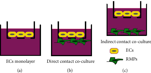
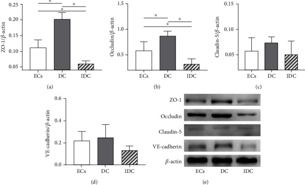
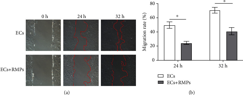
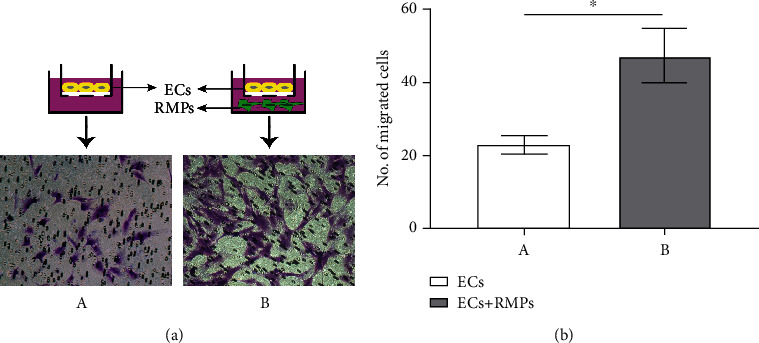
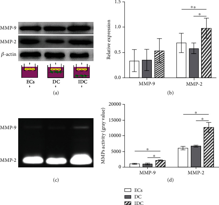
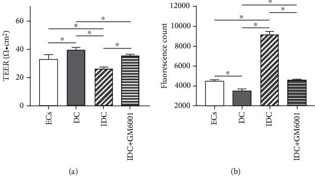
Inner blood-retina barrier (iBRB) is primarily formed of retinal microvascular endothelial cells (ECs) with tight junctions, which are surrounded and supported by retinal microvascular pericytes (RMPs) and basement membrane. Pericytes are believed to be critically involved in the physiology and pathology of iBRB. However, the underlying mechanism remains to be fully elucidated. We developed a novel iBRB model which was composed of primary cultures of rat retinal ECs and RMPs based on Transwell system. We tested the involvement of pericytes in the migration and invasion of ECs, examined the expression and activity of matrix metalloproteinase- (MMP-) 2/MMP-9 in the culture, evaluated the TEER and permeability of iBRB, and assessed the expression of ZO-1, occludin, claudin-5, and VE-cadherin of endothelial junctions. We found that RMPs with indirect contact of ECs can increase the expression of MMP-2 and upgrade the activity of MMP-2/9 in the coculture, which subsequently decreased TJ protein abundance of ZO-1 and occludin in ECs, promoted the migration of ECs, and finally reduced the integrity of iBRB. Taken together, our data show that RMP relative location with ECs is involved in the integrity of iBRB via MMP-2/9 and has important implications for treating diabetic retinopathy and other retinal disorders involving iBRB dysfunction.

摘要

内血视网膜屏障(iBRB)主要由具有紧密连接的视网膜微血管内皮细胞(ECs)形成,其被视网膜微血管周细胞(RMPs)和基膜所包围和支持。周细胞被认为在 iBRB 的生理学和病理学中起着至关重要的作用。然而,其潜在的机制仍有待充分阐明。我们开发了一种基于 Transwell 系统的新型 iBRB 模型,由大鼠视网膜 ECs 和 RMPs 的原代培养物组成。我们测试了周细胞在 EC 迁移和侵袭中的作用,检查了培养物中基质金属蛋白酶-(MMP-)2/MMP-9 的表达和活性,评估了 iBRB 的 TEER 和通透性,并评估了内皮连接的 ZO-1、occludin、claudin-5 和 VE-cadherin 的表达。我们发现,与 ECs 间接接触的 RMPs 可以增加共培养物中 MMP-2 的表达,并提高 MMP-2/9 的活性,随后降低 ECs 中 TJ 蛋白 ZO-1 和 occludin 的丰度,促进 ECs 的迁移,最终降低 iBRB 的完整性。总之,我们的数据表明,RMP 与 ECs 的相对位置通过 MMP-2/9 参与了 iBRB 的完整性,这对于治疗糖尿病性视网膜病变和其他涉及 iBRB 功能障碍的视网膜疾病具有重要意义。

https://cdn.ncbi.nlm.nih.gov/pmc/blobs/183d/8494570/a5d73506d524/DM2021-7124835.006.jpg
https://cdn.ncbi.nlm.nih.gov/pmc/blobs/183d/8494570/4b85b442661b/DM2021-7124835.001.jpg
https://cdn.ncbi.nlm.nih.gov/pmc/blobs/183d/8494570/b02628e85db0/DM2021-7124835.002.jpg
https://cdn.ncbi.nlm.nih.gov/pmc/blobs/183d/8494570/e35e09f829be/DM2021-7124835.003.jpg
https://cdn.ncbi.nlm.nih.gov/pmc/blobs/183d/8494570/63c9d33e3cd4/DM2021-7124835.004.jpg
https://cdn.ncbi.nlm.nih.gov/pmc/blobs/183d/8494570/723b7185b7c5/DM2021-7124835.005.jpg
https://cdn.ncbi.nlm.nih.gov/pmc/blobs/183d/8494570/a5d73506d524/DM2021-7124835.006.jpg

相似文献

1
Pericytes of Indirect Contact Coculture Decrease Integrity of Inner Blood-Retina Barrier Model by Upgrading MMP-2/9 Activity.间接接触共培养的周细胞通过上调 MMP-2/9 活性降低内血视网膜屏障模型的完整性。
Dis Markers. 2021 Sep 29;2021:7124835. doi: 10.1155/2021/7124835. eCollection 2021.
2
Microglia increase tight-junction permeability in coordination with Müller cells under hypoxic condition in an in vitro model of inner blood-retinal barrier.在体外血视网膜内屏障模型中,缺氧条件下小胶质细胞与 Müller 细胞协调增加紧密连接通透性。
Exp Eye Res. 2021 Apr;205:108490. doi: 10.1016/j.exer.2021.108490. Epub 2021 Feb 16.
3
Size-selective and in vitro assessment of inner blood retina barrier permeability.视网膜内血液屏障通透性的大小选择性及体外评估
Methods Mol Biol. 2011;763:355-67. doi: 10.1007/978-1-61779-191-8_24.
4
A novel co-culture model of the blood-retinal barrier based on primary retinal endothelial cells, pericytes and astrocytes.基于原代视网膜血管内皮细胞、周细胞和星形胶质细胞的新型血视网膜屏障共培养模型。
Exp Eye Res. 2012 Mar;96(1):181-90. doi: 10.1016/j.exer.2011.12.003. Epub 2011 Dec 16.
5
The impact of pericytes on the blood-brain barrier integrity depends critically on the pericyte differentiation stage.周细胞对血脑屏障完整性的影响取决于周细胞的分化阶段。
Int J Biochem Cell Biol. 2011 Sep;43(9):1284-93. doi: 10.1016/j.biocel.2011.05.002. Epub 2011 May 12.
6
Regulation of the blood-brain barrier integrity by pericytes via matrix metalloproteinases mediated activation of vascular endothelial growth factor in vitro.体外周细胞通过基质金属蛋白酶介导的血管内皮生长因子激活调节血脑屏障完整性。
Brain Res. 2010 Aug 6;1347:1-10. doi: 10.1016/j.brainres.2010.05.096. Epub 2010 Jun 8.
7
C1q/TNF-Related Protein 3 Prevents Diabetic Retinopathy via AMPK-Dependent Stabilization of Blood-Retinal Barrier Tight Junctions.C1q/TNF 相关蛋白 3 通过 AMPK 依赖途径稳定血视网膜屏障紧密连接来预防糖尿病视网膜病变。
Cells. 2022 Feb 23;11(5):779. doi: 10.3390/cells11050779.
8
Melatonin protects blood-brain barrier integrity and permeability by inhibiting matrix metalloproteinase-9 via the NOTCH3/NF-κB pathway.褪黑素通过NOTCH3/NF-κB途径抑制基质金属蛋白酶-9,从而保护血脑屏障的完整性和通透性。
Aging (Albany NY). 2019 Dec 7;11(23):11391-11415. doi: 10.18632/aging.102537.
9
Erythropoietin protects the inner blood-retinal barrier by inhibiting microglia phagocytosis via Src/Akt/cofilin signalling in experimental diabetic retinopathy.促红细胞生成素通过抑制Src/Akt/丝切蛋白信号通路抑制小胶质细胞吞噬作用来保护实验性糖尿病视网膜病变中的血视网膜内屏障。
Diabetologia. 2021 Jan;64(1):211-225. doi: 10.1007/s00125-020-05299-x. Epub 2020 Oct 26.
10
Inhibition of advanced glycation and absence of galectin-3 prevent blood-retinal barrier dysfunction during short-term diabetes.抑制晚期糖基化和缺乏半乳糖凝集素-3可预防短期糖尿病期间的血视网膜屏障功能障碍。
Exp Diabetes Res. 2007;2007:51837. doi: 10.1155/2007/51837.

引用本文的文献

1
Retinal and choroidal changes following corneal collagen cross-linking in keratoconus: a systematic review and meta-analysis of OCT and OCTA studies.圆锥角膜患者角膜胶原交联术后的视网膜和脉络膜变化:一项关于光学相干断层扫描(OCT)和光学相干断层扫描血管造影(OCTA)研究的系统评价和荟萃分析
Int J Retina Vitreous. 2025 Aug 26;11(1):97. doi: 10.1186/s40942-025-00726-w.
2
Release of Pro-Inflammatory/Angiogenic Factors by Retinal Microvascular Cells Is Mediated by Extracellular Vesicles Derived from M1-Activated Microglia.M1 活化小胶质细胞来源的细胞外囊泡介导视网膜微血管细胞促炎/血管生成因子的释放。
Int J Mol Sci. 2023 Dec 19;25(1):15. doi: 10.3390/ijms25010015.
3

本文引用的文献

1
Pericyte mechanics and mechanobiology.血管周细胞的力学和机械生物学。
J Cell Sci. 2021 Mar 22;134(6):jcs240226. doi: 10.1242/jcs.240226.
2
Assessment of Blood-Brain Barrier Permeability Using Miniaturized Fluorescence Microscopy in Freely Moving Rats.在自由活动大鼠中使用小型荧光显微镜评估血脑屏障通透性
Methods Mol Biol. 2021;2367:123-135. doi: 10.1007/7651_2020_315.
3
Pericyte morphology and function.周细胞的形态与功能。
The role of TET2-mediated ROBO4 hypomethylation in the development of diabetic retinopathy.
TET2 介导的 ROBO4 低甲基化在糖尿病视网膜病变发展中的作用。
J Transl Med. 2023 Jul 10;21(1):455. doi: 10.1186/s12967-023-04310-4.
4
Reduced folate carrier 1 is present in retinal microvessels and crucial for the inner blood retinal barrier integrity.还原叶酸载体 1 存在于视网膜微血管中,对于维持内血液视网膜屏障的完整性至关重要。
Fluids Barriers CNS. 2023 Jun 16;20(1):47. doi: 10.1186/s12987-023-00442-3.
5
A New Pharmacological Vitreolysis through the Supplement of Mixed Fruit Enzymes for Patients with Ocular Floaters or Vitreous Hemorrhage-Induced Floaters.通过补充混合水果酶对飞蚊症或玻璃体出血引起的飞蚊症患者进行新型药理玻璃体溶解术。
J Clin Med. 2022 Nov 13;11(22):6710. doi: 10.3390/jcm11226710.
6
Pathophysiological mechanisms of chronic compressive spinal cord injury due to vascular events.血管事件所致慢性压迫性脊髓损伤的病理生理机制
Neural Regen Res. 2023 Apr;18(4):790-796. doi: 10.4103/1673-5374.353485.
Histol Histopathol. 2021 Jun;36(6):633-643. doi: 10.14670/HH-18-314. Epub 2021 Feb 17.
4
Pericyte-Endothelial Interactions in the Retinal Microvasculature.周细胞-内皮细胞相互作用在视网膜微血管中的作用。
Int J Mol Sci. 2020 Oct 8;21(19):7413. doi: 10.3390/ijms21197413.
5
A New Human Blood-Retinal Barrier Model Based on Endothelial Cells, Pericytes, and Astrocytes.基于内皮细胞、周细胞和星形胶质细胞的新型人血视网膜屏障模型。
Int J Mol Sci. 2020 Feb 27;21(5):1636. doi: 10.3390/ijms21051636.
6
Inner Blood-Retinal Barrier Regulation in Retinopathies.视网膜病变中的内血-视网膜屏障调节。
Adv Exp Med Biol. 2019;1185:329-333. doi: 10.1007/978-3-030-27378-1_54.
7
Effect of gene knockout on retinal vascular form and function.基因敲除对视网膜血管形态和功能的影响。
Physiol Genomics. 2019 Dec 1;51(12):613-622. doi: 10.1152/physiolgenomics.00041.2019. Epub 2019 Nov 11.
8
MMP targeting in the battle for vision: Recent developments and future prospects in the treatment of diabetic retinopathy.靶向 MMP 在视力争夺战中的应用:糖尿病性视网膜病变治疗的最新进展和未来前景。
Life Sci. 2019 Jul 15;229:149-156. doi: 10.1016/j.lfs.2019.05.038. Epub 2019 May 15.
9
The inner blood-retinal barrier: Cellular basis and development.血视网膜内屏障:细胞基础与发育
Vision Res. 2017 Oct;139:123-137. doi: 10.1016/j.visres.2017.05.009. Epub 2017 Jun 27.
10
Accelerated differentiation of human induced pluripotent stem cells to blood-brain barrier endothelial cells.人诱导多能干细胞向血脑屏障内皮细胞的加速分化。
Fluids Barriers CNS. 2017 Apr 13;14(1):9. doi: 10.1186/s12987-017-0059-0.