Department of Neurosurgery, Azienda Ospedaliera Universitaria Pisana (AOUP), University of Pisa, 56100 Pisa, Italy.
Department of Information Engineering, University of Pisa, 56100 Pisa, Italy.
Int J Environ Res Public Health. 2021 Sep 22;18(19):9955. doi: 10.3390/ijerph18199955.
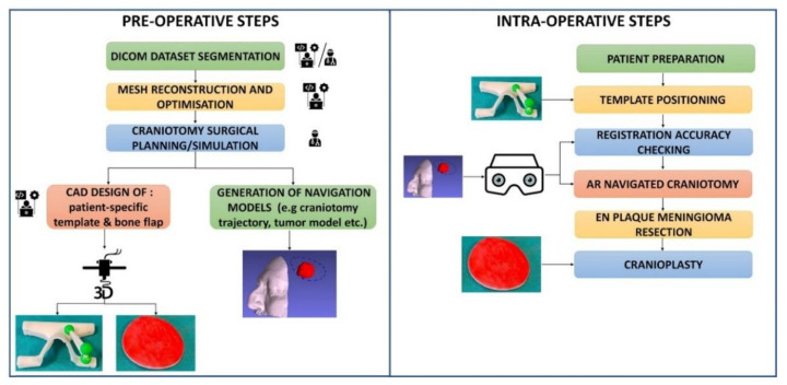
This report discusses the utility of a wearable augmented reality platform in neurosurgery for parasagittal and convexity en plaque meningiomas with bone flap removal and custom-made cranioplasty.
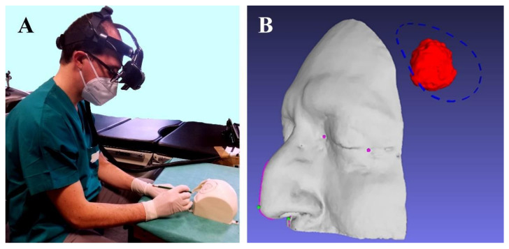
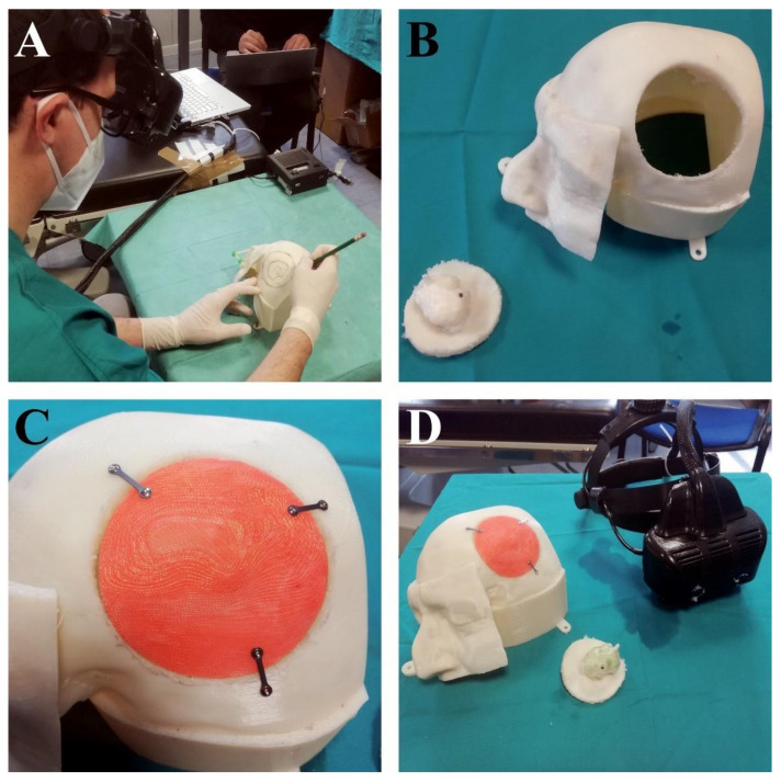
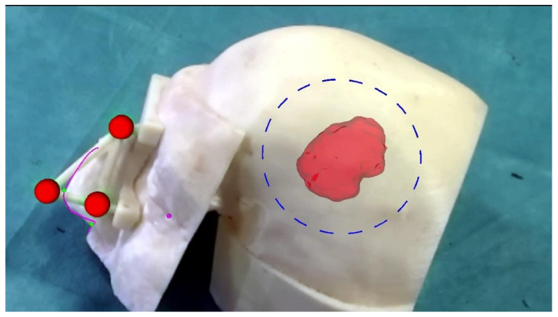
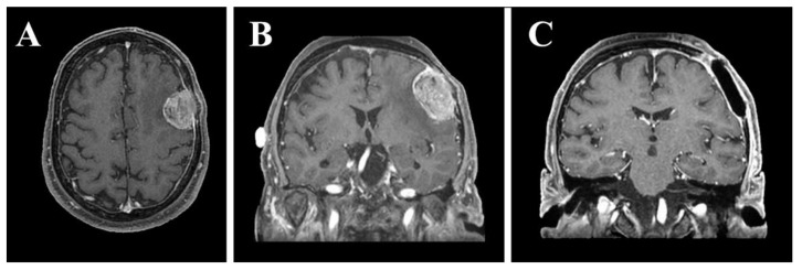
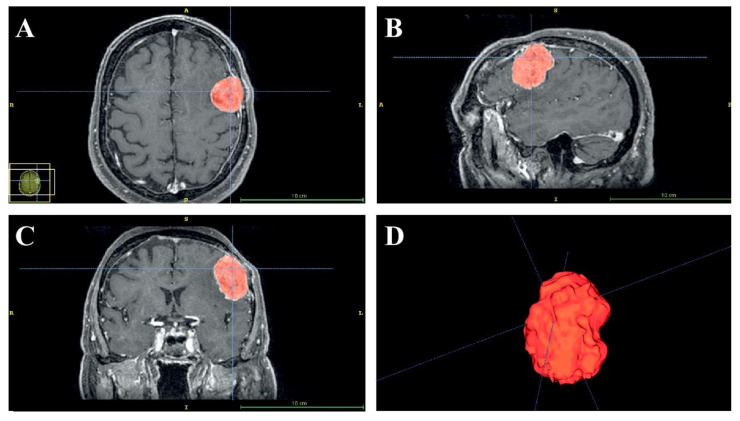
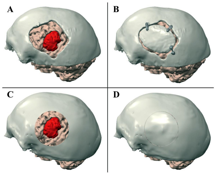
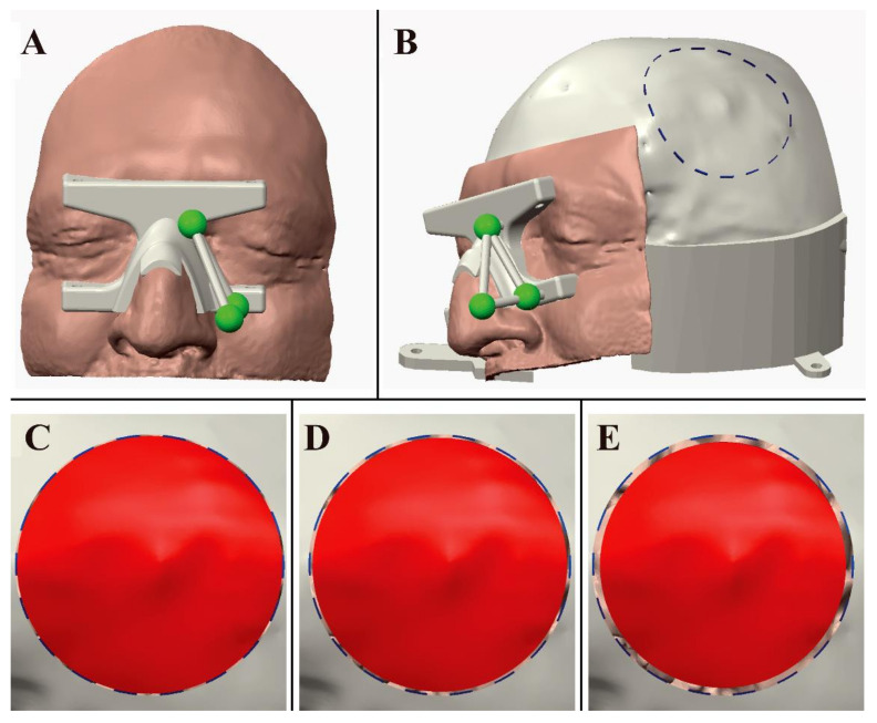
A real patient with en plaque cranial vault meningioma with diffuse and extensive dural involvement, extracranial extension into the calvarium, and homogeneous contrast enhancement on gadolinium-enhanced T1-weighted MRI, was selected for this case study. A patient-specific manikin was designed starting with the segmentation of the patient's preoperative MRI images to simulate a craniotomy procedure. Surgical planning was performed according to the segmented anatomy, and customized bone flaps were designed accordingly. During the surgical simulation stage, the VOSTARS head-mounted display was used to accurately display the planned craniotomy trajectory over the manikin skull. The precision of the craniotomy was assessed based on the evaluation of previously prepared custom-made bone flaps.
A bone flap with a radius 0.5 mm smaller than the radius of an ideal craniotomy fitted perfectly over the performed craniotomy, demonstrating an error of less than ±1 mm in the task execution. The results of this laboratory-based experiment suggest that the proposed augmented reality platform helps in simulating convexity en plaque meningioma resection and custom-made cranioplasty, as carefully planned in the preoperative phase.
Augmented reality head-mounted displays have the potential to be a useful adjunct in tumor surgical resection, cranial vault lesion craniotomy and also skull base surgery, but more study with large series is needed.
本报告讨论了可穿戴式增强现实平台在神经外科中的应用,用于切除带骨瓣的矢状窦和凸面贴骨脑膜瘤,并进行定制颅骨成形术。
选择一名患有颅骨贴骨脑膜瘤的真实患者,该患者肿瘤广泛累及硬脑膜,颅外延伸至颅骨,钆增强 T1 加权 MRI 呈均匀对比增强。本病例研究从患者术前 MRI 图像的分割开始设计患者特异性的人体模型,以模拟开颅手术。根据分割的解剖结构进行手术规划,并相应设计定制骨瓣。在手术模拟阶段,使用 VOSTARS 头戴式显示器准确显示人体模型颅骨上的计划开颅轨迹。根据预先准备的定制骨瓣的评估来评估开颅的精度。
比理想开颅半径小 0.5 毫米的骨瓣完美贴合在完成的开颅上,表明在执行任务时误差小于±1 毫米。该实验室实验的结果表明,所提出的增强现实平台有助于模拟凸面贴骨脑膜瘤切除术和定制颅骨成形术,如同术前精心规划的那样。
增强现实头戴式显示器有可能成为肿瘤切除术、颅顶病变开颅术和颅底手术的有用辅助手段,但需要更多的大系列研究。