Mathew Shibu, Saboukh Islam, Singh Parminder, Fries Bastian, Johnson Victoria, Schneider Nikita, Fraebel Christian, Chasan Ritvan, Hamm Christian W, Schmitt Jörn
Department of Cardiology, University Hospital of Giessen, 35392 Giessen, Germany.
J Clin Med. 2021 Sep 28;10(19):4478. doi: 10.3390/jcm10194478.
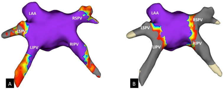
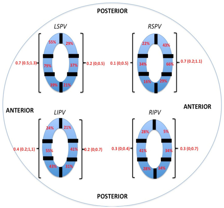
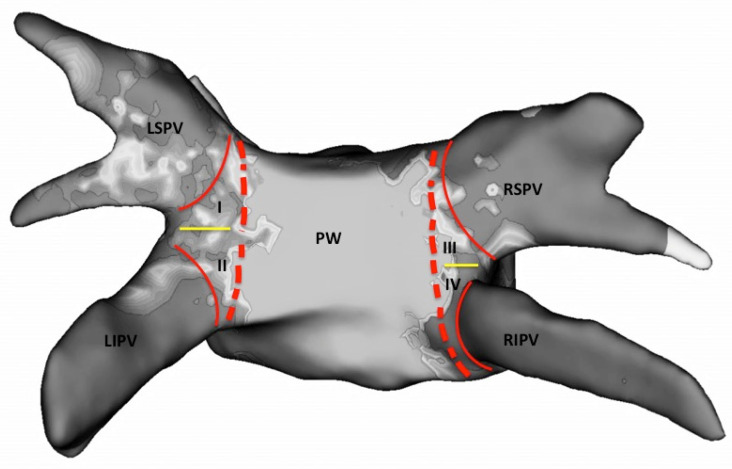
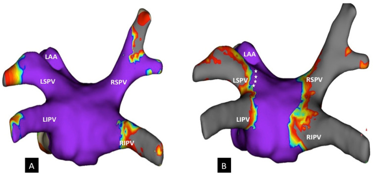
Cryoballoon (CB)-based pulmonary vein isolation (PVI) is an effective treatment modality for patients with atrial fibrillation (AF) with encouraging acute and long-term outcome data. However, the size of collaterally created lesion sets adjacent to the pulmonary veins (PVs) remains unclear, especially when CB ablation is performed with individualized time-to-isolation (TTI) protocols. This study seeks to investigate the extension of lesions at the posterior wall and the roof of the left atrium (LA). Thirty patients with paroxysmal or persistent AF underwent ablation with a fourth-generation CB. The individual freeze-cycle duration was set at TTI + 120 s. A total of 120 PVs were identified, and all were successfully isolated. A three-dimensional electroanatomical high-density (HD) mapping of the LA was performed in every patient before and after PVI. The surface areas of the posterior wall and LA roof were measured and compared with lesion extension after PVI. After CB ablation, 65.6 ± 16.9% of the posterior wall and 75.4 ± 18.4% of the LA roof remained unablated. In addition, non-antral lesion formation was observed in every patient in at least one PV. After CB ablation, anterior antral parts of the superior PVs showed the greatest unablated areas compared with the other antral areas. HD re-mapping after CB-based PVI demonstrated that major regions of the posterior wall and roof remained electrically normal and unaffected. Unablated antral areas were localized predominantly in the anterior segments of the superior PVs and may be partly responsible for AF recurrence.
基于冷冻球囊(CB)的肺静脉隔离(PVI)是治疗心房颤动(AF)患者的一种有效治疗方式,其急性和长期疗效数据令人鼓舞。然而,与肺静脉(PVs)相邻的侧支形成的损伤灶大小仍不明确,尤其是在采用个体化隔离时间(TTI)方案进行CB消融时。本研究旨在调查左心房(LA)后壁和顶部的损伤范围。30例阵发性或持续性AF患者接受了第四代CB消融。个体冷冻周期持续时间设定为TTI + 120秒。共识别出120条PVs,均成功隔离。在PVI前后,对每位患者进行了LA的三维电解剖高密度(HD)标测。测量后壁和LA顶部的表面积,并与PVI后的损伤范围进行比较。CB消融后,后壁的65.6±16.9%和LA顶部的75.4±18.4%未被消融。此外,在每位患者的至少一条PV中观察到非窦周损伤形成。CB消融后,与其他窦周区域相比,上PVs的前窦周部分显示出最大的未消融区域。基于CB的PVI后的HD重新标测显示,后壁和顶部的主要区域保持电活动正常且未受影响。未消融的窦周区域主要位于上PVs的前段,可能是AF复发的部分原因。