Penn Statistics in Imaging and Visualization Endeavor (PennSIVE) Center, Department of Biostatistics, Epidemiology, and Informatics, University of Pennsylvania, Philadelphia, PA, USA.
Translational Neuroradiology Section, National Institute of Neurological Disorders and Stroke (NINDS), National Institutes of Health (NIH), Bethesda, MD, USA; Department of Neurology, Cedars-Sinai Medical Center, Los Angeles, CA, USA.
Neuroimage Clin. 2021;32:102796. doi: 10.1016/j.nicl.2021.102796. Epub 2021 Aug 27.
The presence of a paramagnetic rim around a white matter lesion has recently been shown to be a hallmark of a particular pathological type of multiple sclerosis lesion. Increased prevalence of these paramagnetic rim lesions is associated with a more severe disease course in MS, but manual identification is time-consuming. We present APRL, a method to automatically detect paramagnetic rim lesions on 3T T2*-phase images.
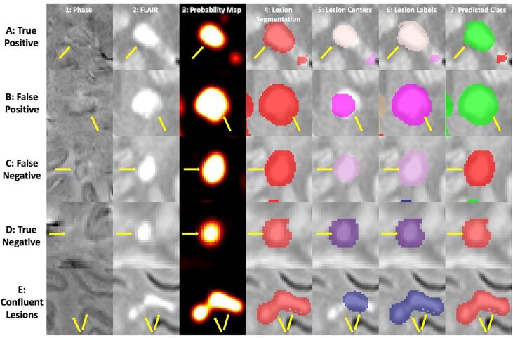
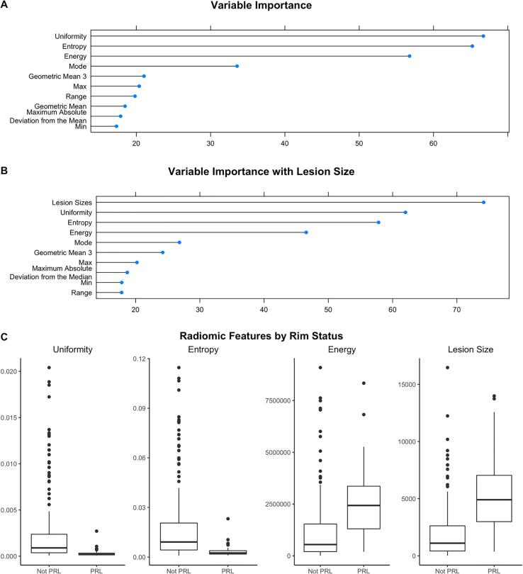
T1-weighted, T2-FLAIR, and T2*-phase MRI of the brain were collected at 3T for 20 subjects with MS. The images were then processed with automated lesion segmentation, lesion center detection, lesion labelling, and lesion-level radiomic feature extraction. A total of 951 lesions were identified, 113 (12%) of which contained a paramagnetic rim. We divided our data into a training set (16 patients, 753 lesions) and a testing set (4 patients, 198 lesions), fit a random forest classification model on the training set, and assessed our ability to classify paramagnetic rim lesions on the test set.
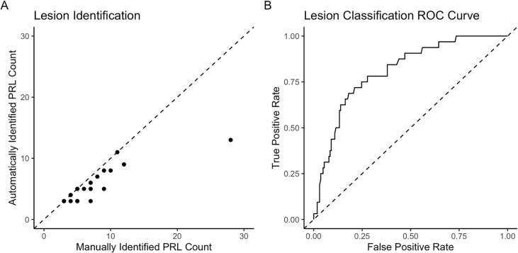
The number of paramagnetic rim lesions per subject identified via our automated lesion labelling method was highly correlated with the gold standard count per subject, r = 0.86 (95% CI [0.68, 0.94]). The classification algorithm using radiomic features classified lesions with an area under the curve of 0.82 (95% CI [0.74, 0.92]).
This study develops a fully automated technique, APRL, for the detection of paramagnetic rim lesions using standard T1 and FLAIR sequences and a T2*phase sequence obtained on 3T MR images.
最近研究表明,在脑白质病变周围存在顺磁性边缘是一种特定的多发性硬化病变的病理类型的标志。这些顺磁性边缘病变的发生率增加与多发性硬化症更严重的病程有关,但手动识别很耗时。我们提出了 APRL,一种用于在 3T T2*-相图像上自动检测顺磁性边缘病变的方法。
对 20 名多发性硬化症患者的大脑进行了 3T 的 T1 加权、T2-FLAIR 和 T2*-相 MRI 采集。然后,对图像进行自动病变分割、病变中心检测、病变标记和病变水平的放射组学特征提取。共识别出 951 个病变,其中 113 个(12%)包含顺磁性边缘。我们将数据分为训练集(16 例患者,753 个病变)和测试集(4 例患者,198 个病变),在训练集上拟合随机森林分类模型,并评估我们在测试集上分类顺磁性边缘病变的能力。
我们的自动病变标记方法识别的每个患者的顺磁性边缘病变数量与金标准计数高度相关,r = 0.86(95%CI[0.68, 0.94])。使用放射组学特征的分类算法对病变的曲线下面积为 0.82(95%CI[0.74, 0.92])。
本研究开发了一种完全自动化的技术,APRL,用于使用标准的 T1 和 FLAIR 序列以及在 3T MRI 图像上获得的 T2*相序列检测顺磁性边缘病变。