Department of Oral Anatomy and Cell Biology, Graduate School of Medical and Dental Sciences, Kagoshima University, 8-35-1 Sakuragaoka, Kagoshima, Kagoshima, 890-8544, Japan.
Department of Psychosomatic Internal Medicine, Graduate School of Medical and Dental Sciences, Kagoshima University, 8-35-1 Sakuragaoka, Kagoshima, Kagoshima, 890-8544, Japan.
J Neuroinflammation. 2021 Oct 13;18(1):227. doi: 10.1186/s12974-021-02283-z.
Macrophages in the peripheral nervous system are key players in the repair of nerve tissue and the development of neuropathic pain due to peripheral nerve injury. However, there is a lack of information on the origin and morphological features of macrophages in sensory ganglia after peripheral nerve injury, unlike those in the brain and spinal cord. We analyzed the origin and morphological features of sensory ganglionic macrophages after nerve ligation or transection using wild-type mice and mice with bone-marrow cell transplants.
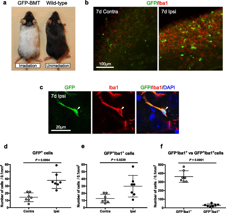
After protecting the head of C57BL/6J mice with lead caps, they were irradiated and transplanted with bone-marrow-derived cells from GFP transgenic mice. The infraorbital nerve of a branch of the trigeminal nerve of wild-type mice was ligated or the infraorbital nerve of GFP-positive bone-marrow-cell-transplanted mice was transected. After immunostaining the trigeminal ganglion, the structures of the ganglionic macrophages, neurons, and satellite glial cells were analyzed using two-dimensional or three-dimensional images.
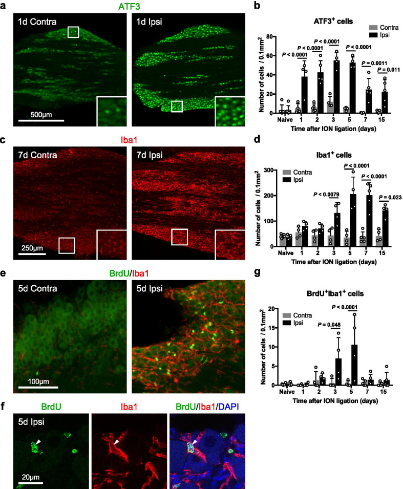
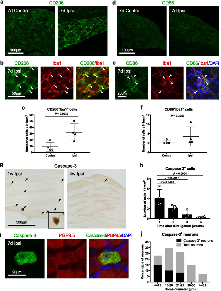
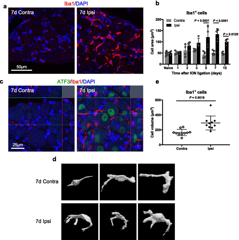
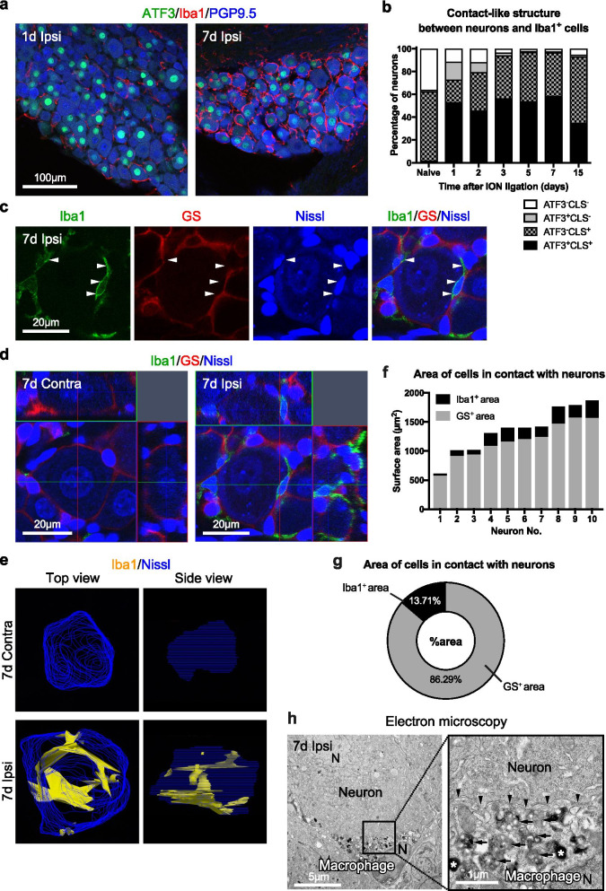
The number of damaged neurons in the trigeminal ganglion increased from day 1 after infraorbital nerve ligation. Ganglionic macrophages proliferated from days 3 to 5. Furthermore, the numbers of macrophages increased from days 3 to 15. Bone-marrow-derived macrophages increased on day 7 after the infraorbital nerve was transected in the trigeminal ganglion of GFP-positive bone-marrow-cell-transplanted mice but most of the ganglionic macrophages were composed of tissue-resident cells. On day 7 after infraorbital nerve ligation, ganglionic macrophages increased in volume, extended their processes between the neurons and satellite glial cells, and contacted these neurons. Most of the ganglionic macrophages showed an M2 phenotype when contact was observed, and little neuronal cell death occurred.
Most of the macrophages that appear after a nerve injury are tissue-resident, and these make direct contact with damaged neurons that act in a tissue-protective manner in the M2 phenotype. These results imply that tissue-resident macrophages signal to neurons directly through physical contact.
在外周神经损伤后,周围神经系统中的巨噬细胞是修复神经组织和发展神经病理性疼痛的关键因素。然而,与大脑和脊髓不同,对于感觉神经节中巨噬细胞的起源和形态特征,我们对外周神经损伤后的信息知之甚少。我们使用野生型小鼠和骨髓细胞移植小鼠分析了神经结扎或横断后感觉神经节中巨噬细胞的起源和形态特征。
用铅帽保护 C57BL/6J 小鼠的头部,然后对其进行辐照并移植 GFP 转基因小鼠的骨髓细胞。结扎野生型小鼠三叉神经分支的眶下神经或横断 GFP 阳性骨髓细胞移植小鼠的眶下神经。对三叉神经节进行免疫染色后,使用二维或三维图像分析神经节巨噬细胞、神经元和卫星神经胶质细胞的结构。
眶下神经结扎后第 1 天,三叉神经节中受损神经元的数量增加。从第 3 天到第 5 天,神经节巨噬细胞增殖。此外,从第 3 天到第 15 天,巨噬细胞的数量增加。在 GFP 阳性骨髓细胞移植小鼠的三叉神经节中,眶下神经横断后第 7 天,骨髓源性巨噬细胞增加,但大多数神经节巨噬细胞由组织驻留细胞组成。眶下神经结扎后第 7 天,神经节巨噬细胞体积增大,其突起在神经元和卫星神经胶质细胞之间延伸,并与这些神经元接触。当观察到接触时,大多数神经节巨噬细胞表现出 M2 表型,很少发生神经元细胞死亡。
大多数在神经损伤后出现的巨噬细胞是组织驻留的,这些巨噬细胞以 M2 表型与受损神经元直接接触,起到组织保护作用。这些结果表明,组织驻留的巨噬细胞通过物理接触直接向神经元发出信号。