Breen Alexander, De Carvalho Diana, Funabashi Martha, Kawchuk Greg, Pagé Isabelle, Wong Arnold Y L, Breen Alan
AECC University College, Bournemouth, United Kingdom.
Division of Community Health and Humanities, Faculty of Medicine, Memorial University of Newfoundland, St. John's, NL, Canada.
Front Bioeng Biotechnol. 2021 Sep 27;9:745837. doi: 10.3389/fbioe.2021.745837. eCollection 2021.
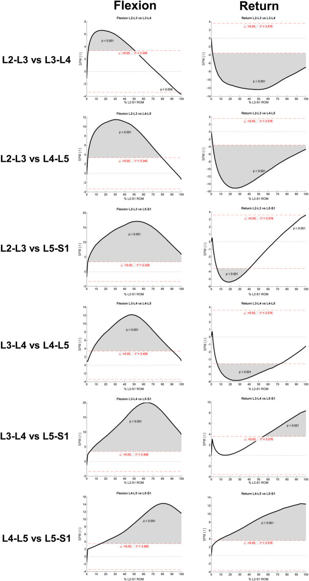
Lumbar instability has long been thought of as the failure of lumbar vertebrae to maintain their normal patterns of displacement. However, it is unknown what these patterns consist of. Research using quantitative fluoroscopy (QF) has shown that continuous lumbar intervertebral patterns of rotational displacement can be reliably measured during standing flexion and return motion using standardised protocols and can be used to assess patients with suspected lumbar spine motion disorders. However, normative values are needed to make individualised comparisons. One hundred and thirty-one healthy asymptomatic participants were recruited and performed guided flexion and return motion by following the rotating arm of an upright motion frame. Fluoroscopic image acquisition at 15fps was performed and individual intervertebral levels from L2-3 to L5-S1 were tracked and analysed during separate outward flexion and return phases. Results were presented as proportional intervertebral motion representing these phases using continuous means and 95%CIs, followed by verification of the differences between levels using Statistical Parametric Mapping (SPM). A secondary analysis of 8 control participants matched to 8 patients with chronic, non-specific low back pain (CNSLBP) was performed for comparison. One hundred and twenty-seven asymptomatic participants' data were analysed. Their ages ranged from 18 to 70 years (mean 38.6) with mean body mass index 23.8 kg/m 48.8% were female. Both the flexion and return phases for each level evidenced continuous change in mean proportional motion share, with narrow confidence intervals, highly significant differences and discrete motion paths between levels as confirmed by SPM. Patients in the secondary analysis evidenced significantly less L5-S1 motion than controls ( < 0.05). A reference database of spinal displacement patterns during lumbar (L2-S1) intersegmental flexion and return motion using a standardised motion protocol using fluoroscopy is presented. Spinal displacement patterns in asymptomatic individuals were found to be distinctive and consistent for each intervertebral level, and to continuously change during bending and return. This database may be used to allow continuous intervertebral kinematics to drive dynamic models of joint and muscular forces as well as reference values against which to make patient-specific comparisons in suspected cases of lumbar spine motion disorders.
长期以来,腰椎不稳一直被认为是腰椎无法维持其正常位移模式。然而,这些模式具体包括哪些内容尚不清楚。使用定量荧光透视法(QF)的研究表明,在站立位屈伸和恢复运动过程中,通过标准化方案可以可靠地测量腰椎连续的椎间旋转位移模式,并且可用于评估疑似腰椎运动障碍的患者。然而,需要规范值来进行个体化比较。招募了131名健康无症状参与者,他们通过跟随直立运动框架的旋转臂进行有指导的屈伸和恢复运动。以每秒15帧的速度进行荧光透视图像采集,并在单独的向外屈伸和恢复阶段跟踪和分析从L2-3到L5-S1的各个椎间水平。结果以代表这些阶段的比例椎间运动呈现,使用连续均值和95%置信区间,随后使用统计参数映射(SPM)验证各水平之间的差异。对8名与8名慢性非特异性下腰痛(CNSLBP)患者匹配的对照参与者进行了二次分析以作比较。分析了127名无症状参与者的数据。他们的年龄在18至70岁之间(平均38.6岁),平均体重指数为23.8kg/m²,48.8%为女性。每个水平的屈伸和恢复阶段均显示平均比例运动份额持续变化,置信区间狭窄,各水平之间存在高度显著差异和离散的运动路径,这一点经SPM证实。二次分析中的患者在L5-S1节段的运动明显少于对照组(P<0.05)。本文给出了一个使用荧光透视法的标准化运动方案,在腰椎(L2-S1)节段间屈伸和恢复运动过程中的脊柱位移模式参考数据库。发现无症状个体的脊柱位移模式在每个椎间水平上都是独特且一致的,并且在弯曲和恢复过程中持续变化。该数据库可用于使连续的椎间运动学驱动关节和肌肉力量的动态模型,以及在疑似腰椎运动障碍病例中进行患者特异性比较的参考值。