• 文献检索
  • 文档翻译
  • 深度研究
  • 学术资讯
  • Suppr Zotero 插件Zotero 插件
  • 邀请有礼
  • 套餐&价格
  • 历史记录
应用&插件
Suppr Zotero 插件Zotero 插件浏览器插件Mac 客户端Windows 客户端微信小程序
定价
高级版会员购买积分包购买API积分包
服务
文献检索文档翻译深度研究API 文档MCP 服务
关于我们
关于 Suppr公司介绍联系我们用户协议隐私条款
关注我们

Suppr 超能文献

核心技术专利:CN118964589B侵权必究
粤ICP备2023148730 号-1Suppr @ 2026

文献检索

告别复杂PubMed语法,用中文像聊天一样搜索,搜遍4000万医学文献。AI智能推荐,让科研检索更轻松。

立即免费搜索

文件翻译

保留排版,准确专业,支持PDF/Word/PPT等文件格式,支持 12+语言互译。

免费翻译文档

深度研究

AI帮你快速写综述,25分钟生成高质量综述,智能提取关键信息,辅助科研写作。

立即免费体验

高频超声诊断颈丛神经肿瘤。

Diagnosis of cervical plexus tumours by high-frequency ultrasonography.

机构信息

Department of Ultrasound, Xijing Hospital, Fourth Military Medical University, No. 127 Changle West Road, Xi'an, 710032, Shaanxi, China.

Department of Special Clinic, Rehabilitation Center, Joint Logistics Support Force of PLA, Lintong, 710600, Shaanxi, China.

出版信息

BMC Med Imaging. 2021 Oct 14;21(1):148. doi: 10.1186/s12880-021-00682-5.

DOI:10.1186/s12880-021-00682-5
PMID:34649505
原文链接:https://pmc.ncbi.nlm.nih.gov/articles/PMC8515767/
Abstract

BACKGROUND

Cervical plexus (CP) tumours are difficult to diagnose because of atypical symptoms. This study aimed to summarize the features of a normal CP and CP tumours observed on high-frequency ultrasonography.

METHODS

The ultrasound data of 11 CP tumour patients and 22 normal volunteers were collected. All 11 patients underwent magnetic resonance imaging (MRI), and 4 patients also underwent computed tomography (CT). The imaging data were compared with surgery and pathology data.

RESULTS

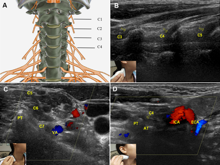
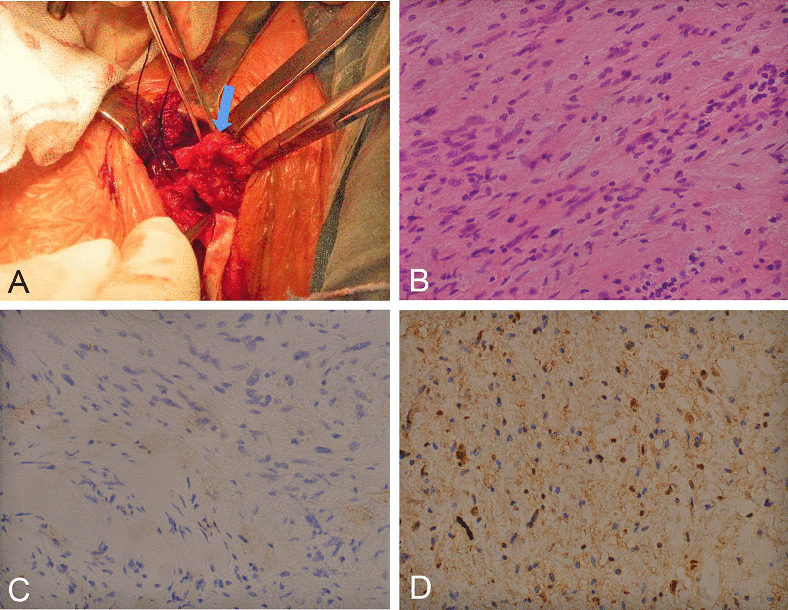
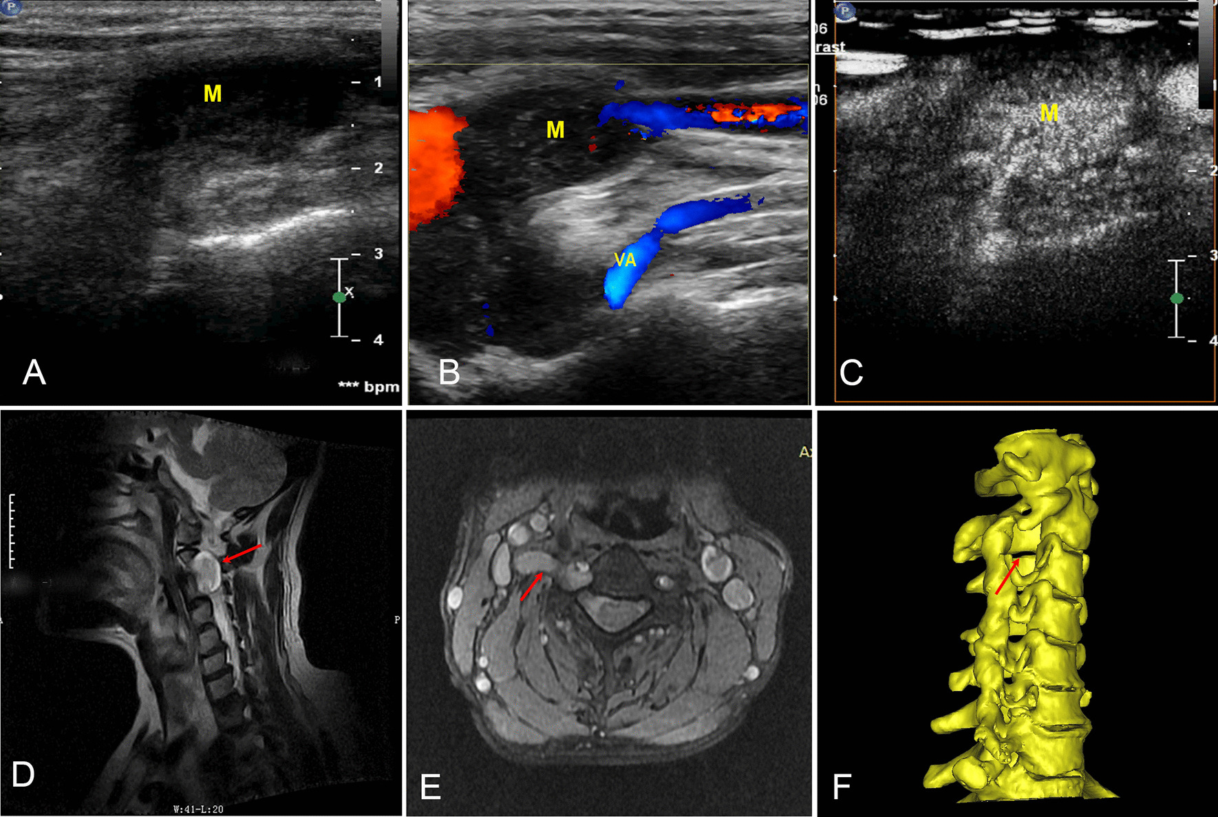
The C7 vertebra and bifurcation of the carotid common artery (CCA) were useful anatomic markers for identifying the CP. In contrast to the C1 nerve (22.7%), the C2-4 nerves were well displayed and thinner than the brachial plexus (P < 0.05). CP tumours were more common in females (72.7%) and generally located at C4 (72.7%) on the right side (81.8%). Additionally, the nerve trunk in tumour patients was obviously wider than that in normal controls (7.49 ± 1.03 mm vs 2.67 ± 0.36 mm, P < 0.01). Compared with pathology, the diagnostic rates of CP tumours by MRI, CT and high-frequency ultrasound were 72.7% (8/11), 25% (1/4) and 90.9% (10/11), respectively.

CONCLUSIONS

The diagnosis of CP neuropathy is accurate and reliable by high-frequency ultrasound, and the C7 vertebra and bifurcation of the CCA are useful anatomic markers in CP ultrasonography.

摘要

背景

由于不典型症状,颈丛(CP)肿瘤的诊断较为困难。本研究旨在总结高频超声观察到的正常 CP 和 CP 肿瘤的特征。

方法

收集了 11 例 CP 肿瘤患者和 22 例正常志愿者的超声数据。所有 11 例患者均行磁共振成像(MRI)检查,4 例患者还行计算机断层扫描(CT)检查。将影像学数据与手术和病理数据进行比较。

结果

C7 椎体和颈总动脉(CCA)分叉是识别 CP 的有用解剖标志物。与 C1 神经(22.7%)相比,C2-4 神经显示得更好,也比臂丛更细(P<0.05)。CP 肿瘤更常见于女性(72.7%),通常位于右侧 C4(72.7%)(81.8%)。此外,肿瘤患者的神经干明显比正常对照组宽(7.49±1.03mm 比 2.67±0.36mm,P<0.01)。与病理相比,MRI、CT 和高频超声诊断 CP 肿瘤的准确率分别为 72.7%(8/11)、25%(1/4)和 90.9%(10/11)。

结论

高频超声对 CP 神经病变的诊断准确可靠,C7 椎体和 CCA 分叉是 CP 超声检查的有用解剖标志物。

https://cdn.ncbi.nlm.nih.gov/pmc/blobs/bbbf/8515767/8e63d105dbf0/12880_2021_682_Fig3_HTML.jpg
https://cdn.ncbi.nlm.nih.gov/pmc/blobs/bbbf/8515767/b5a78b797659/12880_2021_682_Fig1_HTML.jpg
https://cdn.ncbi.nlm.nih.gov/pmc/blobs/bbbf/8515767/b8a8aa96580c/12880_2021_682_Fig2_HTML.jpg
https://cdn.ncbi.nlm.nih.gov/pmc/blobs/bbbf/8515767/8e63d105dbf0/12880_2021_682_Fig3_HTML.jpg
https://cdn.ncbi.nlm.nih.gov/pmc/blobs/bbbf/8515767/b5a78b797659/12880_2021_682_Fig1_HTML.jpg
https://cdn.ncbi.nlm.nih.gov/pmc/blobs/bbbf/8515767/b8a8aa96580c/12880_2021_682_Fig2_HTML.jpg
https://cdn.ncbi.nlm.nih.gov/pmc/blobs/bbbf/8515767/8e63d105dbf0/12880_2021_682_Fig3_HTML.jpg

相似文献

1
Diagnosis of cervical plexus tumours by high-frequency ultrasonography.高频超声诊断颈丛神经肿瘤。
BMC Med Imaging. 2021 Oct 14;21(1):148. doi: 10.1186/s12880-021-00682-5.
2
[Ultrasound-guided intermediate cervical plexus block. Anatomical study].[超声引导下颈丛神经中支阻滞。解剖学研究]
Anaesthesist. 2015 Jun;64(6):446-50. doi: 10.1007/s00101-015-0018-6. Epub 2015 May 27.
3
[Computerized tomography and magnetic resonance assessment of multiple bilateral neurinomas of the cervical plexus and the phrenic nerve].[颈丛和膈神经多发性双侧神经鞘瘤的计算机断层扫描和磁共振评估]
Radiol Med. 1991 May;81(5):735-7.
4
Perineural Spread of Melanoma to the Brachial Plexus: Identifying the Anatomic Pathway(s).黑色素瘤沿神经向臂丛神经的扩散:确定解剖路径
World Neurosurg. 2018 Mar;111:e921-e926. doi: 10.1016/j.wneu.2018.01.031. Epub 2018 Jan 8.
5
Cystic schwannoma of the cervical plexus masquerading as a type II second branchial cleft cyst.伪装成II型第二鳃裂囊肿的颈丛囊性神经鞘瘤。
Eur Arch Otorhinolaryngol. 2009 Mar;266(3):459-62. doi: 10.1007/s00405-008-0678-9. Epub 2008 Apr 17.
6
Schwannoma of the brachial plexus: cross-sectional imaging diagnosis using CT, sonography, and MR imaging.臂丛神经鞘瘤:使用CT、超声和磁共振成像的横断面影像诊断
Eur Radiol. 2003 Aug;13(8):1872-5. doi: 10.1007/s00330-002-1680-z. Epub 2002 Oct 26.
7
Ultrasound of the Brachial Plexus.臂丛神经超声检查
Semin Musculoskelet Radiol. 2018 Jul;22(3):323-333. doi: 10.1055/s-0038-1645862. Epub 2018 May 23.
8
Diagnosis of closed injury and neoplasm of the brachial plexus by ultrasonography.超声检查对臂丛神经闭合性损伤及肿瘤的诊断
J Clin Ultrasound. 2014 Sep;42(7):417-22. doi: 10.1002/jcu.22155. Epub 2014 Mar 26.
9
An anatomical basis for blocking of the deep cervical plexus and cervical sympathetic tract using an ultrasound-guided technique.超声引导下阻滞颈深丛和颈交感干的解剖学基础。
Anesth Analg. 2010 Mar 1;110(3):964-8. doi: 10.1213/ANE.0b013e3181c91ea0. Epub 2009 Dec 15.
10
High-Resolution Ultrasonography of the Transverse Cervical Nerve.颈椎横突超声检查
Ultrasound Med Biol. 2020 Jul;46(7):1599-1607. doi: 10.1016/j.ultrasmedbio.2020.02.003. Epub 2020 Apr 9.

引用本文的文献

1
Diagnosis of obstetric brachial plexus injury in a 2-year-old girl using high‑frequency ultrasonography.高频超声在 2 岁女孩产瘫诊断中的应用
Pediatr Radiol. 2024 Feb;54(2):362-366. doi: 10.1007/s00247-023-05835-y. Epub 2023 Dec 28.
2
Diagnosis of thoracic outlet syndrome with the lower trunk compression of brachial plexus by high-frequency ultrasonography.高频超声检查下干臂丛神经受压诊断胸廓出口综合征。
BMC Musculoskelet Disord. 2023 Aug 29;24(1):690. doi: 10.1186/s12891-023-06762-7.
3
Early Diagnosis of Herpes Zoster Neuralgia: A Narrative Review.

本文引用的文献

1
Superb microvascular imaging technique in depicting vascularity in focal liver lesions: more hypervascular supply patterns were depicted in hepatocellular carcinoma.高超的微血管成像技术在描绘局灶性肝脏病变的血管生成中的应用:肝细胞癌中更多的高血管供应模式被描绘出来。
Cancer Imaging. 2019 Dec 30;19(1):92. doi: 10.1186/s40644-019-0277-6.
2
Ultra-high-frequency Ultrasound to Assess Nerve Fascicles in Median Nerve Traumatic Neuroma.超高频率超声评估正中神经创伤性神经瘤中的神经束
Cureus. 2019 Jun 10;11(6):e4871. doi: 10.7759/cureus.4871.
3
Primary Application of Micro-Flow Imaging Technology in the Diagnosis of Hepatic Tumors.
带状疱疹后神经痛的早期诊断:一篇叙述性综述
Pain Ther. 2023 Aug;12(4):893-901. doi: 10.1007/s40122-023-00510-4. Epub 2023 May 2.
微流成像技术在肝脏肿瘤诊断中的主要应用
Ultrasound Med Biol. 2019 Feb;45(2):395-401. doi: 10.1016/j.ultrasmedbio.2018.09.025. Epub 2018 Nov 5.
4
Brachial plexus schwannoma mimicking cervical lymphadenopathy: A case report with emphasis on imaging features.酷似颈部淋巴结病的臂丛神经鞘瘤:1例报告并重点讨论影像特征
Medicine (Baltimore). 2018 Oct;97(42):e12880. doi: 10.1097/MD.0000000000012880.
5
Surgical Approach to Injuries of the Cervical Plexus and Its Peripheral Nerve Branches.颈部丛及其周围神经分支损伤的手术入路。
Plast Reconstr Surg. 2018 Apr;141(4):1021-1025. doi: 10.1097/PRS.0000000000004240.
6
A Cervical Schwannoma Masquerading as a Thyroid Nodule.一例伪装成甲状腺结节的颈部神经鞘瘤。
Eur Thyroid J. 2017 Jul;6(4):216-220. doi: 10.1159/000454877. Epub 2017 Jan 21.
7
Diagnosis of closed injury and neoplasm of the brachial plexus by ultrasonography.超声检查对臂丛神经闭合性损伤及肿瘤的诊断
J Clin Ultrasound. 2014 Sep;42(7):417-22. doi: 10.1002/jcu.22155. Epub 2014 Mar 26.
8
Anatomy of the neck.颈部解剖学。
Clin Plast Surg. 2014 Jan;41(1):1-6. doi: 10.1016/j.cps.2013.09.016.
9
High-resolution ultrasonography in the diagnosis and intraoperative management of peripheral nerve lesions.高频超声在周围神经病变的诊断和术中处理中的应用。
J Neurosurg. 2011 Jan;114(1):206-11. doi: 10.3171/2010.2.JNS091324. Epub 2010 Mar 12.
10
Extracranial head and neck schwannomas--a 10-year review.颅外头颈部神经鞘瘤——一项为期10年的回顾
Auris Nasus Larynx. 2007 Sep;34(3):353-9. doi: 10.1016/j.anl.2007.01.006. Epub 2007 Mar 21.