General Medical Research Service, James J. Peters Department of Veterans Affairs Medical Center, 130 West Kingsbridge Road, Bronx, NY, 10468, USA.
Department of Psychiatry, Icahn School of Medicine at Mount Sinai, One Gustave Levy Place, New York, NY, 10029, USA.
Acta Neuropathol Commun. 2021 Oct 15;9(1):167. doi: 10.1186/s40478-021-01269-5.
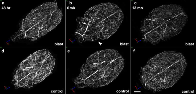
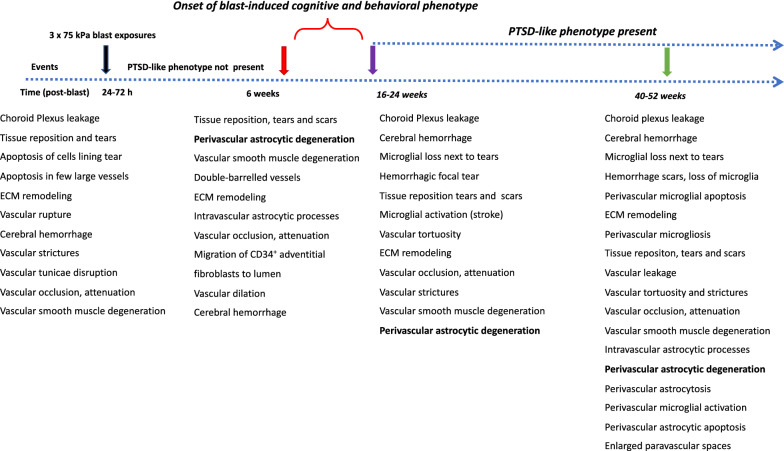
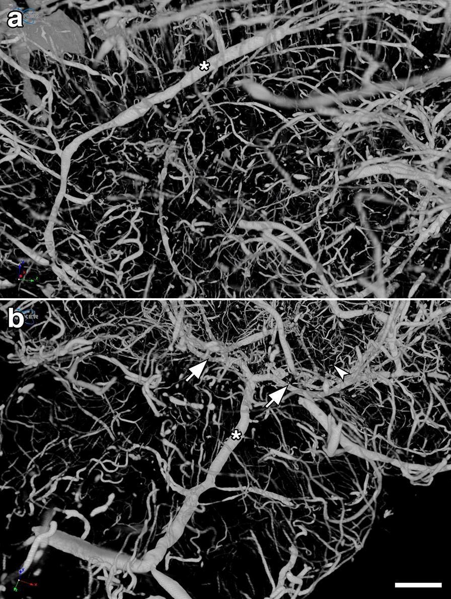
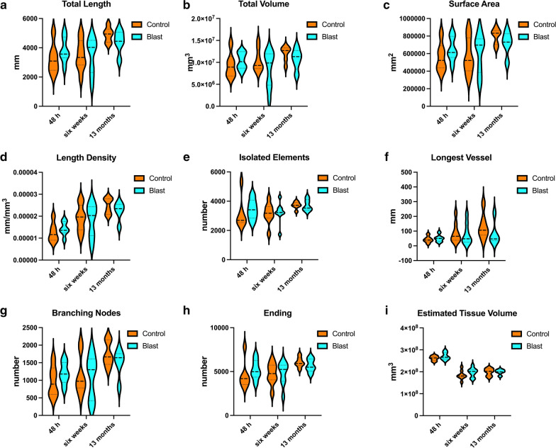
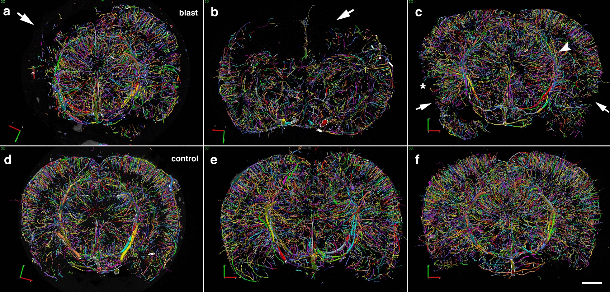
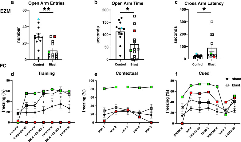
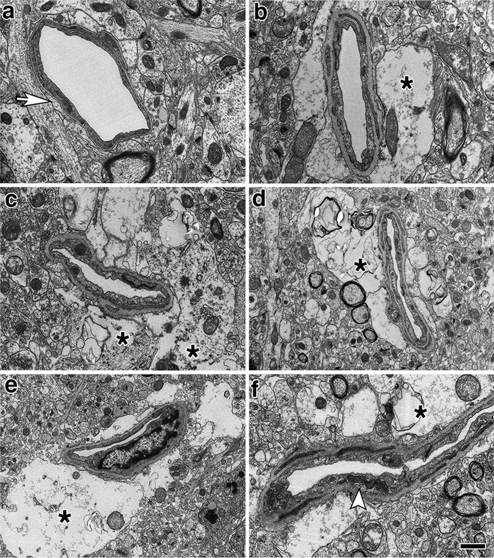
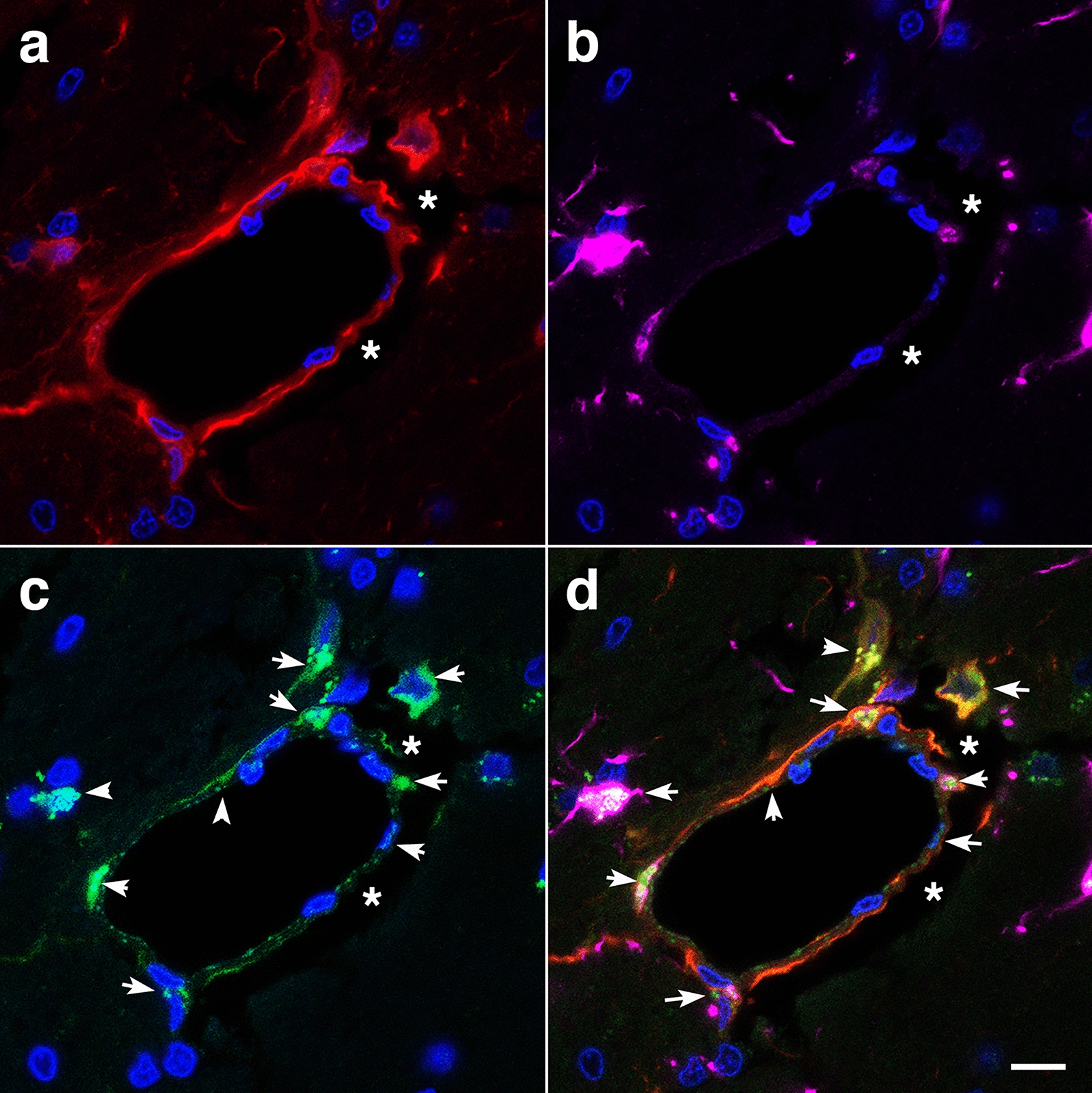
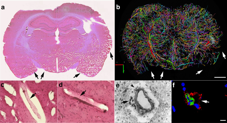
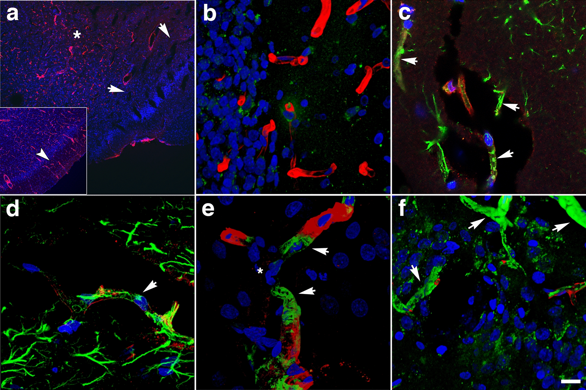
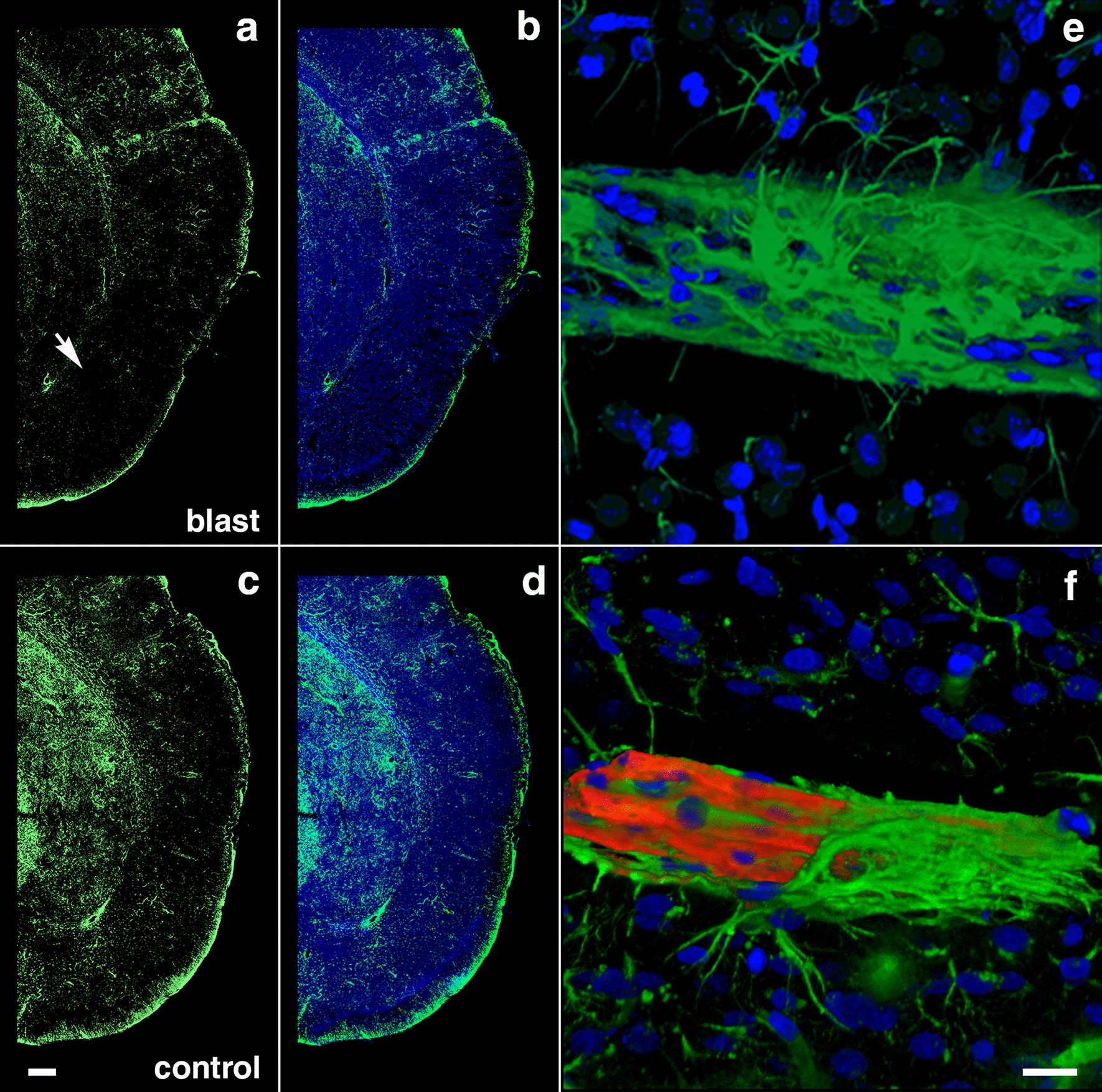
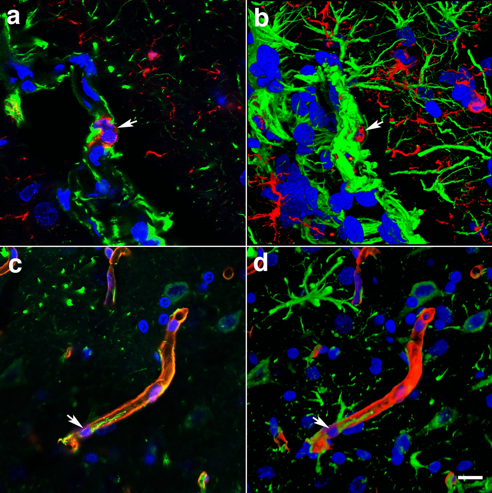
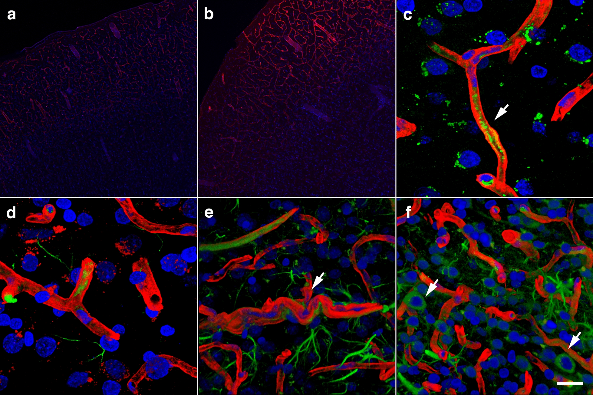
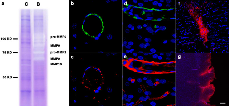
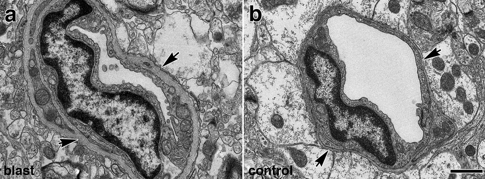
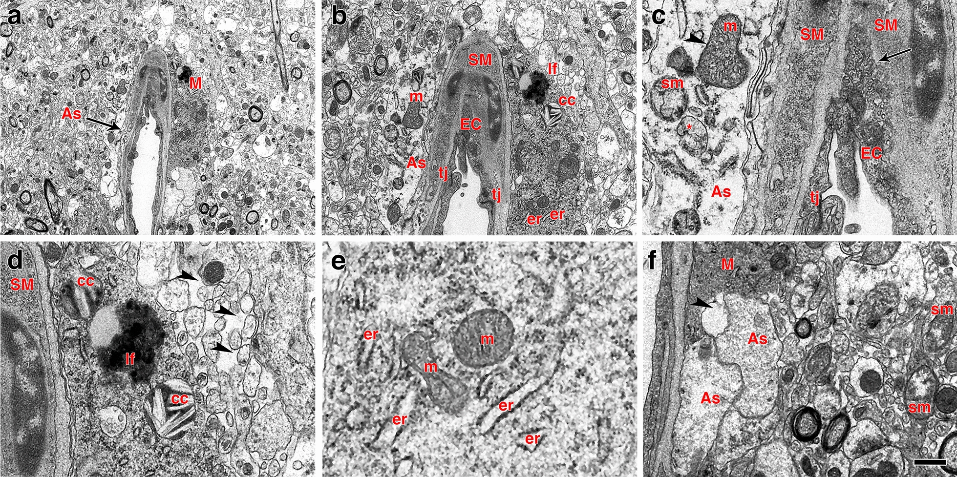
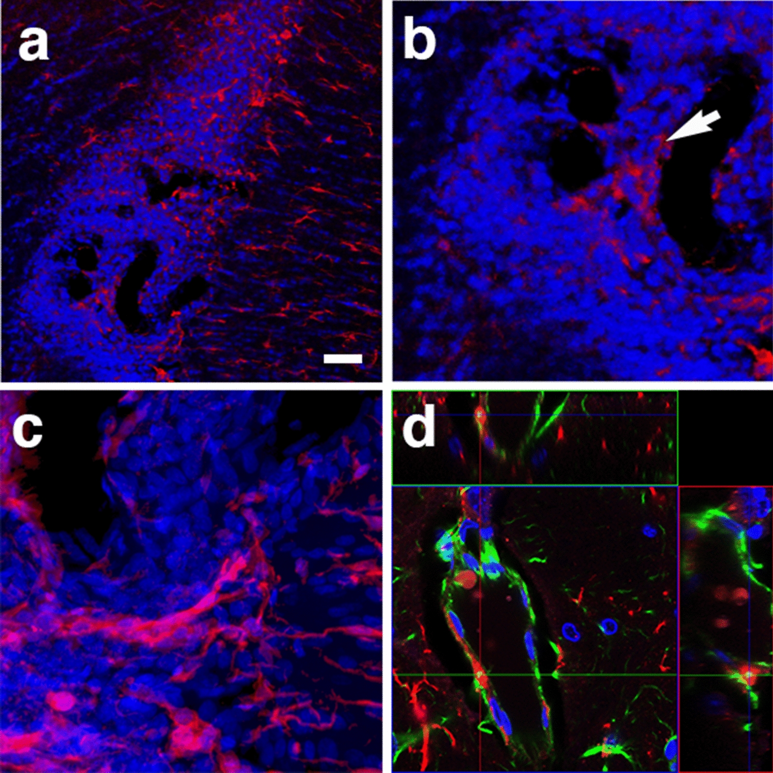
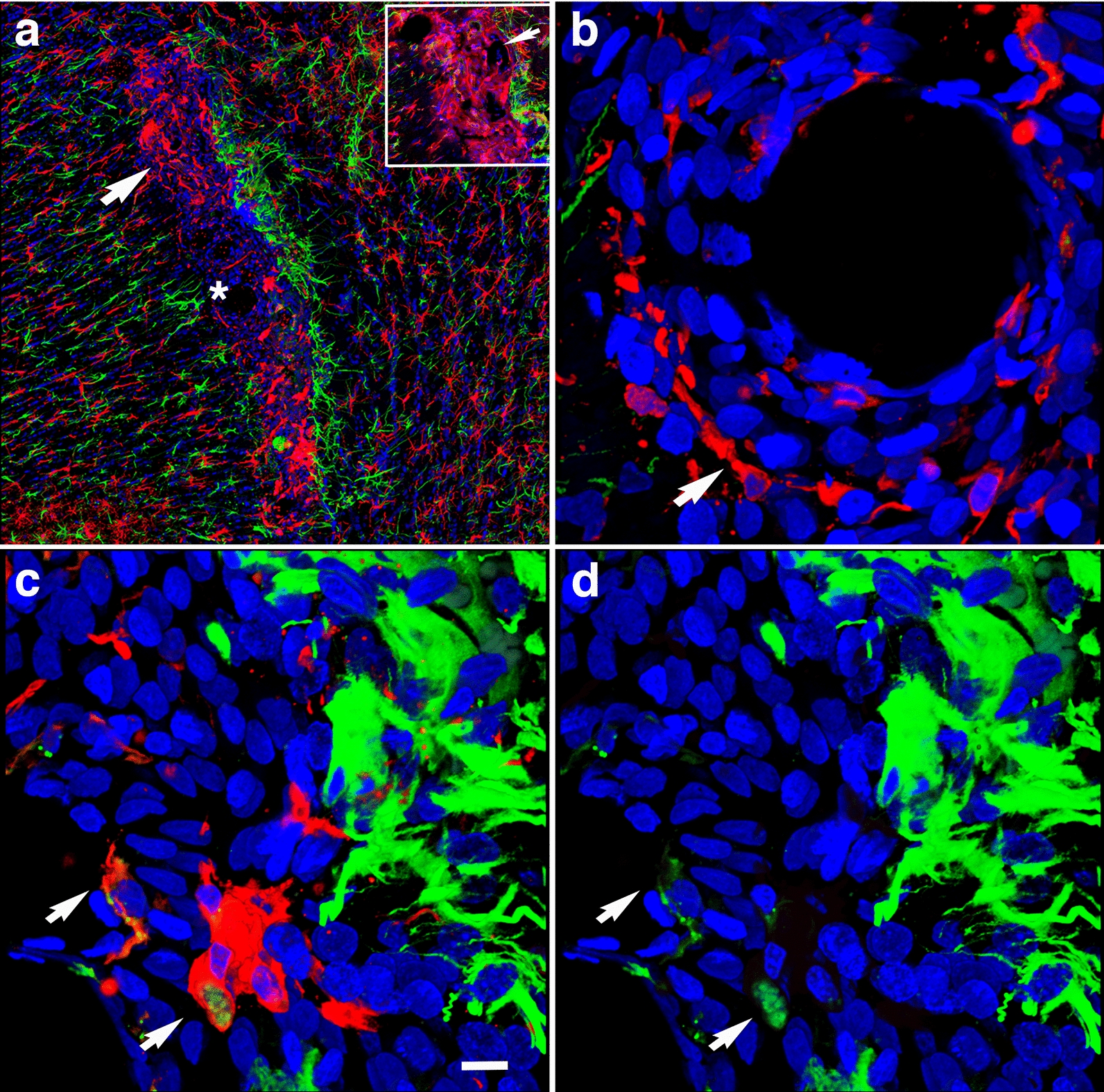
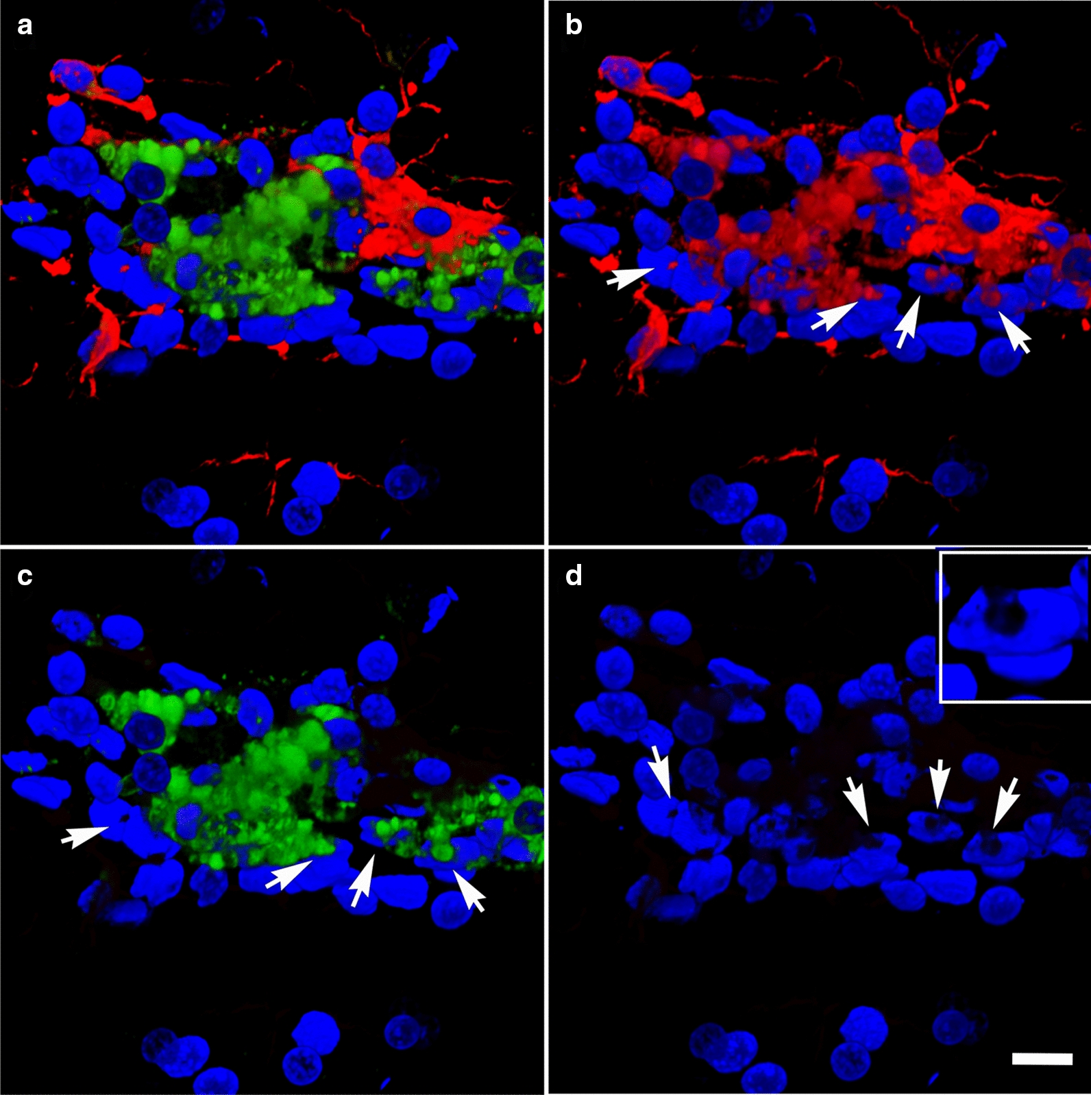
Cerebral vascular injury as a consequence of blast-induced traumatic brain injury is primarily the result of blast wave-induced mechanical disruptions within the neurovascular unit. In rodent models of blast-induced traumatic brain injury, chronic vascular degenerative processes are associated with the development of an age-dependent post-traumatic stress disorder-like phenotype. To investigate the evolution of blast-induced chronic vascular degenerative changes, Long-Evans rats were blast-exposed (3 × 74.5 kPa) and their brains analyzed at different times post-exposure by X-ray microcomputed tomography, immunohistochemistry and electron microscopy. On microcomputed tomography scans, regional cerebral vascular attenuation or occlusion was observed as early as 48 h post-blast, and cerebral vascular disorganization was visible at 6 weeks and more accentuated at 13 months post-blast. Progression of the late-onset pathology was characterized by detachment of the endothelial and smooth muscle cellular elements from the neuropil due to degeneration and loss of arteriolar perivascular astrocytes. Development of this pathology was associated with vascular remodeling and neuroinflammation as increased levels of matrix metalloproteinases (MMP-2 and MMP-9), collagen type IV loss, and microglial activation were observed in the affected vasculature. Blast-induced chronic alterations within the neurovascular unit should affect cerebral blood circulation, glymphatic flow and intramural periarterial drainage, all of which may contribute to development of the blast-induced behavioral phenotype. Our results also identify astrocytic degeneration as a potential target for the development of therapies to treat blast-induced brain injury.
爆炸导致的创伤性脑损伤会引起脑血管损伤,其主要是由神经血管单元内的爆震波引起的机械性破坏所致。在爆炸诱导的创伤性脑损伤的啮齿动物模型中,慢性血管退行性病变与年龄依赖性创伤后应激障碍样表型的发展有关。为了研究爆炸引起的慢性血管退行性变化的演变,对长耳大鼠进行了爆炸暴露(3×74.5 kPa),并在暴露后不同时间通过 X 射线微计算机断层扫描、免疫组织化学和电子显微镜对其大脑进行了分析。在微计算机断层扫描扫描中,早在爆炸后 48 小时就观察到区域性脑血管衰减或闭塞,并且在 6 周时可以看到脑血管紊乱,在爆炸后 13 个月时更加明显。晚期发病机制的进展特征为由于小动脉周围星形胶质细胞的变性和丧失,内皮和平滑肌细胞元素从神经胶质分离。这种病理学的发展与血管重塑和神经炎症有关,因为在受影响的血管中观察到基质金属蛋白酶(MMP-2 和 MMP-9)、IV 型胶原丧失和小胶质细胞激活水平升高。神经血管单元内的爆炸诱导的慢性改变会影响脑血液循环、糖质流和壁内动脉周围引流,所有这些都可能导致爆炸诱导的行为表型的发展。我们的结果还表明,星形胶质细胞变性可能是治疗爆炸诱导性脑损伤的潜在治疗靶点。