General Medical Research Service, James J. Peters Department of Veterans Affairs Medical Center, 130 West Kingsbridge Road, Bronx, NY, 10468, USA.
Department of Psychiatry, Icahn School of Medicine at Mount Sinai, One Gustave Levy Place, New York, NY, 10029, USA.
Acta Neuropathol Commun. 2019 Jan 9;7(1):6. doi: 10.1186/s40478-018-0647-5.
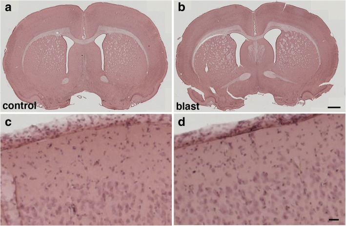
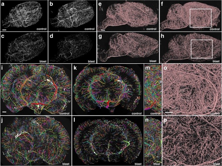
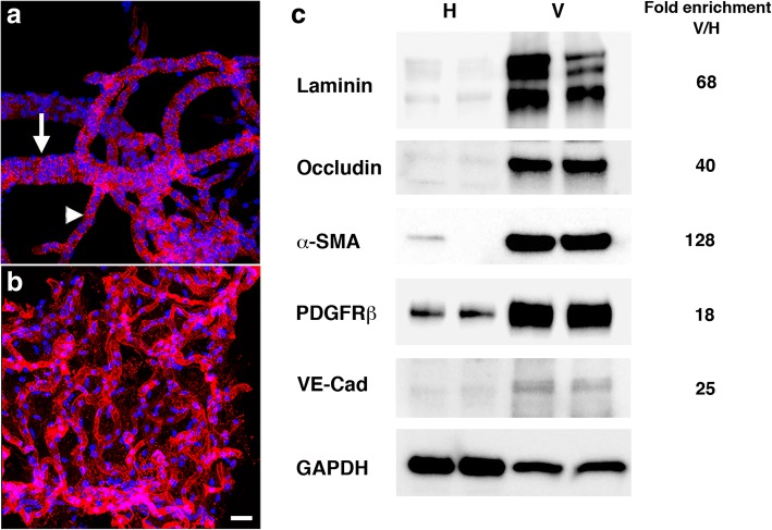
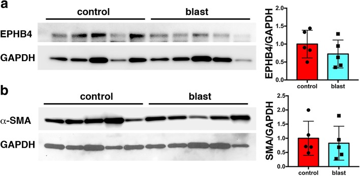
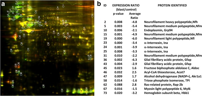
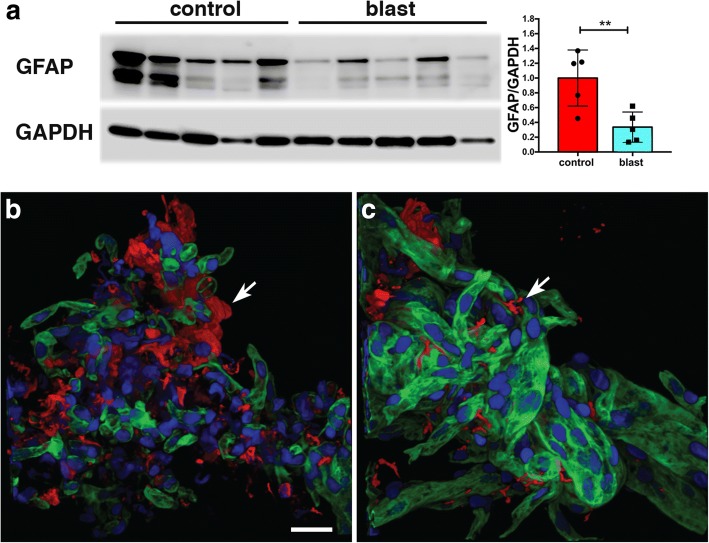
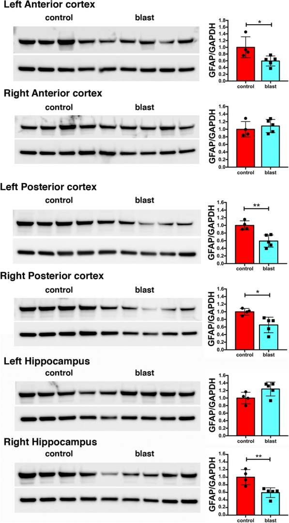
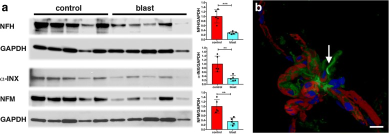
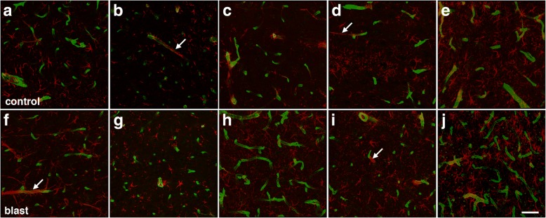
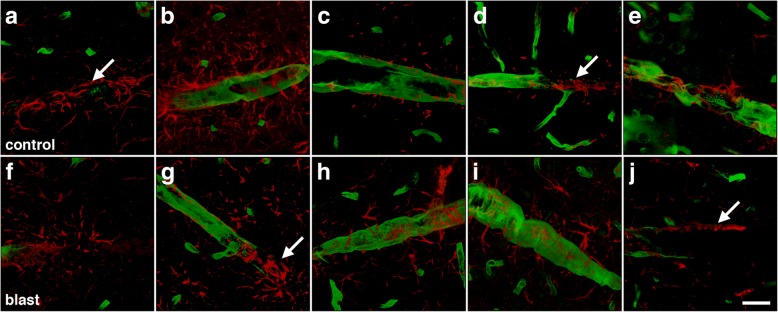
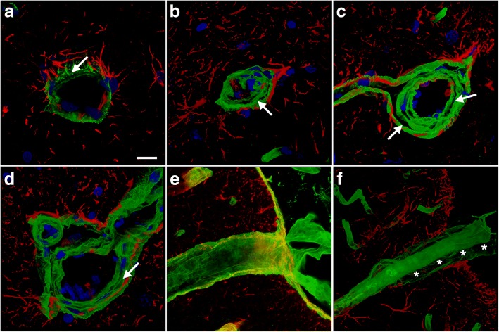
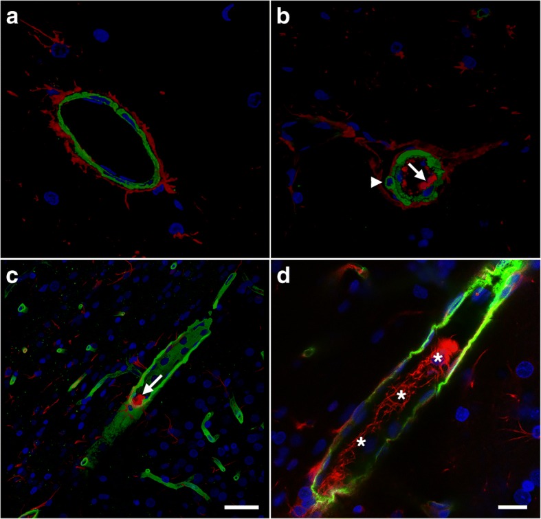
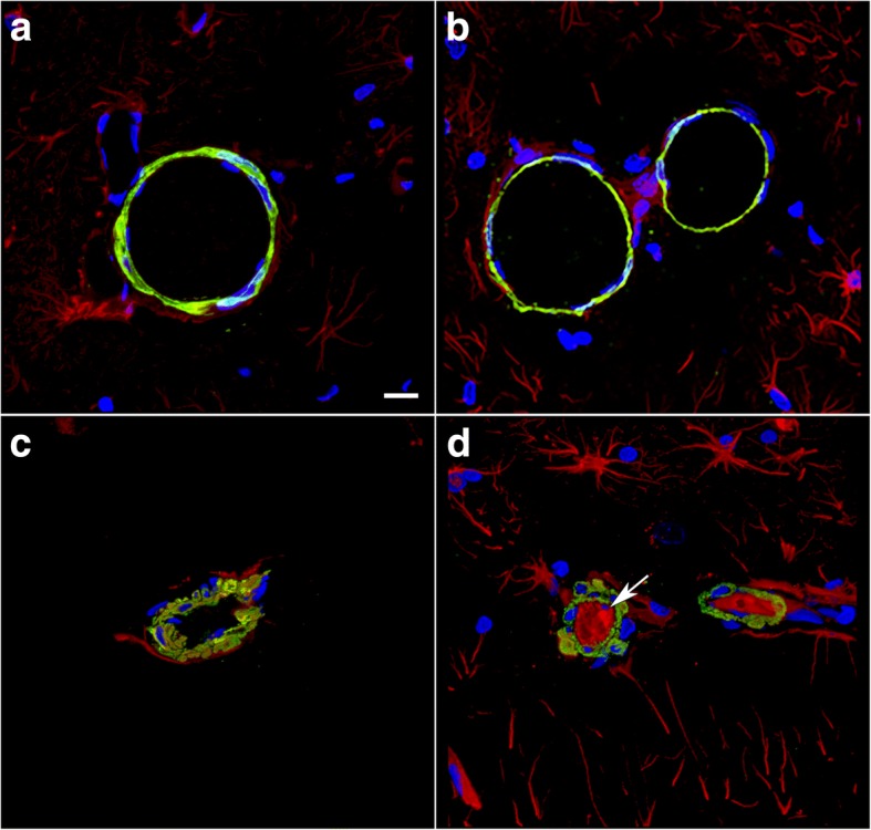
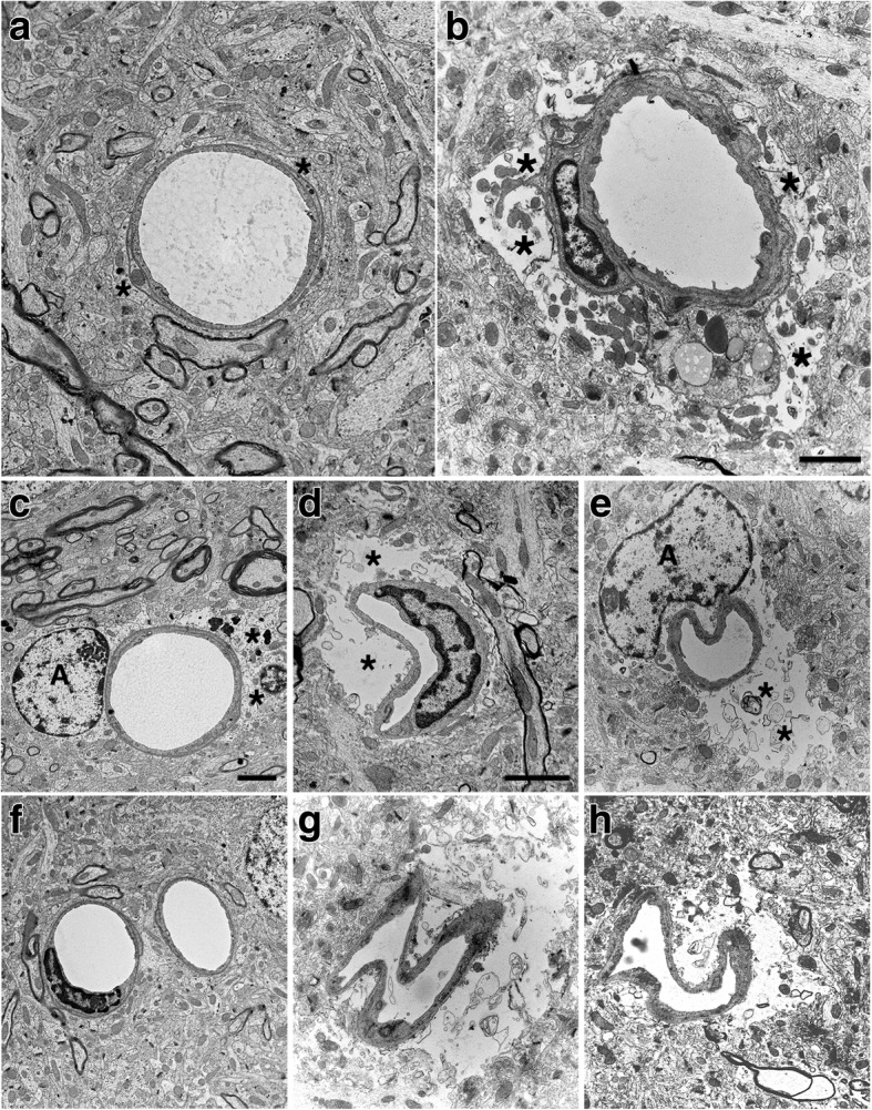
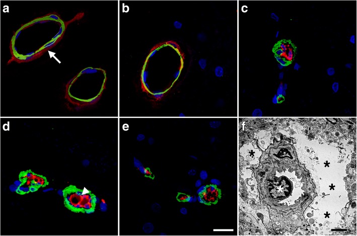
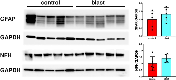
Much concern exists over the role of blast-induced traumatic brain injury (TBI) in the chronic cognitive and mental health problems that develop in veterans and active duty military personnel. The brain vasculature is particularly sensitive to blast injury. The aim of this study was to characterize the evolving molecular and histologic alterations in the neurovascular unit induced by three repetitive low-energy blast exposures (3 × 74.5 kPa) in a rat model mimicking human mild TBI or subclinical blast exposure. High-resolution two-dimensional differential gel electrophoresis (2D-DIGE) and matrix-assisted laser desorption/ionization (MALDI) mass spectrometry of purified brain vascular fractions from blast-exposed animals 6 weeks post-exposure showed decreased levels of vascular-associated glial fibrillary acidic protein (GFAP) and several neuronal intermediate filament proteins (α-internexin and the low, middle, and high molecular weight neurofilament subunits). Loss of these proteins suggested that blast exposure disrupts gliovascular and neurovascular interactions. Electron microscopy confirmed blast-induced effects on perivascular astrocytes including swelling and degeneration of astrocytic endfeet in the brain cortical vasculature. Because the astrocyte is a major sensor of neuronal activity and regulator of cerebral blood flow, structural disruption of gliovascular integrity within the neurovascular unit should impair cerebral autoregulation. Disrupted neurovascular connections to pial and parenchymal blood vessels might also affect brain circulation. Blast exposures also induced structural and functional alterations in the arterial smooth muscle layer. Interestingly, by 8 months after blast exposure, GFAP and neuronal intermediate filament expression had recovered to control levels in isolated brain vascular fractions. However, despite this recovery, a widespread vascular pathology was still apparent at 10 months after blast exposure histologically and on micro-computed tomography scanning. Thus, low-level blast exposure disrupts gliovascular and neurovascular connections while inducing a chronic vascular pathology.
人们非常关注爆炸引起的创伤性脑损伤(TBI)在退伍军人和现役军人中出现的慢性认知和心理健康问题中的作用。脑血管系统对爆炸伤特别敏感。本研究的目的是描述在模拟人类轻度 TBI 或亚临床爆炸暴露的大鼠模型中,三次重复低能量爆炸暴露(3×74.5kPa)对神经血管单元中不断演变的分子和组织学改变进行特征描述。暴露于爆炸后的动物的纯化脑血管部分的高分辨率二维差异凝胶电泳(2D-DIGE)和基质辅助激光解吸/电离(MALDI)质谱分析显示,血管相关的胶质纤维酸性蛋白(GFAP)和几种神经元中间丝蛋白(α-中间丝蛋白和低、中、高分子量神经丝亚单位)的水平降低。这些蛋白质的丧失表明,爆炸暴露破坏了血管周围星形胶质细胞和神经血管的相互作用。电子显微镜证实了爆炸对血管周围星形胶质细胞的影响,包括大脑皮质血管中星形胶质细胞终足的肿胀和退化。由于星形胶质细胞是神经元活动的主要传感器和大脑血流的调节剂,因此神经血管单元内血管完整性的结构破坏应损害脑自动调节。神经血管连接到脑膜和实质血管的中断也可能影响脑循环。爆炸暴露还诱导动脉平滑肌层的结构和功能改变。有趣的是,在爆炸暴露 8 个月后,分离的脑血管部分中 GFAP 和神经元中间丝表达已恢复到对照水平。然而,尽管有这种恢复,在爆炸暴露 10 个月后,在组织学和微计算机断层扫描上仍明显存在广泛的血管病理学。因此,低水平的爆炸暴露破坏了血管周围胶质细胞和神经血管的连接,同时诱导了慢性血管病理学。