Karim Lamya, Kwaczala Andrea, Vashishth Deepak, Judex Stefan
Department of Bioengineering, University of Massachusetts Dartmouth, Dartmouth, MA, USA.
Department of Biomedical Engineering, Western New England University, Springfield, MA, USA.
Bone Rep. 2021 Oct 1;15:101137. doi: 10.1016/j.bonr.2021.101137. eCollection 2021 Dec.
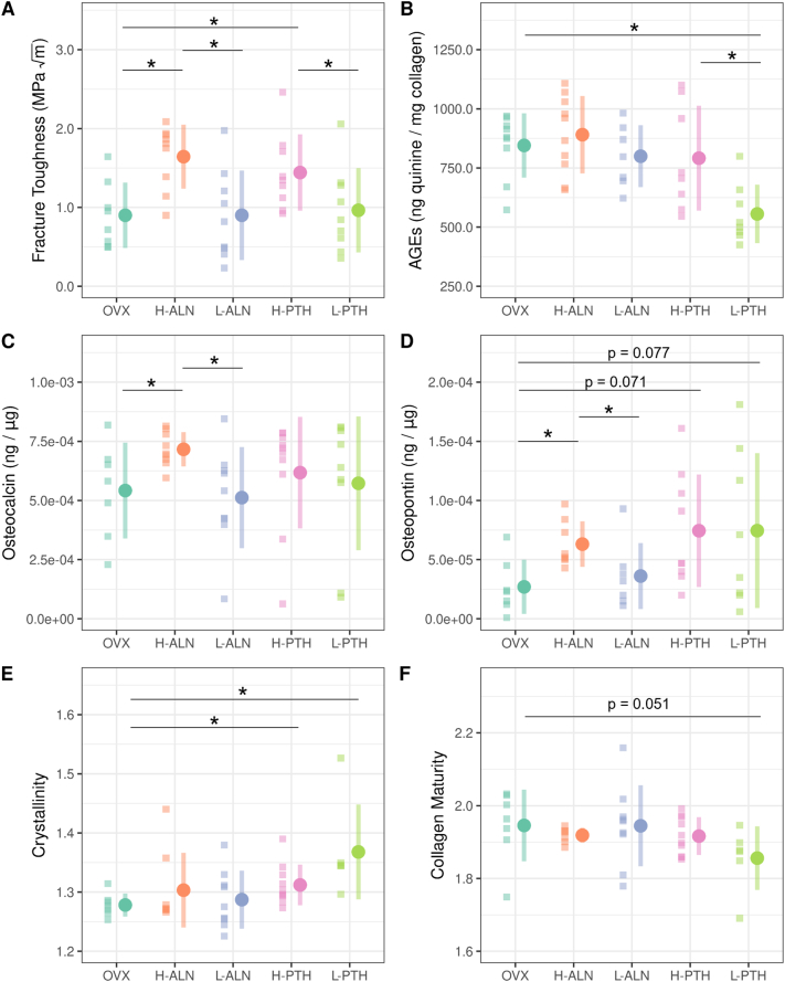
As both anabolic and anti-catabolic osteoporosis drugs affect bone formation and resorption processes, they may contribute to bone's overall mechanical behavior by altering the quality of the bone matrix. We used an ovariectomized rat model and a novel fracture mechanics approach to investigate whether treatment with an anabolic (parathyroid hormone) or anti-catabolic (alendronate) osteoporosis drugs will alter the organic and mineral matrix components and consequently cortical bone fracture toughness. Ovariectomized (at 5 months age) rats were treated with either parathyroid hormone or alendronate at low and high doses for 6 months (age 6-12 months). Specifically, treatment groups included untreated ovariectomized controls (n = 9), high-dose alendronate (n = 10), low-dose alendronate (n = 9), high-dose parathyroid hormone (n = 10), and low-dose parathyroid hormone (n = 9). After euthanasia, cortical microbeams from the lateral quadrant were extracted, notched, and tested in 3-point bending to measure fracture toughness. Portions of the bone were used to measure changes in the 1) organic matrix through quantification of advanced glycation end-products (AGEs) and non-collagenous proteins, and 2) mineral matrix through assessment of mineral crystallinity. Compared to the ovariectomized group, rats treated with high doses of parathyroid hormone and alendronate had significantly increased cortical bone fracture toughness, which corresponded primarily to increased non-collagenous proteins while there was no change in AGEs. Additionally, low-dose PTH treatment increased matrix crystallinity and decreased AGE levels. In summary, ovariectomized rats treated with pharmaceutical drugs had increased non-collagenous matrix proteins and improved fracture toughness compared to controls. Further investigation is required for different doses and longer treatment periods.
由于合成代谢和抗分解代谢的骨质疏松症药物都会影响骨形成和骨吸收过程,它们可能通过改变骨基质的质量来影响骨骼的整体力学行为。我们使用去卵巢大鼠模型和一种新颖的断裂力学方法来研究用合成代谢药物(甲状旁腺激素)或抗分解代谢药物(阿仑膦酸盐)治疗骨质疏松症是否会改变有机和矿物质基质成分,进而影响皮质骨的断裂韧性。对5个月大的去卵巢大鼠,分别用低剂量和高剂量的甲状旁腺激素或阿仑膦酸盐治疗6个月(6至12个月龄)。具体而言,治疗组包括未治疗的去卵巢对照组(n = 9)、高剂量阿仑膦酸盐组(n = 10)、低剂量阿仑膦酸盐组(n = 9)、高剂量甲状旁腺激素组(n = 10)和低剂量甲状旁腺激素组(n = 9)。安乐死后,从外侧象限提取皮质微梁,刻痕并进行三点弯曲测试以测量断裂韧性。取部分骨骼用于测量以下变化:1)通过定量晚期糖基化终产物(AGEs)和非胶原蛋白来测量有机基质的变化;2)通过评估矿物结晶度来测量矿物基质的变化。与去卵巢组相比,高剂量甲状旁腺激素和阿仑膦酸盐治疗的大鼠皮质骨断裂韧性显著增加,这主要对应于非胶原蛋白的增加,而AGEs没有变化。此外,低剂量甲状旁腺激素治疗增加了基质结晶度并降低了AGE水平。总之,与对照组相比,用药物治疗的去卵巢大鼠非胶原蛋白基质蛋白增加,断裂韧性提高。不同剂量和更长治疗期还需要进一步研究。