• 文献检索
  • 文档翻译
  • 深度研究
  • 学术资讯
  • Suppr Zotero 插件Zotero 插件
  • 邀请有礼
  • 套餐&价格
  • 历史记录
应用&插件
Suppr Zotero 插件Zotero 插件浏览器插件Mac 客户端Windows 客户端微信小程序
定价
高级版会员购买积分包购买API积分包
服务
文献检索文档翻译深度研究API 文档MCP 服务
关于我们
关于 Suppr公司介绍联系我们用户协议隐私条款
关注我们

Suppr 超能文献

核心技术专利:CN118964589B侵权必究
粤ICP备2023148730 号-1Suppr @ 2026

文献检索

告别复杂PubMed语法,用中文像聊天一样搜索,搜遍4000万医学文献。AI智能推荐,让科研检索更轻松。

立即免费搜索

文件翻译

保留排版,准确专业,支持PDF/Word/PPT等文件格式,支持 12+语言互译。

免费翻译文档

深度研究

AI帮你快速写综述,25分钟生成高质量综述,智能提取关键信息,辅助科研写作。

立即免费体验

PROCEM 研究方案:术前对比增强乳腺摄影在恶性乳腺病变分期中的附加价值 - 一项前瞻性随机多中心研究。

The PROCEM study protocol: Added value of preoperative contrast-enhanced mammography in staging of malignant breast lesions - a prospective randomized multicenter study.

机构信息

Department of Surgery, Halland Hospital, 301 85, Halmstad, Sweden.

Institution of Clinical Sciences, Department of Surgery, Lund University, Lund, Sweden.

出版信息

BMC Cancer. 2021 Oct 18;21(1):1115. doi: 10.1186/s12885-021-08832-2.

DOI:10.1186/s12885-021-08832-2
PMID:34663236
原文链接:https://pmc.ncbi.nlm.nih.gov/articles/PMC8521511/
Abstract

BACKGROUND

Correct preoperative estimation of the malignant extent is crucial for optimal planning of breast cancer surgery. The sensitivity of mammography is lower in dense breasts, and additional imaging techniques are sometimes warranted. Contrast-enhanced mammography (CEM) has shown similar sensitivity and in some cases better specificity, than magnetic resonance imaging (MRI) in small, observational studies. CEM may be more cost-effective than MRI, and may provide better identification of the tumor extent, however, no randomized trials have been performed to date to investigate the added value of CEM. In a feasibility study, we found that the treatment was changed in 10/47 (21%) cases after additional CEM. The purpose of the present study is to evaluate the added value of CEM in preoperative staging of breast cancer in a randomized study.

METHOD

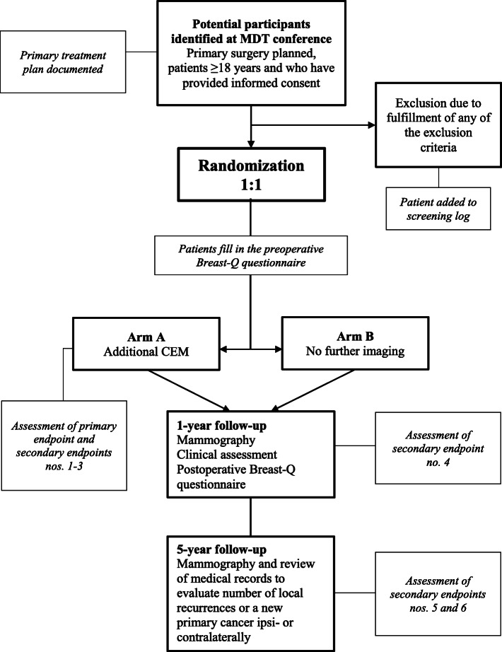
This prospective randomized study will include 440 patients with strongly suspected or established diagnosis of breast malignancy, based on assessment with mammography, ultrasound and core biopsy/cytology, and for whom primary surgery is planned. Patients will be randomized 1:1 using a web-based randomization tool to additional investigation with CEM or no further imaging. The CEM findings will be taken into consideration, which may lead to changes in primary treatment, which is the primary endpoint of this study. Secondary endpoints include rate of reoperation and number of avoidable mastectomies, as well as a cost-benefit analysis of additional CEM. Patient-reported health-related quality of life will be investigated at 1 year with the validated Breast-Q™ questionnaire. The rate of local recurrence or new cancer ipsi- or contralaterally within 5 years will be assessed from medical records and pathology reports.

DISCUSSION

The aim of this trial is to explore the added value of CEM in preoperative staging of breast cancer. The results obtained from this study will contribute to our knowledge on CEM as an additional imaging method to standard investigation with digital mammography and ultrasound. The findings may also provide additional information on which patient groups would benefit from CEM, and on the economic aspects of CEM in standard preoperative practice.

TRIAL REGISTRATION

This trial is registered at clinicaltrials.gov , registration no: NCT04437602 , date of registration: June 18, 2020.

摘要

背景

正确的术前恶性程度评估对于乳腺癌手术的最佳规划至关重要。在致密乳房中,乳房 X 线摄影的敏感性较低,有时需要额外的成像技术。与磁共振成像(MRI)相比,对比增强乳腺摄影(CEM)在小型观察性研究中具有相似的敏感性,在某些情况下具有更好的特异性。CEM 可能比 MRI 更具成本效益,并且可以更好地识别肿瘤范围,但是,迄今为止,尚无随机试验来研究 CEM 的附加价值。在一项可行性研究中,我们发现 47 例(21%)患者在进行额外的 CEM 后改变了治疗方案。本研究的目的是在一项随机研究中评估 CEM 在乳腺癌术前分期中的附加价值。

方法

本前瞻性随机研究将包括 440 例强烈怀疑或确诊为乳腺癌的患者,这些患者基于乳房 X 线摄影、超声和核心活检/细胞学评估,计划进行原发性手术。患者将使用基于网络的随机化工具以 1:1 的比例随机分为 CEM 检查组或无进一步影像学检查组。将考虑 CEM 的检查结果,这可能导致原发性治疗的改变,这是本研究的主要终点。次要终点包括再次手术的发生率和可避免的乳房切除术的数量,以及 CEM 的成本效益分析。将使用经过验证的 Breast-Q™问卷在 1 年时调查患者报告的健康相关生活质量。将从病历和病理报告中评估 5 年内同侧或对侧局部复发或新发癌症的发生率。

讨论

本试验的目的是探讨 CEM 在乳腺癌术前分期中的附加价值。该研究的结果将有助于我们了解 CEM 作为数字乳房 X 线摄影和超声标准检查的附加成像方法。这些发现还可能提供有关哪些患者群体将从 CEM 中受益以及 CEM 在标准术前实践中的经济方面的更多信息。

试验注册

该试验在 clinicaltrials.gov 注册,注册号:NCT04437602,注册日期:2020 年 6 月 18 日。

https://cdn.ncbi.nlm.nih.gov/pmc/blobs/6fde/8524950/202c6ebe67c1/12885_2021_8832_Fig1_HTML.jpg

相似文献

1
The PROCEM study protocol: Added value of preoperative contrast-enhanced mammography in staging of malignant breast lesions - a prospective randomized multicenter study.PROCEM 研究方案:术前对比增强乳腺摄影在恶性乳腺病变分期中的附加价值 - 一项前瞻性随机多中心研究。
BMC Cancer. 2021 Oct 18;21(1):1115. doi: 10.1186/s12885-021-08832-2.
2
Added value of contrast-enhanced mammography (CEM) in staging of malignant breast lesions-a feasibility study.对比增强乳腺摄影(CEM)在恶性乳腺病变分期中的附加价值——一项可行性研究。
World J Surg Oncol. 2020 May 21;18(1):100. doi: 10.1186/s12957-020-01865-0.
3
Contrast-Enhanced Mammography in Local Staging of Screen-Detected Breast Cancer.对比增强乳腺摄影在筛查性乳腺癌局部分期中的应用。
Ann Surg Oncol. 2024 Oct;31(10):6820-6830. doi: 10.1245/s10434-024-15848-y. Epub 2024 Jul 24.
4
Rapid Access to Contrast-Enhanced spectral mammogRaphy in women recalled from breast cancer screening: the RACER trial study design.从乳腺癌筛查中召回的女性的快速获取对比增强光谱 mammogRaphy:RACER 试验研究设计。
Trials. 2019 Dec 23;20(1):759. doi: 10.1186/s13063-019-3867-5.
5
Contrast-enhanced mammography (CEM) versus MRI for breast cancer staging: detection of additional malignant lesions not seen on conventional imaging.对比增强乳腺摄影(CEM)与 MRI 用于乳腺癌分期:检测常规影像未见的额外恶性病变。
Eur Radiol Exp. 2023 Feb 13;7(1):8. doi: 10.1186/s41747-022-00318-5.
6
Diagnostic Performance of MRI, Molecular Breast Imaging, and Contrast-enhanced Mammography in Women with Newly Diagnosed Breast Cancer.MRI、分子乳腺成像和对比增强乳腺摄影在新诊断乳腺癌女性中的诊断性能。
Radiology. 2019 Dec;293(3):531-540. doi: 10.1148/radiol.2019190887. Epub 2019 Oct 29.
7
Comparison of Performance in Diagnosis and Characterization of Breast Lesions: Contrast-Enhanced Mammography Versus Breast Magnetic Resonance Imaging.对比增强乳腺摄影与乳腺磁共振成像在乳腺病变诊断和特征描述中的性能比较。
Clin Breast Cancer. 2024 Aug;24(6):481-493. doi: 10.1016/j.clbc.2024.04.007. Epub 2024 Apr 15.
8
Contrast enhanced mammography (CEM) versus magnetic resonance imaging (MRI) for staging of breast cancer: The pro CEM perspective.对比增强乳腺摄影术(CEM)与磁共振成像(MRI)在乳腺癌分期中的应用:支持 CEM 的观点。
Eur J Radiol. 2021 Sep;142:109883. doi: 10.1016/j.ejrad.2021.109883. Epub 2021 Jul 30.
9
The role of contrast-enhanced mammography in the preoperative evaluation of invasive lobular carcinoma of the breast.对比增强乳腺摄影在乳腺浸润性小叶癌术前评估中的作用。
Clin Radiol. 2024 Jun;79(6):e799-e806. doi: 10.1016/j.crad.2024.01.035. Epub 2024 Feb 12.
10
Multireader comparison of contrast-enhanced mammography versus the combination of digital mammography and digital breast tomosynthesis in the preoperative assessment of breast cancer.多阅读器对比增强乳腺摄影与数字乳腺摄影和数字乳腺断层合成在乳腺癌术前评估中的联合应用。
Radiol Med. 2021 Nov;126(11):1407-1414. doi: 10.1007/s11547-021-01400-5. Epub 2021 Jul 24.

引用本文的文献

1
Evaluation of contrast-enhanced mammography (CEM) in the preoperative staging of breast cancer: large-scale single center experience, update to 1005 cases.对比增强乳腺钼靶摄影(CEM)在乳腺癌术前分期中的评估:大规模单中心经验,病例更新至1005例
Radiol Med. 2025 Mar 28. doi: 10.1007/s11547-025-02009-8.
2
Contrast-enhanced mammography improves patient access to functional breast imaging.对比增强乳腺钼靶摄影改善了患者获得功能性乳腺成像的机会。
J Med Imaging Radiat Oncol. 2025 Feb;69(1):46-61. doi: 10.1111/1754-9485.13789. Epub 2024 Oct 31.
3
Performance of dual-energy subtraction in contrast-enhanced mammography for three different manufacturers: a phantom study.

本文引用的文献

1
Added value of contrast-enhanced mammography (CEM) in staging of malignant breast lesions-a feasibility study.对比增强乳腺摄影(CEM)在恶性乳腺病变分期中的附加价值——一项可行性研究。
World J Surg Oncol. 2020 May 21;18(1):100. doi: 10.1186/s12957-020-01865-0.
2
Breast Sonography and Mammography: Complementarity and Correlation.乳腺超声检查与乳腺钼靶摄影:互补性与相关性
Radiol Technol. 2017 Sep;89(1):45M-64M.
3
Breast Radiation Dose With CESM Compared With 2D FFDM and 3D Tomosynthesis Mammography.对比二维全视野数字化乳腺摄影(2D FFDM)和三维乳腺断层合成摄影,乳腺增强合成钼靶(CESM)的乳腺辐射剂量
双能减影在三种不同厂家对比增强乳腺摄影中的性能:一项体模研究。
Eur Radiol Exp. 2024 Oct 14;8(1):113. doi: 10.1186/s41747-024-00516-3.
4
Screening and diagnostic breast MRI: how do they impact surgical treatment? Insights from the MIPA study.乳腺 MRI 的筛查和诊断:它们如何影响手术治疗?来自 MIPA 研究的见解。
Eur Radiol. 2023 Sep;33(9):6213-6225. doi: 10.1007/s00330-023-09600-5. Epub 2023 May 3.
5
Invasive Lobular Carcinoma: A Review of Imaging Modalities with Special Focus on Pathology Concordance.浸润性小叶癌:影像学检查方法综述,特别关注病理一致性
Healthcare (Basel). 2023 Mar 3;11(5):746. doi: 10.3390/healthcare11050746.
6
Barriers to Implementation of Contrast-Enhanced Mammography in Clinical Practice: Expert Panel Narrative Review.临床实施对比增强乳腺摄影的障碍:专家小组叙述性综述。
AJR Am J Roentgenol. 2023 Jul;221(1):3-6. doi: 10.2214/AJR.22.28567. Epub 2022 Nov 30.
AJR Am J Roentgenol. 2017 Feb;208(2):362-372. doi: 10.2214/AJR.16.16743.
4
Contrast-enhanced spectral mammography vs. mammography and MRI - clinical performance in a multi-reader evaluation.对比增强光谱乳腺摄影术与乳腺摄影术和磁共振成像的多阅片者评估中的临床性能
Eur Radiol. 2017 Jul;27(7):2752-2764. doi: 10.1007/s00330-016-4650-6. Epub 2016 Nov 28.
5
Mammography: an update of the EUSOBI recommendations on information for women.乳腺钼靶检查:欧洲乳腺影像学会(EUSOBI)关于女性信息的建议更新
Insights Imaging. 2017 Feb;8(1):11-18. doi: 10.1007/s13244-016-0531-4. Epub 2016 Nov 16.
6
The potential of multiparametric MRI of the breast.乳腺多参数磁共振成像的潜力。
Br J Radiol. 2017 Jan;90(1069):20160715. doi: 10.1259/bjr.20160715. Epub 2016 Nov 2.
7
Contrast-Enhanced Digital Mammography in the Surgical Management of Breast Cancer.对比增强数字化乳腺摄影在乳腺癌手术治疗中的应用
Ann Surg Oncol. 2016 Dec;23(Suppl 5):649-655. doi: 10.1245/s10434-016-5567-7. Epub 2016 Sep 15.
8
Added Value of Contrast-Enhanced Spectral Mammography in Postscreening Assessment.对比增强光谱乳腺摄影在筛查后评估中的附加价值
Breast J. 2016 Sep;22(5):520-8. doi: 10.1111/tbj.12627. Epub 2016 Jun 27.
9
Contrast-enhanced spectral mammography improves diagnostic accuracy in the symptomatic setting.对比增强光谱乳腺摄影术可提高有症状情况下的诊断准确性。
Clin Radiol. 2016 Nov;71(11):1148-55. doi: 10.1016/j.crad.2016.05.009. Epub 2016 Jun 11.
10
The use of ultrasonography and digital mammography in women under 40 years with symptomatic breast cancer: a 7-year Irish experience.超声检查和数字化乳腺摄影在40岁以下有症状乳腺癌女性中的应用:爱尔兰7年经验
Ir J Med Sci. 2017 Feb;186(1):63-67. doi: 10.1007/s11845-016-1472-0. Epub 2016 Jun 7.