Centre for Discovery Brain Sciences and the Euan MacDonald Centre for Motor Neurone Disease Research, University of Edinburgh, George Square, Edinburgh EH8 9XD, UK.
Public Health England, National Infection Service, Porton Down, Salisbury SP4 0JG, UK.
Biomolecules. 2021 Oct 12;11(10):1499. doi: 10.3390/biom11101499.
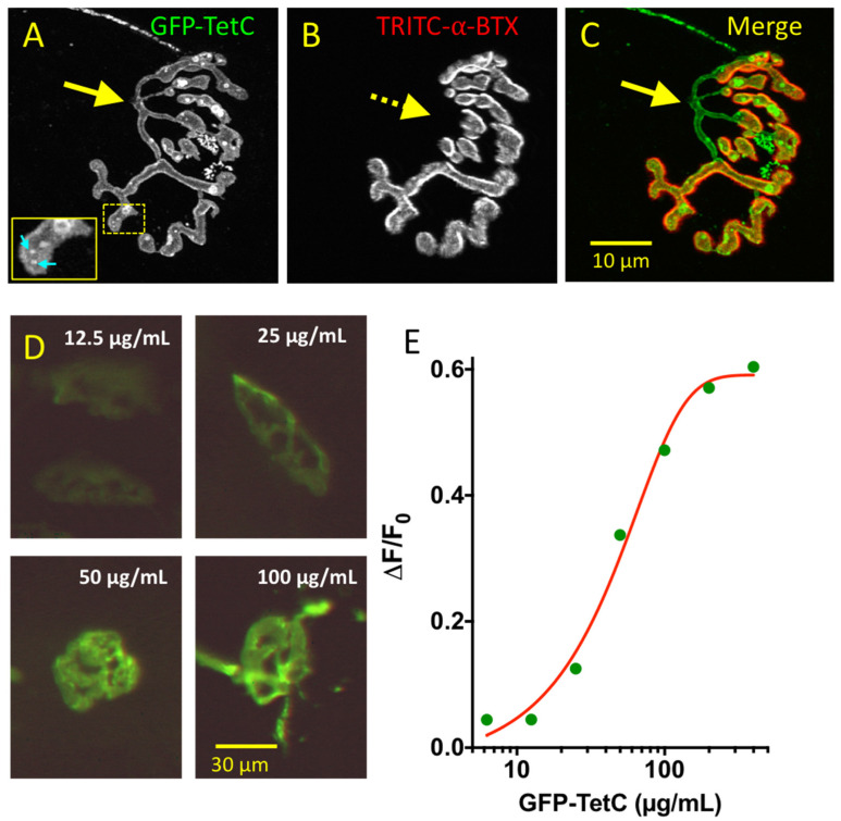
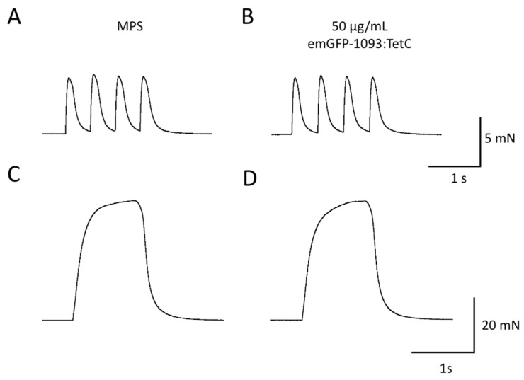
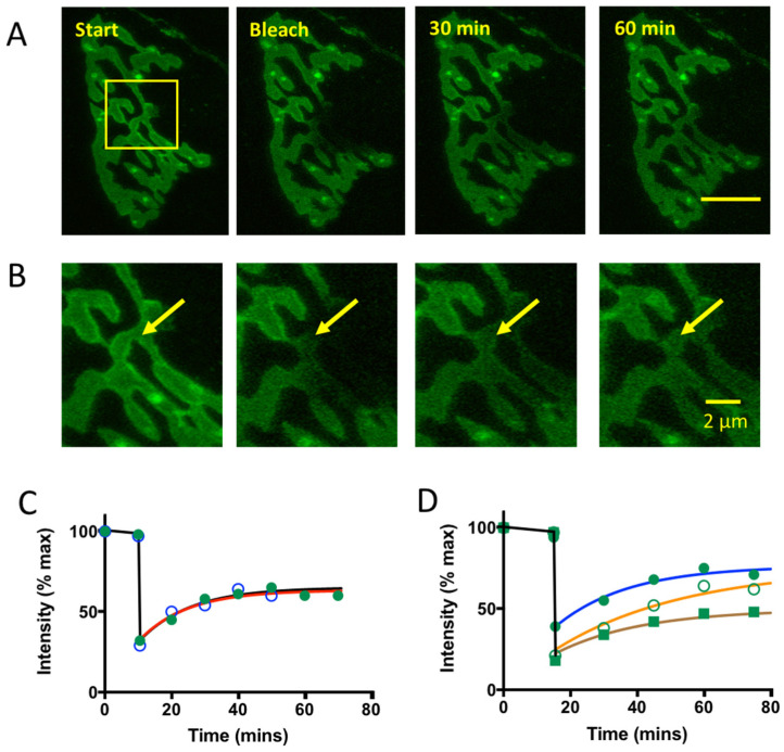
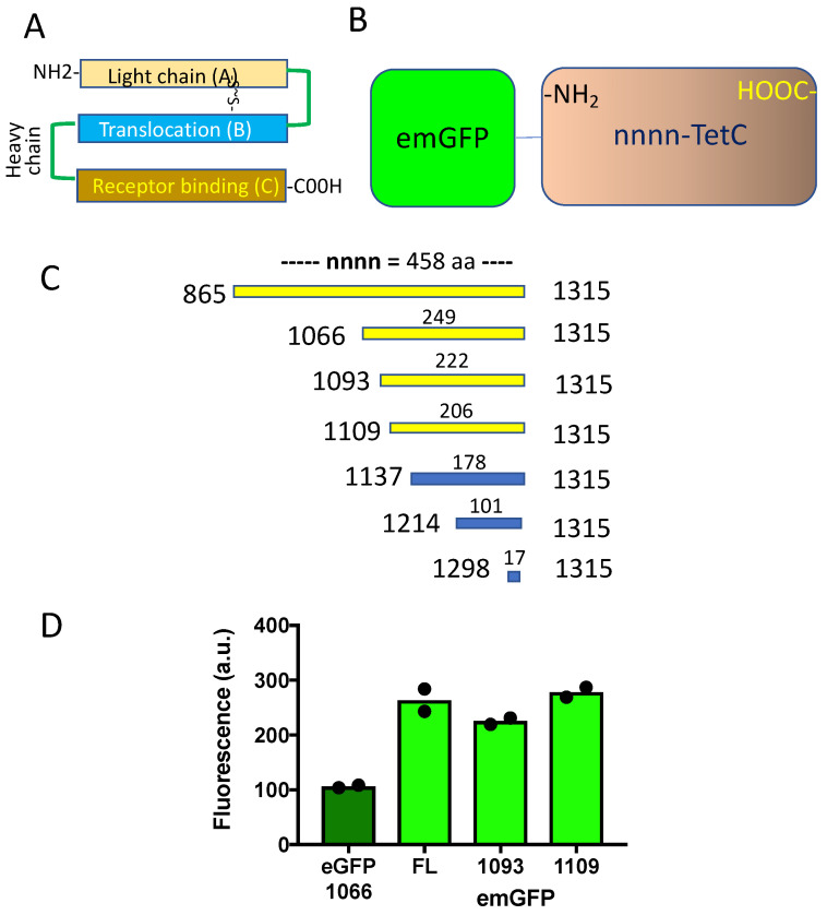
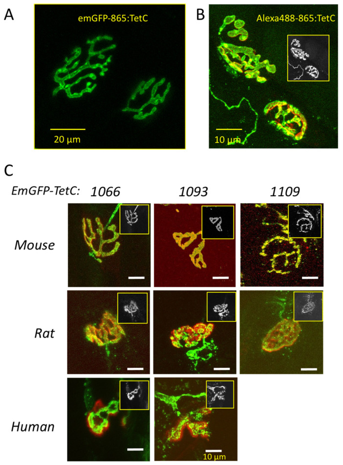
Live imaging of neuromuscular junctions (NMJs) in situ has been constrained by the suitability of ligands for inert vital staining of motor nerve terminals. Here, we constructed several truncated derivatives of the tetanus toxin C-fragment (TetC) fused with Emerald Fluorescent Protein (emGFP). Four constructs, namely full length emGFP-TetC (emGFP-865:TetC) or truncations comprising amino acids 1066-1315 (emGFP-1066:TetC), 1093-1315 (emGFP-1093:TetC) and 1109-1315 (emGFP-1109:TetC), produced selective, high-contrast staining of motor nerve terminals in rodent or human muscle explants. Isometric tension and intracellular recordings of endplate potentials from mouse muscles indicated that neither full-length nor truncated emGFP-TetC constructs significantly impaired NMJ function or transmission. Motor nerve terminals stained with emGFP-TetC constructs were readily visualised in situ or in isolated preparations using fibre-optic confocal endomicroscopy (CEM). emGFP-TetC derivatives and CEM also visualised regenerated NMJs. Dual-waveband CEM imaging of preparations co-stained with fluorescent emGFP-TetC constructs and Alexa647-α-bungarotoxin resolved innervated from denervated NMJs in axotomized mouse muscle and degenerating NMJs in transgenic SOD1G93A mouse muscle. Our findings highlight the region of the TetC fragment required for selective binding and visualisation of motor nerve terminals and show that fluorescent derivatives of TetC are suitable for in situ morphological and physiological characterisation of healthy, injured and diseased NMJs.
活体成像神经肌肉接头(NMJ)一直受到适合惰性活染运动神经末梢配体的限制。在这里,我们构建了几个与翡翠荧光蛋白(emGFP)融合的破伤风毒素 C 片段(TetC)的截断衍生物。四个构建体,即全长 emGFP-TetC(emGFP-865:TetC)或截断包含氨基酸 1066-1315(emGFP-1066:TetC),1093-1315(emGFP-1093:TetC)和 1109-1315(emGFP-1109:TetC),产生了选择性、高对比度的啮齿动物或人类肌肉外植体运动神经末梢染色。等长张力和来自小鼠肌肉的内板电位的细胞内记录表明,全长或截断的 emGFP-TetC 构建体均未显著损害 NMJ 功能或传递。使用光纤共聚焦内窥显微镜(CEM),可以在原位或在分离的制剂中轻松观察用 emGFP-TetC 构建体染色的运动神经末梢。emGFP-TetC 衍生物和 CEM 还可观察到再生的 NMJ。用荧光 emGFP-TetC 构建体和 Alexa647-α-箭毒蛋白共染色的制剂的双波段 CEM 成像可分辨出在轴突切断的小鼠肌肉中被神经支配的和去神经支配的 NMJ 以及在转基因 SOD1G93A 小鼠肌肉中变性的 NMJ。我们的发现强调了 TetC 片段中用于选择性结合和可视化运动神经末梢的区域,并表明 TetC 的荧光衍生物适合于健康、受损和患病 NMJ 的原位形态和生理学特征。