Division of Neurology, Department of Medicine, University of British Columbia, British Columbia, Canada.
Computational Neuroimaging Group, Biomedical Sciences Institute, Trinity College Dublin, Dublin, Ireland.
Brain Behav. 2021 Dec;11(12):e2403. doi: 10.1002/brb3.2403. Epub 2021 Oct 28.
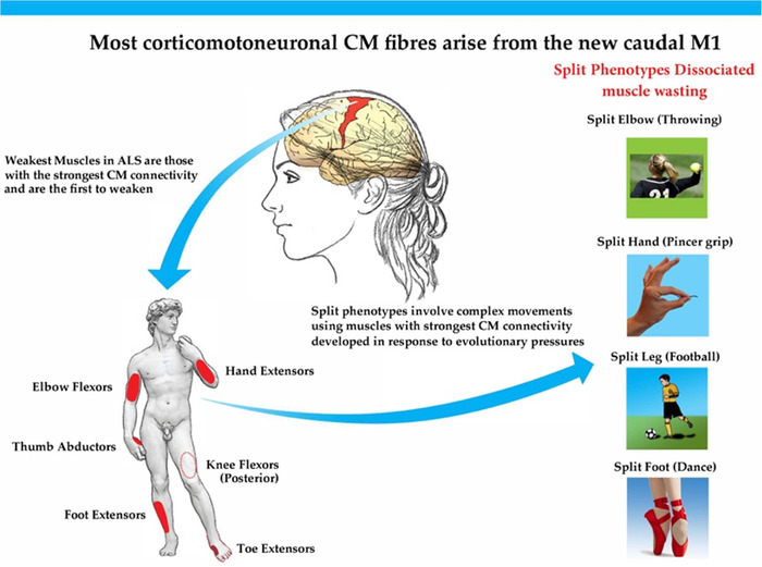
Split phenotypes, (split hand, elbow, leg, and foot), are probably unique to ALS, and are characterized by having a shared peripheral input of both affected and unaffected muscles. This implies an anatomical origin rostral to the spinal cord, primarily within the cerebral cortex. Therefore, split phenotypes are a potential marker of ALS upper motor neuron pathology. However, to date, reports documenting upper motor neuron dysfunction in split phenotypes have been limited to using transcranial magnetic stimulation and cortical threshold tracking techniques. Here, we consider several other potential methodologies that could confirm a primary upper motor neuron pathology in split phenotypes.
We review the potential of: 1. measuring the compound excitatory post-synaptic potential recorded from a single activated motor unit, 2. cortical-muscular coherence, and 3. new advanced modalities of neuroimaging (high-resolution imaging protocols, ultra-high field MRI platforms [7T], and novel Non-Gaussian diffusion models).
We propose that muscles involved in split phenotypes are those functionally involved in the human motor repertoire used particularly in complex activities. Their anterior horn cells receive the strongest corticomotoneuronal input. This is also true of the weakest muscles that are the earliest to be affected in ALS. Descriptions of split hand in non-ALS cases and proposals that peripheral nerve or muscle dysfunction may be causative are contentious. Only a few carefully controlled cases of each form of split phenotype, using upper motor neuron directed methodologies, are necessary to prove our postulate.
分裂表型(分裂手、肘、腿和足)可能是 ALS 所特有的,其特征是受影响和未受影响的肌肉都有共同的外周输入。这意味着其解剖学起源于脊髓上方,主要位于大脑皮层。因此,分裂表型是 ALS 上运动神经元病理的一个潜在标志物。然而,迄今为止,记录分裂表型中上运动神经元功能障碍的报告仅限于使用经颅磁刺激和皮质阈值跟踪技术。在这里,我们考虑了其他几种潜在的方法,这些方法可以证实分裂表型中的主要上运动神经元病理学。
我们回顾了以下几种方法的潜力:1. 测量单个激活运动单位记录的复合兴奋性突触后电位;2. 皮质-肌肉相干性;3. 新的高级神经影像学模式(高分辨率成像方案、超高场 MRI 平台[7T]和新的非高斯扩散模型)。
我们提出,参与分裂表型的肌肉是那些在人类运动 repertoire 中功能上参与复杂活动的肌肉。它们的前角细胞接收到最强的皮质运动神经元输入。这也适用于最早在 ALS 中受到影响的最弱肌肉。非 ALS 病例中分裂手的描述以及认为周围神经或肌肉功能障碍可能是病因的观点存在争议。只有使用上运动神经元定向方法,对每种形式的分裂表型进行少数精心控制的病例,才能证明我们的假设。