Sakamoto Asuka, Watanabe Goro, Morito Tsuyoshi, Katayama Kimio, Kumagai Hajime, Gamada Kazuyoshi
Faculty of rehabilitation sciences, Nishikyusyu university, 4490-9 Osaki, Kanzaki-machi, Kanzaki-shi, Saga-ken, 842-8585, Japan.
Department of physical therapy, Kawahara medical science institute, 3-6 Hanazono-cho, Matsuyama-shi, Ehime-ken, 790-0005, Japan.
Radiol Case Rep. 2021 Oct 19;16(12):3955-3960. doi: 10.1016/j.radcr.2021.09.053. eCollection 2021 Dec.
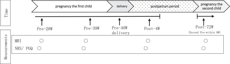
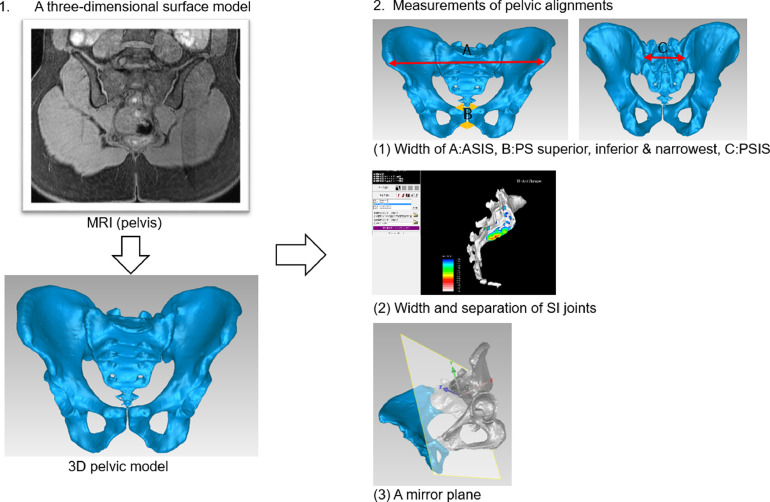
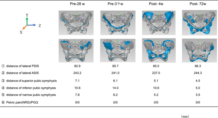
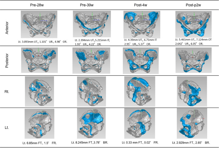
3-dimensional pelvic models based on magnetic resonance images (MRI) can be used to investigate accuracy and specifics of changing pelvic alignment during pregnancy and after childbirth. Few studies have investigated changes of pelvic alignment during pregnancy and after childbirth using three-dimensional pelvic models. This case report documents the changes of pelvic alignment during late pregnancy and after childbirth using MRI-based three-dimensional (3D) pelvic models. This was a longitudinal observation case report. A woman was imaged with MRI at 28 and 39 gestational weeks, as well as 4 and 72 weeks after childbirth. Greater internal, anterior, and downward rotation of both innominates at week 39 was observed from that at gestation week 28. Decreased internal, anterior, and downward rotation of both innominates at week 4 after child birth was observed compared with that at gestation week 39. We report the first case in Japan of changes of pelvic alignment measured using an MRI-based 3D pelvic alignment model during pregnancy and after child birth. This case suggests that the small changes of pubic area and greater separation of anterior portions of sacroiliac joints. Internal, anterior, and downward rotation of both innominates was observed in a Japanese primipara woman having no pelvic pain.
基于磁共振成像(MRI)的三维骨盆模型可用于研究妊娠期间及产后骨盆排列变化的准确性和具体情况。很少有研究使用三维骨盆模型来研究妊娠期间及产后骨盆排列的变化。本病例报告记录了使用基于MRI的三维(3D)骨盆模型观察到的妊娠晚期及产后骨盆排列的变化。这是一份纵向观察病例报告。一名女性在妊娠28周和39周时以及产后4周和72周时接受了MRI检查。观察到与妊娠28周时相比,在39周时双侧无名骨的内旋、前旋和向下旋转增加。与妊娠39周时相比,产后4周时双侧无名骨的内旋、前旋和向下旋转减少。我们报告了日本首例使用基于MRI的3D骨盆排列模型测量妊娠期间及产后骨盆排列变化的病例。该病例表明耻骨区域有微小变化,骶髂关节前部间距增大。在一名无骨盆疼痛的日本初产妇中观察到双侧无名骨的内旋、前旋和向下旋转。