Vinci Ramona, Pedicino Daniela, Bonanni Alice, D'Aiello Alessia, Severino Anna, Pisano Eugenia, Ponzo Myriana, Canonico Francesco, Ciampi Pellegrino, Russo Giulio, Di Sario Marianna, Montone Rocco Antonio, Trani Carlo, Conte Cristina, Grimaldi Maria Chiara, Cribari Francesco, Massetti Massimo, Crea Filippo, Liuzzo Giovanna
Department of Cardiovascular and Pneumological Sciences, Catholic University of the Sacred Heart, Rome, Italy.
Department of Cardiovascular Sciences, Fondazione Policlinico Universitario A. Gemelli IRCCS, Rome, Italy.
Front Cell Dev Biol. 2021 Oct 12;9:753223. doi: 10.3389/fcell.2021.753223. eCollection 2021.
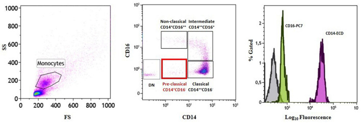
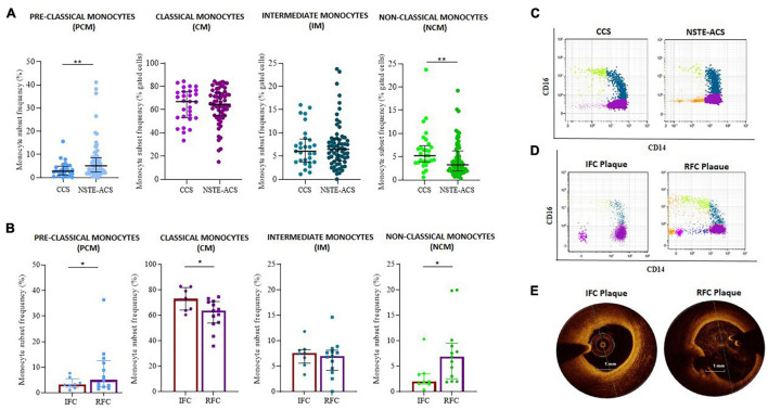
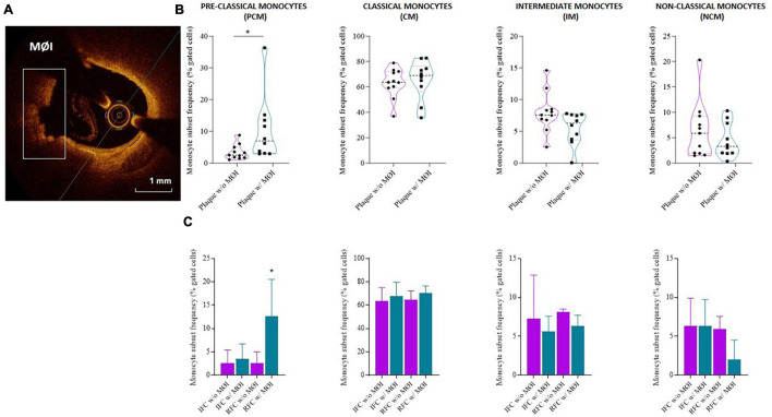
The evaluation of monocyte subset distribution among acute coronary syndrome (ACS) patients according to culprit coronary plaque morphology has never been explored. We evaluated whether there were significant differences in frequency of circulating monocyte subsets isolated from ACS patients according to optical coherence tomography (OCT) investigation of plaque erosion and rupture. We enrolled 74 patients with non-ST-elevation ACS (NSTE-ACS), 21 of them underwent OCT investigation of the culprit coronary plaque and local macrophage infiltration (MØI) assessment. As control, we enrolled 30 chronic coronary syndrome (CCS) patients. We assessed the frequency of monocyte subsets in the whole study population, in reliance on their CD14 and CD16 expression (classical, CM: CD14CD16; intermediates, IM: CD14CD16; non-classical, NCM: CD14CD16). Then, we tested the effect of lipopolysaccharide (LPS) (a CD14 ligand) on peripheral blood mononuclear cells (PBMCs) of NSTE-ACS patients, quantifying the inflammatory cytokine levels in cell-culture supernatants. Our data proved that monocyte subsets isolated from NSTE-ACS patients represent a peculiar biological signature of the pathophysiological mechanism lying beneath atherosclerotic plaque with a ruptured fibrous cap (RFC) as compared with plaque erosion. Moreover, the magnitude of LPS-mediated effects on IL-1β, IL-6, and IL-10 cytokine release in cell-culture supernatants appeared to be greater in NSTE-ACS patients with RFC. Finally, we described a fourth monocyte population never explored before in this clinical setting (pre-classical monocytes, PCM: CD14CD16) that was prevalent in NSTE-ACS patients as compared with CCS and, especially, in patients with RFC and culprit plaque with MØI.
根据罪犯冠状动脉斑块形态对急性冠状动脉综合征(ACS)患者的单核细胞亚群分布进行评估的研究从未开展过。我们评估了根据光学相干断层扫描(OCT)对斑块侵蚀和破裂情况的检查,从ACS患者中分离出的循环单核细胞亚群频率是否存在显著差异。我们纳入了74例非ST段抬高型ACS(NSTE-ACS)患者,其中21例接受了罪犯冠状动脉斑块的OCT检查及局部巨噬细胞浸润(MØI)评估。作为对照,我们纳入了30例慢性冠状动脉综合征(CCS)患者。我们依据单核细胞的CD14和CD16表达情况(经典型,CM:CD14⁺CD16⁻;中间型,IM:CD14⁺CD16⁺;非经典型,NCM:CD14⁻CD16⁺),评估了整个研究人群中单核细胞亚群的频率。然后,我们检测了脂多糖(LPS)(一种CD14配体)对NSTE-ACS患者外周血单个核细胞(PBMCs)的作用,对细胞培养上清液中的炎性细胞因子水平进行了定量分析。我们的数据证明,与斑块侵蚀相比,从NSTE-ACS患者中分离出的单核细胞亚群代表了具有纤维帽破裂(RFC)的动脉粥样硬化斑块潜在病理生理机制的一种特殊生物学特征。此外,LPS对细胞培养上清液中IL-1β、IL-6和IL-10细胞因子释放的介导作用在患有RFC的NSTE-ACS患者中似乎更大。最后,我们描述了在这种临床情况下以前从未探索过的第四种单核细胞群体(前经典单核细胞,PCM:CD14⁺CD16⁺),与CCS患者相比,该群体在NSTE-ACS患者中更为普遍,尤其是在患有RFC和伴有MØI的罪犯斑块的患者中。