Department of Otolaryngology, Head and Neck Surgery, Medical School OWL, Bielefeld University, Campus Klinikum Bielefeld Mitte, Teutoburgerstr.50, 33604, Bielefeld, Germany.
Department of Radiology, Medical School OWL, Bielefeld University, Campus Klinikum Bielefeld Mitte, Bielefeld, Germany.
Sci Rep. 2021 Oct 29;11(1):21298. doi: 10.1038/s41598-021-00824-3.
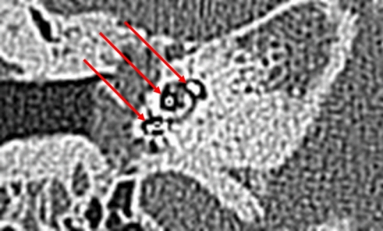
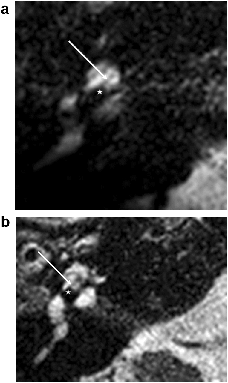
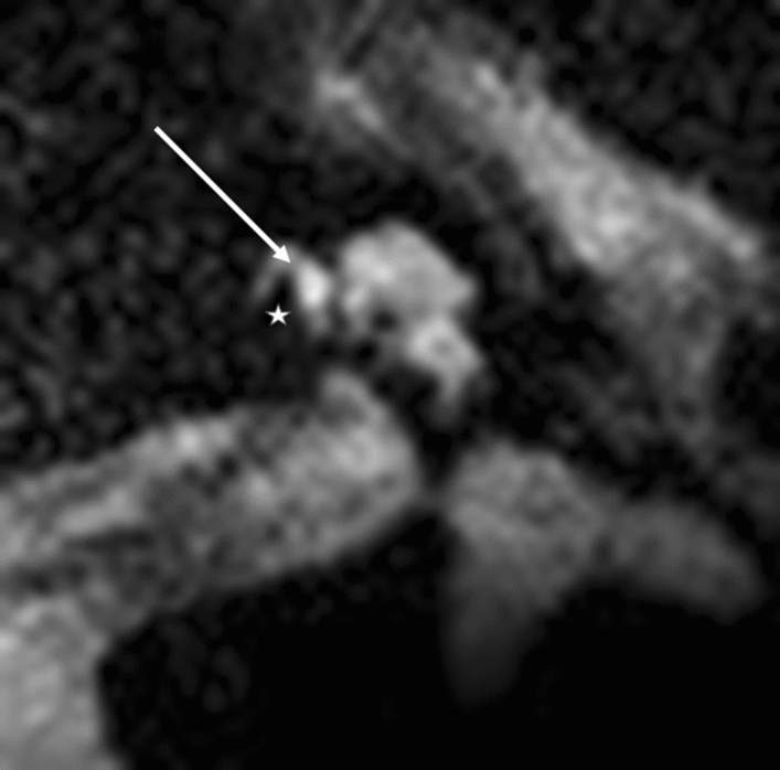
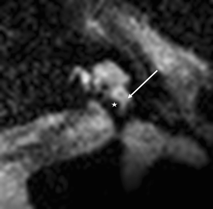
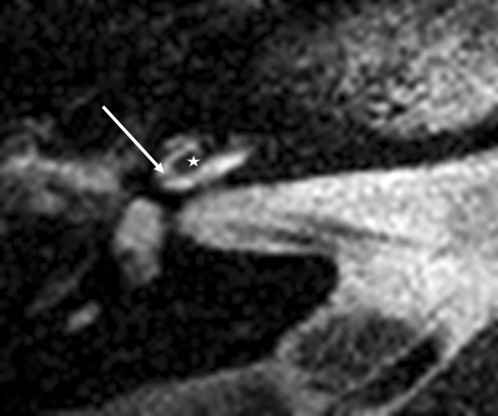
The estimation of scalar electrode position is a central point of quality control during the cochlear implant procedure. Ionic radiation is a disadvantage of commonly used radiologic estimation of electrode position. Recent developments in the field of cochlear implant magnets, implant receiver magnet position, and MRI sequence usage allow the postoperative evaluation of inner ear changes after cochlear implantation. The aim of the present study was to evaluate the position of lateral wall and modiolar cochlear implant electrodes using 3 T MRI scanning. In a prospective study, we evaluated 20 patients (10× Med-El Flex 28; 5× HFMS AB and 5× SlimJ AB) with a 3 T MRI and a T2 2D Drive MS sequence (voxel size: 0.3 × 0.3 × 0.9 mm) for the estimation of the intracochlear position of the cochlear implant electrode. In all cases, MRI allowed a determination of the electrode position in relation to the basilar membrane. This observation made the estimation of 19 scala tympani electrode positions and a single case of electrode translocation possible. 3 T MRI scanning allows the estimation of lateral wall and modiolar electrode intracochlear scalar positions.
在人工耳蜗植入手术过程中,对电极位置的标测是质量控制的一个关键点。离子辐射是目前常用的电极位置放射学评估方法的一个缺点。在人工耳蜗磁铁、植入体接收器磁铁位置以及 MRI 序列应用方面的最新进展,使得术后可以对人工耳蜗植入后内耳变化进行评估。本研究的目的是使用 3T MRI 扫描评估外侧壁和耳蜗内植入体电极的位置。在一项前瞻性研究中,我们使用 3T MRI 和 T2 二维驱动 MS 序列(体素大小:0.3×0.3×0.9mm)评估了 20 例患者(10×Med-El Flex 28;5×HFMS AB 和 5×SlimJ AB)的耳蜗内植入体电极位置,以评估耳蜗内植入体电极的位置。在所有病例中,MRI 都可以确定电极与基底膜的关系。这一观察结果使得可以对 19 个耳蜗鼓阶电极位置和 1 个电极移位病例进行估计。3T MRI 扫描可以估计外侧壁和耳蜗内植入体的电极位置。