The Sidney Kimmel Comprehensive Cancer Center, Johns Hopkins University School of Medicine, Baltimore, MD, USA.
Department of Oncology, Johns Hopkins University School of Medicine, 1650 Orleans Street, CRB1 Room 488, Baltimore, MD, 21287, USA.
J Hematol Oncol. 2021 Nov 2;14(1):184. doi: 10.1186/s13045-021-01203-1.
Metastasis occurs in the majority of pancreatic ductal adenocarcinoma (PDAC) patients at diagnosis or following resection. Patients with liver metastasis and those with lung metastasis have significantly different prognosis. Here, we sought to understand how cancer-associated fibroblasts (CAFs) play roles in the development of organ-specific metastasis.
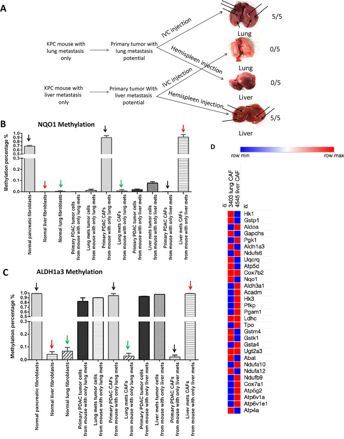
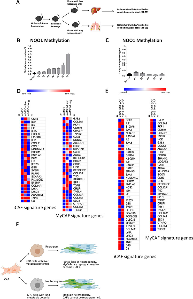
PDAC tumor cell lines established from the primary tumors with liver and lung metastasis potentials, respectively, in Kras/p53 mutation conditional knock-in (KPC) mice were co-cultured with matched CAFs or mouse mesenchymal stem cells. CAFs were isolated from metastases and subjected to DNA methylation and whole transcriptomic RNA sequencing analysis.
The ability of mouse PDAC tumor cell lines in developing liver or lung-specific metastases was demonstrated in orthotopic models. Tumor cells associated with liver metastasis potential, but not those associated with lung metastasis potential, induced the methylation of metabolism genes including NQO1 and ALDH1a3 and subsequent downregulated mRNA expression of a broader group of metabolism genes in CAFs. DNA methylation and downregulation of metabolism genes in CAFs in liver metastasis, but not those in lung metastasis, appeared to be regulated by DNA methyltransferase. Tumor cells associated with liver metastasis potential, but not those associated with lung metastasis potential, induce inflammatory CAF (iCAF) signatures. CAFs from liver metastasis demonstrated a more homogenous iCAF phenotype, whereas CAFs from lung metastasis maintained the heterogeneity.
PDAC with organ-specific metastatic potentials has different capacities in inducing methylation of metabolism genes in CAFs, modulating CAF phenotypes, and resulting in different levels of heterogeneity of CAFs in different metastatic niches.
在大多数胰腺导管腺癌(PDAC)患者中,转移发生在诊断时或切除后。肝转移患者和肺转移患者的预后有显著差异。在这里,我们试图了解癌症相关成纤维细胞(CAF)如何在器官特异性转移的发展中发挥作用。
分别从 Kras/p53 突变条件敲入(KPC)小鼠的原发肿瘤中建立具有肝转移和肺转移潜力的 PDAC 肿瘤细胞系,与匹配的 CAF 或小鼠间充质干细胞共培养。从转移灶中分离 CAF,并进行 DNA 甲基化和全转录组 RNA 测序分析。
在原位模型中证实了小鼠 PDAC 肿瘤细胞系在形成肝或肺特异性转移的能力。与肺转移潜力相关的肿瘤细胞,但与肝转移潜力相关的肿瘤细胞,诱导 CAF 中代谢基因(包括 NQO1 和 ALDH1a3)的甲基化,随后下调更广泛的代谢基因的 mRNA 表达。肝转移中 CAF 的 DNA 甲基化和代谢基因的下调,而不是肺转移中 CAF 的 DNA 甲基化和代谢基因的下调,似乎受 DNA 甲基转移酶调节。与肺转移潜力相关的肿瘤细胞,但与肝转移潜力相关的肿瘤细胞,诱导炎症性 CAF(iCAF)特征。与肝转移相关的 CAF 表现出更同质的 iCAF 表型,而与肺转移相关的 CAF 保持异质性。
具有器官特异性转移潜力的 PDAC 具有不同的能力,可诱导 CAF 中代谢基因的甲基化,调节 CAF 表型,并导致不同转移部位 CAF 的异质性水平不同。