• 文献检索
  • 文档翻译
  • 深度研究
  • 学术资讯
  • Suppr Zotero 插件Zotero 插件
  • 邀请有礼
  • 套餐&价格
  • 历史记录
应用&插件
Suppr Zotero 插件Zotero 插件浏览器插件Mac 客户端Windows 客户端微信小程序
定价
高级版会员购买积分包购买API积分包
服务
文献检索文档翻译深度研究API 文档MCP 服务
关于我们
关于 Suppr公司介绍联系我们用户协议隐私条款
关注我们

Suppr 超能文献

核心技术专利:CN118964589B侵权必究
粤ICP备2023148730 号-1Suppr @ 2026

文献检索

告别复杂PubMed语法,用中文像聊天一样搜索,搜遍4000万医学文献。AI智能推荐,让科研检索更轻松。

立即免费搜索

文件翻译

保留排版,准确专业,支持PDF/Word/PPT等文件格式,支持 12+语言互译。

免费翻译文档

深度研究

AI帮你快速写综述,25分钟生成高质量综述,智能提取关键信息,辅助科研写作。

立即免费体验

光体积描记术用于划定皮肤鳞状细胞癌边界。

Photoplethysmography for demarcation of cutaneous squamous cell carcinoma.

机构信息

Department of Otolaryngology at the Southdanish University Hospital, 7100, Vejle, Denmark.

Department of Electrical and Computer Engineering, Aarhus University, 8000, Aarhus N, Denmark.

出版信息

Sci Rep. 2021 Nov 2;11(1):21467. doi: 10.1038/s41598-021-00645-4.

DOI:10.1038/s41598-021-00645-4
PMID:34728637
原文链接:https://pmc.ncbi.nlm.nih.gov/articles/PMC8563950/
Abstract

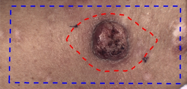
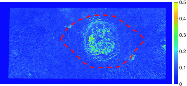
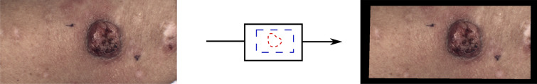
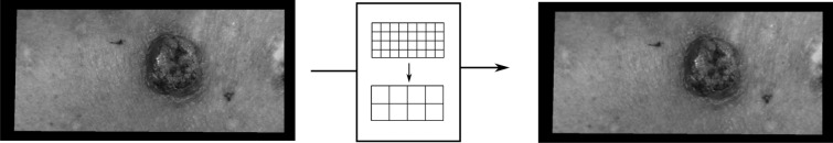
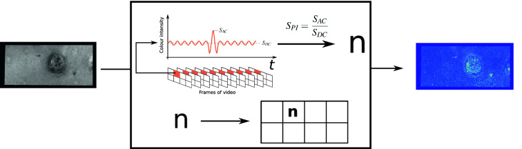
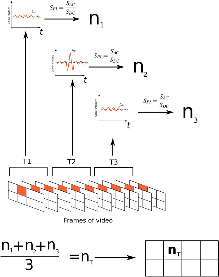
A video processing algorithm designed to identify cancer suspicious skin areas is presented here. It is based on video recordings of squamous cell carcinoma in the skin. Squamous cell carcinoma is a common malignancy, normally treated by surgical removal. The surgeon should always balance sufficient tissue removal against unnecessary mutilation, and therefore methods for distinction of cancer boundaries are wanted. Squamous cell carcinoma has angiogenesis and increased blood supply. Remote photoplethysmography is an evolving technique for analysis of signal variations in video recordings in order to extract vital signs such as pulsation. We hypothesize that the remote photoplethysmography signal inside the area of a squamous cell carcinoma is significantly different from the surrounding healthy skin. Based on high speed video recordings of 13 patients with squamous cell carcinoma, we have examined temporal signal differences in cancer areas versus healthy skin areas. A significant difference in temporal signal changes between cancer areas and healthy areas was found. Our video processing algorithm showed promising results encouraging further investigation to clarify how detailed distinctions can be made.

摘要

这里提出了一种用于识别可疑皮肤癌区域的视频处理算法。它基于皮肤鳞状细胞癌的视频记录。鳞状细胞癌是一种常见的恶性肿瘤,通常通过手术切除治疗。外科医生应始终在足够的组织切除和不必要的畸形之间取得平衡,因此需要区分癌症边界的方法。鳞状细胞癌有血管生成和血液供应增加。远程光体积描记术是一种用于分析视频记录中信号变化以提取脉动等生命体征的新兴技术。我们假设在鳞状细胞癌区域内的远程光体积描记术信号与周围健康皮肤有显著差异。基于 13 名鳞状细胞癌患者的高速视频记录,我们检查了癌症区域与健康皮肤区域之间的时间信号差异。发现癌症区域和健康区域之间的时间信号变化存在显著差异。我们的视频处理算法取得了有希望的结果,鼓励进一步研究以阐明如何进行更详细的区分。

https://cdn.ncbi.nlm.nih.gov/pmc/blobs/9157/8563950/4bae611ad357/41598_2021_645_Fig7_HTML.jpg
https://cdn.ncbi.nlm.nih.gov/pmc/blobs/9157/8563950/76719a18ed47/41598_2021_645_Fig1_HTML.jpg
https://cdn.ncbi.nlm.nih.gov/pmc/blobs/9157/8563950/e32d5bb3fcb1/41598_2021_645_Fig2_HTML.jpg
https://cdn.ncbi.nlm.nih.gov/pmc/blobs/9157/8563950/ef53d95f8f84/41598_2021_645_Fig3_HTML.jpg
https://cdn.ncbi.nlm.nih.gov/pmc/blobs/9157/8563950/3c384ca507d4/41598_2021_645_Fig4_HTML.jpg
https://cdn.ncbi.nlm.nih.gov/pmc/blobs/9157/8563950/3df259e9abd9/41598_2021_645_Fig5_HTML.jpg
https://cdn.ncbi.nlm.nih.gov/pmc/blobs/9157/8563950/a7b86a7f8c10/41598_2021_645_Fig6_HTML.jpg
https://cdn.ncbi.nlm.nih.gov/pmc/blobs/9157/8563950/4bae611ad357/41598_2021_645_Fig7_HTML.jpg

相似文献

1
Photoplethysmography for demarcation of cutaneous squamous cell carcinoma.光体积描记术用于划定皮肤鳞状细胞癌边界。
Sci Rep. 2021 Nov 2;11(1):21467. doi: 10.1038/s41598-021-00645-4.
2
Spatial analysis of photoplethysmography in cutaneous squamous cell carcinoma.光体积描记术在皮肤鳞状细胞癌中的空间分析。
Sci Rep. 2022 May 5;12(1):7318. doi: 10.1038/s41598-022-10924-3.
3
Living-Skin Classification via Remote-PPG.基于远程 PPG 的活体皮肤分类。
IEEE Trans Biomed Eng. 2017 Dec;64(12):2781-2792. doi: 10.1109/TBME.2017.2676160. Epub 2017 Mar 1.
4
Video-based heart rate monitoring across a range of skin pigmentations during an acute hypoxic challenge.急性低氧挑战期间对一系列皮肤色素沉着情况进行基于视频的心率监测。
J Clin Monit Comput. 2018 Oct;32(5):871-880. doi: 10.1007/s10877-017-0076-1. Epub 2017 Nov 9.
5
Identification and Tracking of Physiological Parameters from Skin using Video Photoplethysmography.使用视频光电容积脉搏波描记法从皮肤识别和跟踪生理参数
Annu Int Conf IEEE Eng Med Biol Soc. 2019 Jul;2019:6822-6825. doi: 10.1109/EMBC.2019.8857938.
6
Heart Rate and Heart Rate Variability From Single-Channel Video and ICA Integration of Multiple Signals.单通道视频和多个信号的 ICA 集成的心率和心率变异性。
IEEE J Biomed Health Inform. 2019 Nov;23(6):2398-2408. doi: 10.1109/JBHI.2018.2880097. Epub 2018 Nov 7.
7
Signal recovery in imaging photoplethysmography.成像光体积描记术中的信号恢复。
Physiol Meas. 2013 Nov;34(11):1499-511. doi: 10.1088/0967-3334/34/11/1499. Epub 2013 Oct 22.
8
Initial evaluation of prospective cardiac triggering using photoplethysmography signals recorded with a video camera compared to pulse oximetry and electrocardiography at 7T MRI.在7T磁共振成像(MRI)中,使用摄像机记录的光电容积脉搏波信号与脉搏血氧饱和度仪和心电图相比,对前瞻性心脏触发进行初步评估。
Biomed Eng Online. 2016 Nov 24;15(1):126. doi: 10.1186/s12938-016-0245-3.
9
New insights on super-high resolution for video-based heart rate estimation with a semi-blind source separation method.基于半盲源分离方法的超高分辨率视频心率估计的新见解。
Comput Biol Med. 2020 Jan;116:103535. doi: 10.1016/j.compbiomed.2019.103535. Epub 2019 Nov 16.
10
Estimation of respiratory rate from photoplethysmographic imaging videos compared to pulse oximetry.从光体积描记影像视频估计呼吸率与脉搏血氧饱和度测定法比较。
IEEE J Biomed Health Inform. 2015 Jul;19(4):1331-8. doi: 10.1109/JBHI.2015.2429746. Epub 2015 May 5.

引用本文的文献

1
Indocyanine Green Fluorescence Imaging in the Surgical Management of Skin Squamous Cell Carcinoma.吲哚菁绿荧光成像在皮肤鳞状细胞癌手术治疗中的应用
Clin Cosmet Investig Dermatol. 2023 Nov 15;16:3309-3320. doi: 10.2147/CCID.S413266. eCollection 2023.
2
Enhancing Skin Cancer Immunotheranostics and Precision Medicine through Functionalized Nanomodulators and Nanosensors: Recent Development and Prospects.通过功能化纳米调节剂和纳米传感器增强皮肤癌免疫诊疗与精准医学:最新进展与展望
Int J Mol Sci. 2023 Feb 9;24(4):3493. doi: 10.3390/ijms24043493.
3
Continuous intraoperative perfusion monitoring of free microvascular anastomosed fasciocutaneous flaps using remote photoplethysmography.

本文引用的文献

1
Association of remote imaging photoplethysmography and cutaneous perfusion in volunteers.志愿者中远程成像光体积描记术与皮肤灌注的相关性。
Sci Rep. 2020 Oct 5;10(1):16464. doi: 10.1038/s41598-020-73531-0.
2
A Broader Look: Camera-Based Vital Sign Estimation across the Spectrum.更广阔的视角:跨频谱的基于摄像头的生命体征估计
Yearb Med Inform. 2019 Aug;28(1):102-114. doi: 10.1055/s-0039-1677914. Epub 2019 Aug 16.
3
Camera-based photoplethysmography in an intraoperative setting.基于摄像头的术中光电容积脉搏波描记法。
应用远程光体积描记术对游离吻合的微小血管筋膜皮瓣进行术中连续灌注监测。
Sci Rep. 2023 Jan 27;13(1):1532. doi: 10.1038/s41598-023-28277-w.
4
Functionalization of Nanomaterials for Skin Cancer Theranostics.用于皮肤癌诊疗的纳米材料功能化
Front Bioeng Biotechnol. 2022 Apr 26;10:887548. doi: 10.3389/fbioe.2022.887548. eCollection 2022.
5
Spatial analysis of photoplethysmography in cutaneous squamous cell carcinoma.光体积描记术在皮肤鳞状细胞癌中的空间分析。
Sci Rep. 2022 May 5;12(1):7318. doi: 10.1038/s41598-022-10924-3.
Biomed Eng Online. 2018 Mar 14;17(1):33. doi: 10.1186/s12938-018-0467-7.
4
Merkel Cell Carcinoma of the Head and Neck: Recommendations for Diagnostics and Treatment.头颈部 Merkel 细胞癌:诊断和治疗建议。
Ann Surg Oncol. 2017 Oct;24(11):3430-3437. doi: 10.1245/s10434-017-5993-1. Epub 2017 Jul 31.
5
Evaluation of epigenetic inactivation of vascular endothelial growth factor receptors in head and neck squamous cell carcinoma.头颈部鳞状细胞癌中血管内皮生长因子受体表观遗传失活的评估
Tumour Biol. 2017 Jul;39(7):1010428317711657. doi: 10.1177/1010428317711657.
6
Margin Analysis: Cutaneous Malignancy of the Head and Neck.边缘分析:头颈部皮肤恶性肿瘤
Oral Maxillofac Surg Clin North Am. 2017 Aug;29(3):341-353. doi: 10.1016/j.coms.2017.04.001.
7
Margin Analysis: Squamous Cell Carcinoma of the Oropharynx.边缘分析:口咽鳞状细胞癌
Oral Maxillofac Surg Clin North Am. 2017 Aug;29(3):269-280. doi: 10.1016/j.coms.2017.03.004.
8
Erratum to: Endonasal Endoscopic Surgery in the Management of Sinonasal and Anterior Skull Base Malignancies.《鼻窦和前颅底恶性肿瘤的鼻内镜手术治疗》勘误
Head Neck Pathol. 2017 Jun;11(2):268. doi: 10.1007/s12105-017-0816-z.
9
Extracting remote photoplethysmogram signal from endoscopy videos for vessel and capillary density recognition.从内窥镜检查视频中提取远程光电容积脉搏波信号以识别血管和毛细血管密度。
Annu Int Conf IEEE Eng Med Biol Soc. 2016 Aug;2016:227-230. doi: 10.1109/EMBC.2016.7590681.
10
Algorithmic Principles of Remote PPG.远程光电容积脉搏波描记法的算法原理
IEEE Trans Biomed Eng. 2017 Jul;64(7):1479-1491. doi: 10.1109/TBME.2016.2609282. Epub 2016 Sep 13.