Molecular Biophysics and Integrated Bioimaging Division, Lawrence Berkeley National Laboratory, Berkeley, CA, USA.
Department of Anatomy, University of California, San Francisco, San Francisco, CA, USA.
Cell Rep Methods. 2021 Nov 22;1(7):100117. doi: 10.1016/j.crmeth.2021.100117. Epub 2021 Oct 28.
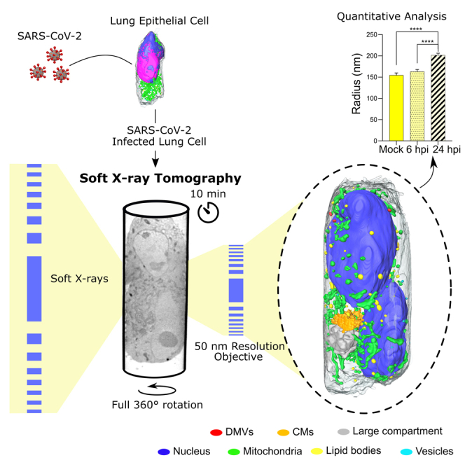
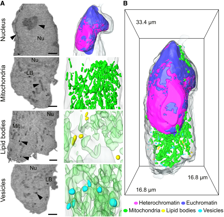
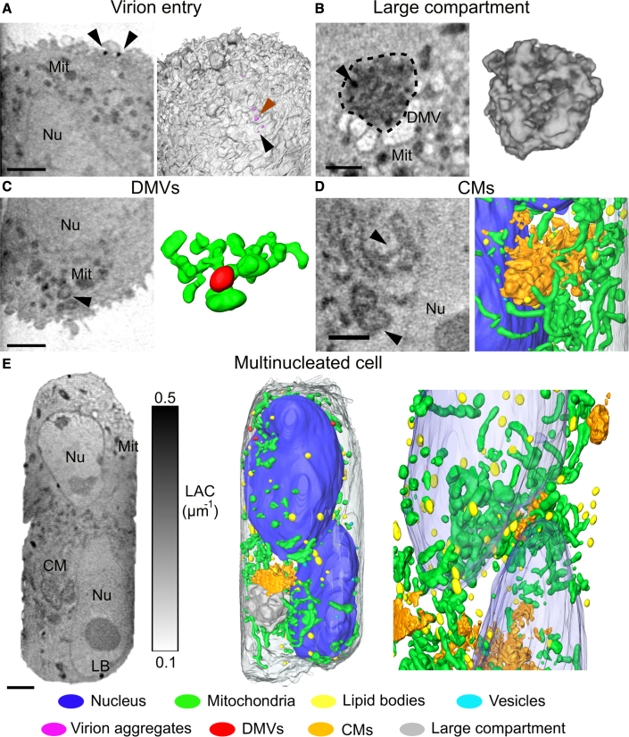
High-resolution and rapid imaging of host cell ultrastructure can generate insights toward viral disease mechanism, for example for a severe acute respiratory syndrome coronavirus-2 (SARS-CoV-2) infection. Here, we employ full-rotation soft X-ray tomography (SXT) to examine organelle remodeling induced by SARS-CoV-2 at the whole-cell level with high spatial resolution and throughput. Most of the current SXT systems suffer from a restricted field of view due to use of flat sample supports and artifacts due to missing data. In this approach using cylindrical sample holders, a full-rotation tomogram of human lung epithelial cells is performed in less than 10 min. We demonstrate the potential of SXT imaging by visualizing aggregates of SARS-CoV-2 virions and virus-induced intracellular alterations. This rapid whole-cell imaging approach allows us to visualize the spatiotemporal changes of cellular organelles upon viral infection in a quantitative manner.
高分辨率和快速成像宿主细胞超微结构可以深入了解病毒疾病的发病机制,例如严重急性呼吸系统综合症冠状病毒 2(SARS-CoV-2)感染。在这里,我们采用全旋转软 X 射线断层扫描(SXT)技术,以高空间分辨率和高通量水平检查 SARS-CoV-2 诱导的细胞器重塑。由于使用平板样品支架和数据缺失引起的伪影,大多数当前的 SXT 系统的视场受到限制。在这种使用圆柱形样品架的方法中,不到 10 分钟即可完成人肺上皮细胞的全旋转断层扫描。我们通过可视化 SARS-CoV-2 病毒粒子聚集体和病毒诱导的细胞内改变,证明了 SXT 成像的潜力。这种快速的全细胞成像方法使我们能够以定量方式可视化病毒感染后细胞细胞器的时空变化。