• 文献检索
  • 文档翻译
  • 深度研究
  • 学术资讯
  • Suppr Zotero 插件Zotero 插件
  • 邀请有礼
  • 套餐&价格
  • 历史记录
应用&插件
Suppr Zotero 插件Zotero 插件浏览器插件Mac 客户端Windows 客户端微信小程序
定价
高级版会员购买积分包购买API积分包
服务
文献检索文档翻译深度研究API 文档MCP 服务
关于我们
关于 Suppr公司介绍联系我们用户协议隐私条款
关注我们

Suppr 超能文献

核心技术专利:CN118964589B侵权必究
粤ICP备2023148730 号-1Suppr @ 2026

文献检索

告别复杂PubMed语法,用中文像聊天一样搜索,搜遍4000万医学文献。AI智能推荐,让科研检索更轻松。

立即免费搜索

文件翻译

保留排版,准确专业,支持PDF/Word/PPT等文件格式,支持 12+语言互译。

免费翻译文档

深度研究

AI帮你快速写综述,25分钟生成高质量综述,智能提取关键信息,辅助科研写作。

立即免费体验

单侧内翻型踝关节骨关节炎中胫骨和腓骨远端退行性模式的可视化和量化。

Visualization and quantification of the degenerative pattern of the distal tibia and fibula in unilateral varus ankle osteoarthritis.

机构信息

Department of Clinical Biomechanics, Keio University, 35 Shinano-machi, Shinjuku-ku, Tokyo, 160-8582, Japan.

Department of Orthopaedic Surgery, Ogikubo Hospital, 3-1-24 Imagawa, Suginami-ku, Tokyo, 167-0035, Japan.

出版信息

Sci Rep. 2021 Nov 3;11(1):21628. doi: 10.1038/s41598-021-00874-7.

DOI:10.1038/s41598-021-00874-7
PMID:34732780
原文链接:https://pmc.ncbi.nlm.nih.gov/articles/PMC8566530/
Abstract

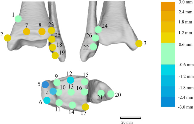
The present study aimed to quantify and visualize the degenerative patterns of the distal tibia and fibula due to ankle osteoarthritis (OA). We analyzed differences in tibial and fibular surface deviation between sides of patients with unilateral varus ankle OA (medial talar tilt > 4°) by registering each surface model to the mirror image of corresponding bone. Computed tomography images of both feet of 33 patients (OA: 22, control: 11) were examined. Statistically significant surface depression of approximately 2.5 mm on the anterior articular surface of the medial malleolus, and surface elevation of approximately 1 mm on the anterodistal edge of the tibiofibular joint and the lateral malleolus were observed in OA patients. These bone degenerations were found to be correlated with those on the other side of the ankle joint, the medial margin of the talar trochlea and the lateral articular surface of the talus, respectively. In contrast, the amount of bone depression on the plafond was smaller than previously anticipated. Such quantitative information about stereotypical patterns of bone degeneration in ankle OA would contribute to better understanding of the development of ankle OA and possible therapeutic interventions.

摘要

本研究旨在定量和可视化踝关节骨关节炎(OA)导致的胫骨和腓骨远端的退行性变化模式。我们通过将每个表面模型与相应骨骼的镜像进行配准,分析了单侧内翻踝关节 OA(距骨内倾斜度>4°)患者两侧胫骨和腓骨表面偏差的差异。检查了 33 名患者(OA:22 名,对照组:11 名)双脚的 CT 图像。在 OA 患者中,观察到距骨内踝前关节面约 2.5mm 的表面凹陷,以及胫腓关节和外踝前远端边缘约 1mm 的表面隆起。这些骨退变分别与踝关节另一侧、距骨滑车的内侧缘和距骨的外侧关节面的骨退变相关。相比之下,距骨底的骨凹陷量小于预期。这种关于踝关节 OA 骨退行性变典型模式的定量信息将有助于更好地理解踝关节 OA 的发展和可能的治疗干预。

https://cdn.ncbi.nlm.nih.gov/pmc/blobs/e60f/8566530/ead3570c18e6/41598_2021_874_Fig6_HTML.jpg
https://cdn.ncbi.nlm.nih.gov/pmc/blobs/e60f/8566530/f177b8dfdfd6/41598_2021_874_Fig1_HTML.jpg
https://cdn.ncbi.nlm.nih.gov/pmc/blobs/e60f/8566530/30fb6902830f/41598_2021_874_Fig2_HTML.jpg
https://cdn.ncbi.nlm.nih.gov/pmc/blobs/e60f/8566530/21c18fd2ca56/41598_2021_874_Fig3a_HTML.jpg
https://cdn.ncbi.nlm.nih.gov/pmc/blobs/e60f/8566530/d349a9c7c2ce/41598_2021_874_Fig4_HTML.jpg
https://cdn.ncbi.nlm.nih.gov/pmc/blobs/e60f/8566530/c3c033b93853/41598_2021_874_Fig5_HTML.jpg
https://cdn.ncbi.nlm.nih.gov/pmc/blobs/e60f/8566530/ead3570c18e6/41598_2021_874_Fig6_HTML.jpg

相似文献

1
Visualization and quantification of the degenerative pattern of the distal tibia and fibula in unilateral varus ankle osteoarthritis.单侧内翻型踝关节骨关节炎中胫骨和腓骨远端退行性模式的可视化和量化。
Sci Rep. 2021 Nov 3;11(1):21628. doi: 10.1038/s41598-021-00874-7.
2
Visualization and quantification of the degenerative pattern of the talus in unilateral varus ankle osteoarthritis.单侧内翻踝骨关节炎中距骨退行性病变的可视化与量化。
Sci Rep. 2019 Nov 25;9(1):17438. doi: 10.1038/s41598-019-53746-6.
3
Morphological features of the non-affected side of the hindfoot in patients with unilateral varus ankle osteoarthritis.单侧踝内翻骨关节炎患者患侧以外后足的形态学特征。
Ann Anat. 2024 Feb;252:152198. doi: 10.1016/j.aanat.2023.152198. Epub 2023 Dec 14.
4
Isolated Syndesmosis Diastasis: Computed Tomography Scan Assessment With Arthroscopic Correlation.孤立性下胫腓联合分离:CT扫描评估与关节镜相关性研究
Arthroscopy. 2017 Apr;33(4):828-834. doi: 10.1016/j.arthro.2017.01.009. Epub 2017 Feb 23.
5
Plain Radiographs Underestimate Varus Deformity of the Tibial Plafond.平片低估了距骨平台的内翻畸形。
J Foot Ankle Surg. 2022 Jul-Aug;61(4):836-840. doi: 10.1053/j.jfas.2021.12.001. Epub 2021 Dec 6.
6
The transfibular approach for ankle arthrodesis.踝关节融合术的经腓骨入路。
Oper Orthop Traumatol. 2005 Oct;17(4-5):361-79. doi: 10.1007/s00064-005-1148-9.
7
Increased Osseous (99m)Tc-DPD Uptake in End-Stage Ankle Osteoarthritis: Correlation Between SPECT-CT Imaging and Histologic Findings.终末期踝关节骨关节炎中骨(99m)锝-二磷酸盐摄取增加:SPECT-CT成像与组织学结果的相关性
Foot Ankle Int. 2015 Dec;36(12):1438-47. doi: 10.1177/1071100715596745. Epub 2015 Jul 31.
8
Investigation of factors related to the occurrence of osteochondral lesions of the talus by 3D bone morphology of the ankle.踝关节 3D 骨骼形态研究距骨骨软骨损伤相关因素。
Bone Joint J. 2018 Nov;100-B(11):1487-1490. doi: 10.1302/0301-620X.100B11.BJJ-2018-0346.R1.
9
Computed Tomographic Evaluation of Joint Geometry in Patients With End-Stage Ankle Osteoarthritis.终末期踝关节骨关节炎患者关节几何形态的计算机断层扫描评估
Foot Ankle Int. 2016 Jun;37(6):644-51. doi: 10.1177/1071100716629777. Epub 2016 Feb 3.
10
Radiographic Assessment of Fibular Length Variance: The Case for "Fibula Minus".腓骨长度差异的影像学评估:“腓骨短缩”的情况
J Foot Ankle Surg. 2018 Jan-Feb;57(1):91-94. doi: 10.1053/j.jfas.2017.08.013.

本文引用的文献

1
Sex- and age-related variations in the three-dimensional orientations and curvatures of the articular surfaces of the human talus.性别和年龄相关性对人体距骨关节面的三维方向和曲率的影响。
Anat Sci Int. 2021 Mar;96(2):258-264. doi: 10.1007/s12565-020-00585-5. Epub 2020 Nov 6.
2
Perioperative axial loading computed tomography findings in varus ankle osteoarthritis: Effect of supramalleolar osteotomy on abnormal internal rotation of the talus.术周轴向加载 CT 影像学在踝内翻骨关节炎中的表现:距骨内旋异常的跗骨上截骨术的影响。
Foot Ankle Surg. 2021 Feb;27(2):217-223. doi: 10.1016/j.fas.2020.04.006. Epub 2020 May 5.
3
Evaluation of Tibiofibular Joint Alignment in Ankle Osteoarthritis Based on 3D Bone Thickness.
基于三维骨厚度评估踝关节骨关节炎中胫腓关节对线情况
Annu Int Conf IEEE Eng Med Biol Soc. 2019 Jul;2019:2123-2126. doi: 10.1109/EMBC.2019.8857763.
4
Visualization and quantification of the degenerative pattern of the talus in unilateral varus ankle osteoarthritis.单侧内翻踝骨关节炎中距骨退行性病变的可视化与量化。
Sci Rep. 2019 Nov 25;9(1):17438. doi: 10.1038/s41598-019-53746-6.
5
Effect of natural full weight-bearing during standing on the rotation of the first metatarsal bone.自然全负重站立对第一跖骨旋转的影响。
Clin Anat. 2019 Jul;32(5):715-721. doi: 10.1002/ca.23385. Epub 2019 Apr 29.
6
Three-dimensional analysis of shape variations and symmetry of the fibula, tibia, calcaneus and talus.腓骨、胫骨、跟骨和距骨的形态变化和对称性的三维分析。
J Anat. 2019 Jan;234(1):132-144. doi: 10.1111/joa.12900. Epub 2018 Nov 4.
7
Three-dimensional innate mobility of the human foot bones under axial loading using biplane X-ray fluoroscopy.使用双平面X线透视检查法研究人体足部骨骼在轴向负荷下的三维固有活动度。
R Soc Open Sci. 2017 Oct 18;4(10):171086. doi: 10.1098/rsos.171086. eCollection 2017 Oct.
8
Effect of tibial coronal inclination on hindfoot kinematics: A biomechanical simulation study.胫骨冠状面倾斜度对后足运动学的影响:一项生物力学模拟研究。
Proc Inst Mech Eng H. 2017 Oct;231(10):952-958. doi: 10.1177/0954411917721941. Epub 2017 Jul 28.
9
Weight-bearing computed tomography findings in varus ankle osteoarthritis: abnormal internal rotation of the talus in the axial plane.内翻型踝关节骨关节炎的负重计算机断层扫描结果:距骨在轴位平面内的异常内旋。
Skeletal Radiol. 2017 Aug;46(8):1071-1080. doi: 10.1007/s00256-017-2655-0. Epub 2017 Apr 22.
10
Rotational Dynamics of the Normal Distal Tibiofibular Joint With Weight-Bearing Computed Tomography.负重计算机断层扫描下正常胫腓远侧关节的旋转动力学
Foot Ankle Int. 2016 Jun;37(6):627-35. doi: 10.1177/1071100716634757. Epub 2016 Feb 27.