• 文献检索
  • 文档翻译
  • 深度研究
  • 学术资讯
  • Suppr Zotero 插件Zotero 插件
  • 邀请有礼
  • 套餐&价格
  • 历史记录
应用&插件
Suppr Zotero 插件Zotero 插件浏览器插件Mac 客户端Windows 客户端微信小程序
定价
高级版会员购买积分包购买API积分包
服务
文献检索文档翻译深度研究API 文档MCP 服务
关于我们
关于 Suppr公司介绍联系我们用户协议隐私条款
关注我们

Suppr 超能文献

核心技术专利:CN118964589B侵权必究
粤ICP备2023148730 号-1Suppr @ 2026

文献检索

告别复杂PubMed语法,用中文像聊天一样搜索,搜遍4000万医学文献。AI智能推荐,让科研检索更轻松。

立即免费搜索

文件翻译

保留排版,准确专业,支持PDF/Word/PPT等文件格式,支持 12+语言互译。

免费翻译文档

深度研究

AI帮你快速写综述,25分钟生成高质量综述,智能提取关键信息,辅助科研写作。

立即免费体验

青光眼微囊型黄斑水肿的危险因素。

Risk factors for microcystic macular oedema in glaucoma.

机构信息

Ophthalmology, Glaucoma, University of California Los Angeles, Los Angeles, CA, USA.

Ophthalmology, Retina, University of California Los Angeles, Los Angeles, CA, USA.

出版信息

Br J Ophthalmol. 2023 Apr;107(4):505-510. doi: 10.1136/bjophthalmol-2021-320137. Epub 2021 Nov 5.

DOI:10.1136/bjophthalmol-2021-320137
PMID:34740886
原文链接:https://pmc.ncbi.nlm.nih.gov/articles/PMC9068828/
Abstract

BACKGROUND/AIMS: To identify clinical characteristics and factors associated with microcystic macular edema (MME) in patients with primary open-angle glaucoma (POAG).

METHODS

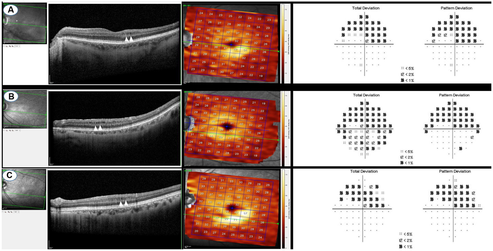
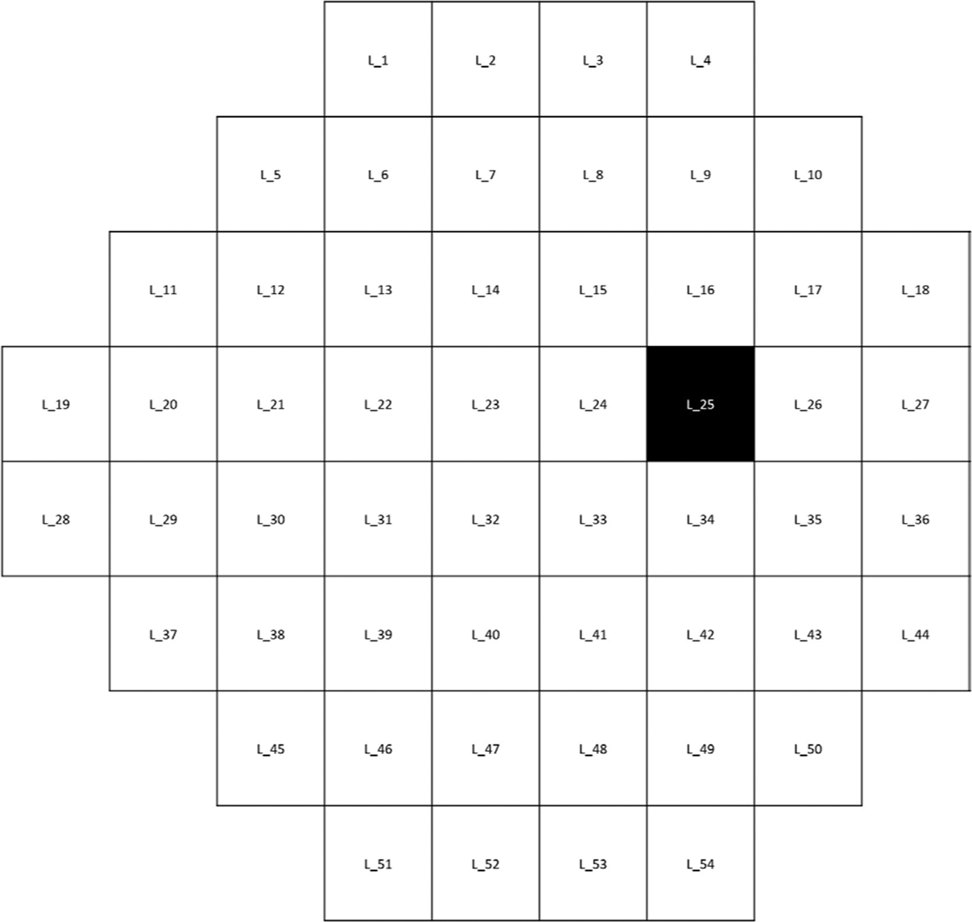
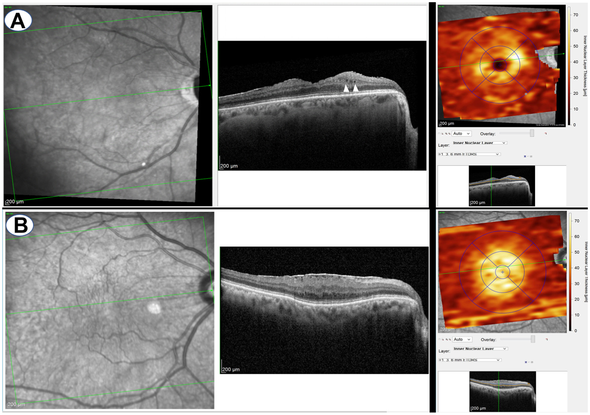
We included 315 POAG eyes between 2010 and 2019 with good-quality macular volume scans that had reliable visual fields (VF) available within 6 months in this observational retrospective cohort study. Eyes with retinal pathologies except for epiretinal membrane (ERM) were excluded. The inner nuclear layer was qualitatively assessed for the presence of MME. Global mean deviation (MD) and Visual Field Index (VFI) decay rates, superior and inferior MD rates and pointwise total deviation rates of change were estimated with linear regression. Logistic regression was performed to identify baseline factors associated with the presence of MME and to determine whether MME is associated with progressive VF loss.

RESULTS

25 out of 315 eyes (7.9%) demonstrated MME. The average (±SD) age and MD in eyes with and without MME was 57.2 (±8.7) versus 62.0 (±9.9) years (p=0.02) and -9.8 (±5.7) versus -4.9 (±5.3) dB (p<0.001), respectively. Worse global MD at baseline (p=0.001) and younger age (p=0.02) were associated with presence of MME. ERM was not associated with the presence of MME (p=0.84) in this cohort. MME was not associated with MD and VFI decay rates (p>0.49).

CONCLUSIONS

More severe glaucoma and younger age were associated with MME. MME was not associated with faster global VF decay in this cohort. MME may confound monitoring of glaucoma with full macular thickness.

摘要

背景/目的:确定原发性开角型青光眼(POAG)患者中与微囊型黄斑水肿(MME)相关的临床特征和因素。

方法

在这项观察性回顾性队列研究中,我们纳入了 2010 年至 2019 年间的 315 只 POAG 眼,这些眼均具有高质量的黄斑容积扫描,并且在 6 个月内可获得可靠的视野(VF)。排除了除视网膜前膜(ERM)以外的视网膜病变的眼睛。定性评估内核层是否存在 MME。使用线性回归估计平均偏差(MD)和视野指数(VFI)衰减率、上下 MD 衰减率和各点总偏差变化率。进行逻辑回归以确定与 MME 存在相关的基线因素,并确定 MME 是否与进行性 VF 损失相关。

结果

在 315 只眼中,有 25 只(7.9%)表现出 MME。在有和没有 MME 的眼中,平均(±SD)年龄和 MD 分别为 57.2(±8.7)岁与 62.0(±9.9)岁(p=0.02)和-9.8(±5.7)dB 与-4.9(±5.3)dB(p<0.001)。基线时更差的全球 MD(p=0.001)和更年轻的年龄(p=0.02)与 MME 的存在相关。在本队列中,ERM 与 MME 的存在无关(p=0.84)。MME 与 MD 和 VFI 衰减率无关(p>0.49)。

结论

更严重的青光眼和更年轻的年龄与 MME 相关。在本队列中,MME 与全球 VF 衰减速度无相关性。MME 可能会混淆对全黄斑厚度的青光眼监测。

https://cdn.ncbi.nlm.nih.gov/pmc/blobs/5a9a/9068828/9bbb5b42853b/nihms-1771037-f0003.jpg
https://cdn.ncbi.nlm.nih.gov/pmc/blobs/5a9a/9068828/3a40f88f3fc3/nihms-1771037-f0001.jpg
https://cdn.ncbi.nlm.nih.gov/pmc/blobs/5a9a/9068828/ce058b273e5a/nihms-1771037-f0002.jpg
https://cdn.ncbi.nlm.nih.gov/pmc/blobs/5a9a/9068828/9bbb5b42853b/nihms-1771037-f0003.jpg
https://cdn.ncbi.nlm.nih.gov/pmc/blobs/5a9a/9068828/3a40f88f3fc3/nihms-1771037-f0001.jpg
https://cdn.ncbi.nlm.nih.gov/pmc/blobs/5a9a/9068828/ce058b273e5a/nihms-1771037-f0002.jpg
https://cdn.ncbi.nlm.nih.gov/pmc/blobs/5a9a/9068828/9bbb5b42853b/nihms-1771037-f0003.jpg

相似文献

1
Risk factors for microcystic macular oedema in glaucoma.青光眼微囊型黄斑水肿的危险因素。
Br J Ophthalmol. 2023 Apr;107(4):505-510. doi: 10.1136/bjophthalmol-2021-320137. Epub 2021 Nov 5.
2
Clinical Characteristics of Microcystic Macular Edema in Chronic Primary Angle-Closure Glaucoma and Primary Open-Angle Glaucoma Patients.慢性原发性闭角型青光眼和原发性开角型青光眼患者的微小囊样黄斑水肿的临床特征。
Ophthalmic Res. 2024;67(1):145-153. doi: 10.1159/000535900. Epub 2024 Jan 19.
3
Retrograde Maculopathy in Patients With Glaucoma.青光眼患者的逆行性黄斑病变
J Glaucoma. 2017 May;26(5):423-429. doi: 10.1097/IJG.0000000000000633.
4
Progression of primary open angle glaucoma in asymmetrically myopic eyes.原发性开角型青光眼在不对称性近视眼中的进展
Graefes Arch Clin Exp Ophthalmol. 2016 Jul;254(7):1331-7. doi: 10.1007/s00417-016-3332-z. Epub 2016 Apr 11.
5
Comparison of the Prevalence and Clinical Characteristics of Epiretinal Membrane in Pseudoexfoliation and Primary Open-angle Glaucoma.比较假性剥脱综合征和原发性开角型青光眼的眼后膜患病率和临床特征。
J Glaucoma. 2021 Sep 1;30(9):859-865. doi: 10.1097/IJG.0000000000001851.
6
Clinical evaluation of microcystic macular edema in patients with glaucoma.青光眼患者微囊性黄斑水肿的临床评估
Eye (Lond). 2016 Nov;30(11):1502-1508. doi: 10.1038/eye.2016.190. Epub 2016 Aug 12.
7
[The function-structure impairment pattern of optic nerves in primary open-angle glaucoma and normal-tension glaucoma].[原发性开角型青光眼和正常眼压性青光眼视神经的功能-结构损害模式]
Zhonghua Yan Ke Za Zhi. 2018 Nov 11;54(11):811-819. doi: 10.3760/cma.j.issn.0412-4081.2018.11.004.
8
Progressive Macular Vessel Density Loss and Visual Field Progression in Open-angle Glaucoma Eyes with Central Visual Field Damage.中心视野损伤的开角型青光眼眼中进行性黄斑血管密度损失和视野进展。
Ophthalmol Glaucoma. 2024 Jan-Feb;7(1):16-29. doi: 10.1016/j.ogla.2023.06.009. Epub 2023 Jun 26.
9
Microcystic macular edema: retrograde maculopathy caused by optic neuropathy.微囊样黄斑水肿:视神经病变引起的逆行性黄斑病变。
Ophthalmology. 2014 Jan;121(1):142-149. doi: 10.1016/j.ophtha.2013.08.045. Epub 2013 Oct 15.
10
The clinical spectrum of microcystic macular edema.微囊型黄斑水肿的临床谱。
Invest Ophthalmol Vis Sci. 2014 Feb 18;55(2):952-61. doi: 10.1167/iovs.13-12912.

引用本文的文献

1
Long-term risk factors for poor visual outcomes in patients with epiretinal membrane and open-angle glaucoma: a retrospective study.视网膜前膜合并开角型青光眼患者视力预后不良的长期危险因素:一项回顾性研究。
Sci Rep. 2024 Nov 19;14(1):28660. doi: 10.1038/s41598-024-80020-1.
2
Thicker Inner Nuclear Layer as a Predictor of Glaucoma Progression and the Impact of Intraocular Pressure Fluctuation.较厚的内核层作为青光眼进展的预测指标及眼压波动的影响
J Clin Med. 2024 Apr 17;13(8):2312. doi: 10.3390/jcm13082312.
3
Associations of smoking and alcohol consumption with the development of open angle glaucoma: a retrospective cohort study.

本文引用的文献

1
Local Macular Thickness Relationships between 2 OCT Devices.两种 OCT 设备的局部黄斑厚度关系。
Ophthalmol Glaucoma. 2021 Mar-Apr;4(2):209-215. doi: 10.1016/j.ogla.2020.08.012. Epub 2020 Aug 29.
2
Distribution and Rates of Visual Field Loss across Different Disease Stages in Primary Open-Angle Glaucoma.原发性开角型青光眼不同疾病阶段视野缺失的分布和发生率。
Ophthalmol Glaucoma. 2018 Jul-Aug;1(1):52-60. doi: 10.1016/j.ogla.2018.05.005. Epub 2018 Jul 3.
3
Rate of visual field decay in glaucomatous eyes with acquired pits of the optic nerve.
吸烟和饮酒与开角型青光眼发展的关联:一项回顾性队列研究。
BMJ Open. 2023 Oct 4;13(10):e072163. doi: 10.1136/bmjopen-2023-072163.
4
The prevalence and presentation patterns of microcystic macular oedema: a systematic review and meta-analysis of 2128 glaucomatous eyes.微囊型黄斑水肿的患病率和表现模式:2128 只青光眼眼中的系统评价和荟萃分析。
Eye (Lond). 2023 Nov;37(16):3322-3333. doi: 10.1038/s41433-023-02524-w. Epub 2023 Apr 18.
5
Impact of Smoking on Visual Field Progression in a Long-term Clinical Follow-up.吸烟对长期临床随访中视野进展的影响。
Ophthalmology. 2022 Nov;129(11):1235-1244. doi: 10.1016/j.ophtha.2022.06.017. Epub 2022 Jun 23.
青光眼伴获得性视神经凹的视野衰退率。
Br J Ophthalmol. 2021 Mar;105(3):381-386. doi: 10.1136/bjophthalmol-2020-315980. Epub 2020 May 13.
4
Risk Factors for Fast Visual Field Progression in Glaucoma.青光眼视野快速进展的危险因素。
Am J Ophthalmol. 2019 Nov;207:268-278. doi: 10.1016/j.ajo.2019.06.019. Epub 2019 Jun 22.
5
PATIENTS WITH EPIRETINAL MEMBRANES DISPLAY RETROGRADE MACULOPATHY AFTER SURGICAL PEELING OF THE INTERNAL LIMITING MEMBRANE.内界膜切开术后,视网膜前膜患者出现逆行性黄斑病变。
Retina. 2019 Nov;39(11):2132-2140. doi: 10.1097/IAE.0000000000002266.
6
Microcystic macular oedema in optic neuropathy: case series and literature review.微囊样黄斑水肿性视神经病变:病例系列及文献复习。
Clin Exp Ophthalmol. 2018 Dec;46(9):1075-1086. doi: 10.1111/ceo.13327. Epub 2018 Jun 20.
7
Asymmetric Patterns of Visual Field Defect in Primary Open-Angle and Primary Angle-Closure Glaucoma.原发性开角型青光眼和原发性闭角型青光眼中视野缺损的不对称模式。
Invest Ophthalmol Vis Sci. 2018 Mar 1;59(3):1279-1287. doi: 10.1167/iovs.17-22980.
8
Retinal putative glial alterations: implication for glaucoma care.视网膜假定的神经胶质改变:对青光眼治疗的意义。
Ophthalmic Physiol Opt. 2018 Jan;38(1):56-65. doi: 10.1111/opo.12425. Epub 2017 Nov 23.
9
Microcystoid Macular Changes in Association With Idiopathic Epiretinal Membranes in Eyes With and Without Glaucoma: Clinical Insights.伴有或不伴有青光眼的眼睛中与特发性视网膜前膜相关的微囊样黄斑改变:临床见解
Am J Ophthalmol. 2017 Sep;181:156-165. doi: 10.1016/j.ajo.2017.06.023. Epub 2017 Jul 1.
10
Frequency of Testing to Detect Visual Field Progression Derived Using a Longitudinal Cohort of Glaucoma Patients.基于青光眼患者纵向队列检测视野进展的检测频率。
Ophthalmology. 2017 Jun;124(6):786-792. doi: 10.1016/j.ophtha.2017.01.027. Epub 2017 Mar 6.