Wiegertjes Kim, Jansen Michelle G, Jolink Wilmar Mt, Duering Marco, Koemans Emma A, Schreuder Floris Hbm, Tuladhar Anil M, Wermer Marieke Jh, Klijn Catharina Jm, de Leeuw Frank-Erik
Department of Neurology, Donders Institute for Brain, Cognition and Behavior, Radboud University Medical Center, Nijmegen, The Netherlands.
Department of Neurology and Neurosurgery, University Medical Center Utrecht, Brain Center, Utrecht University, Utrecht, The Netherlands.
Eur Stroke J. 2021 Sep;6(3):236-244. doi: 10.1177/23969873211031753. Epub 2021 Aug 25.
It is unclear why cerebral small vessel disease (SVD) leads to lacunar stroke in some and to non-lobar intracerebral hemorrhage (ICH) in others. We investigated differences in MRI markers of SVD in patients with lacunar stroke or non-lobar ICH.
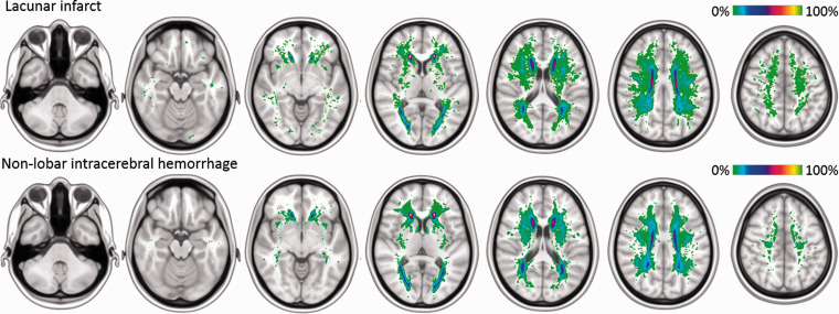
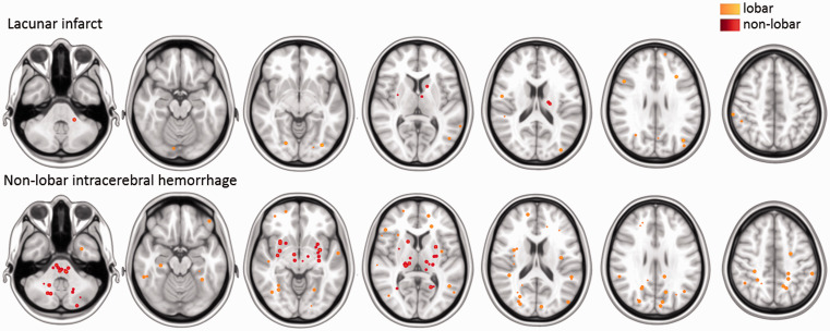
We included patients from two prospective cohort studies with either lacunar stroke (RUN DMC) or non-lobar ICH (FETCH). Differences in SVD markers (white matter hyperintensities [WMH], lacunes, cerebral microbleeds [CMB]) between groups were investigated with univariable tests; multivariable logistic regression analysis, adjusted for age, sex, and vascular risk factors; spatial correlation analysis and voxel-wise lesion symptom mapping.
We included 82 patients with lacunar stroke (median age 63, IQR 57-72) and 54 with non-lobar ICH (66, 59-75). WMH volumes and distribution were not different between groups. Lacunes were more frequent in patients with a lacunar stroke (44% vs. 17%, adjusted odds ratio [aOR] 5.69, 95% CI [1.66-22.75]) compared to patients with a non-lobar ICH. CMB were more frequent in patients with a non-lobar ICH (71% vs. 23%, aOR for lacunar stroke vs non-lobar ICH 0.08 95% CI [0.02-0.26]), and more often located in non-lobar regions compared to CMB in lacunar stroke.
Although we obserd different types of MRI markers of SVD within the same patient, ischemic markers of SVD were more frequent in the ischemic type of lacunar stroke, and hemorrhagic markers were more prevalent in the hemorrhagic phenotype of non-lobar ICH.
There are differences between MRI markers of SVD between patients with a lacunar stroke and those with a non-lobar ICH.
目前尚不清楚为什么脑小血管病(SVD)在一些患者中会导致腔隙性卒中,而在另一些患者中会导致非叶性脑出血(ICH)。我们研究了腔隙性卒中和非叶性ICH患者SVD的MRI标志物差异。
我们纳入了两项前瞻性队列研究的患者,分别为腔隙性卒中(RUN DMC)或非叶性ICH(FETCH)。通过单变量检验研究两组之间SVD标志物(白质高信号[WMH]、腔隙、脑微出血[CMB])的差异;进行多变量逻辑回归分析,并对年龄、性别和血管危险因素进行校正;进行空间相关性分析和体素级病变症状映射。
我们纳入了82例腔隙性卒中患者(中位年龄63岁,四分位间距57 - 72岁)和54例非叶性ICH患者(66岁,59 - 75岁)。两组之间WMH体积和分布无差异。与非叶性ICH患者相比,腔隙性卒中患者的腔隙更常见(44%对17%,校正比值比[aOR]为5.69,95%置信区间[1.66 - 22.75])。非叶性ICH患者的CMB更常见(71%对23%,腔隙性卒中与非叶性ICH的aOR为0.08,95%置信区间[0.02 - 0.26]),并且与腔隙性卒中的CMB相比,更常位于非叶区域。
尽管我们在同一患者中观察到了不同类型的SVD的MRI标志物,但SVD的缺血性标志物在缺血性腔隙性卒中类型中更常见,而出血性标志物在非叶性ICH的出血性表型中更普遍。
腔隙性卒中和非叶性ICH患者之间SVD的MRI标志物存在差异。