Innovative Technology Of Radiotherapy Computations and Hardware (iTORCH) Laboratory, Department of Radiation Oncology, University of Texas Southwestern Medical Center, Dallas, TX, United States of America.
Phys Med Biol. 2021 Dec 6;66(24). doi: 10.1088/1361-6560/ac37fc.
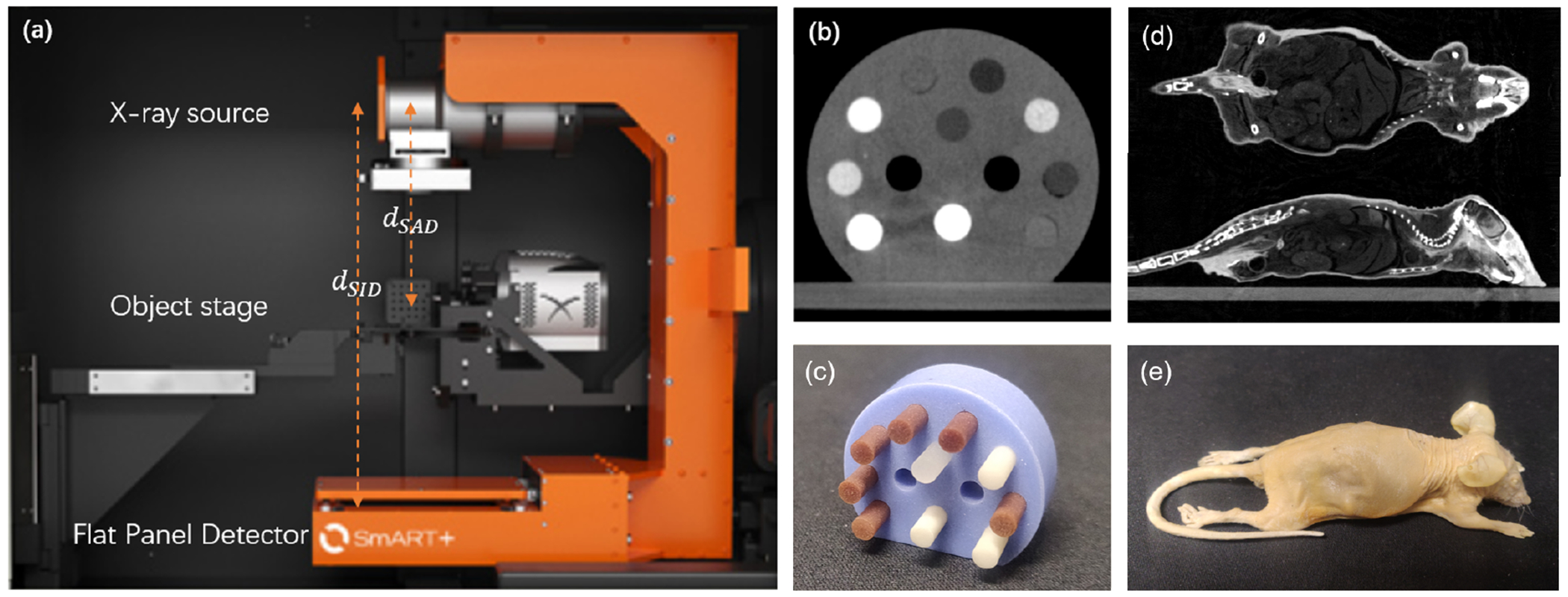
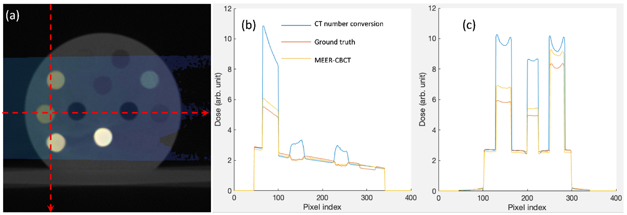
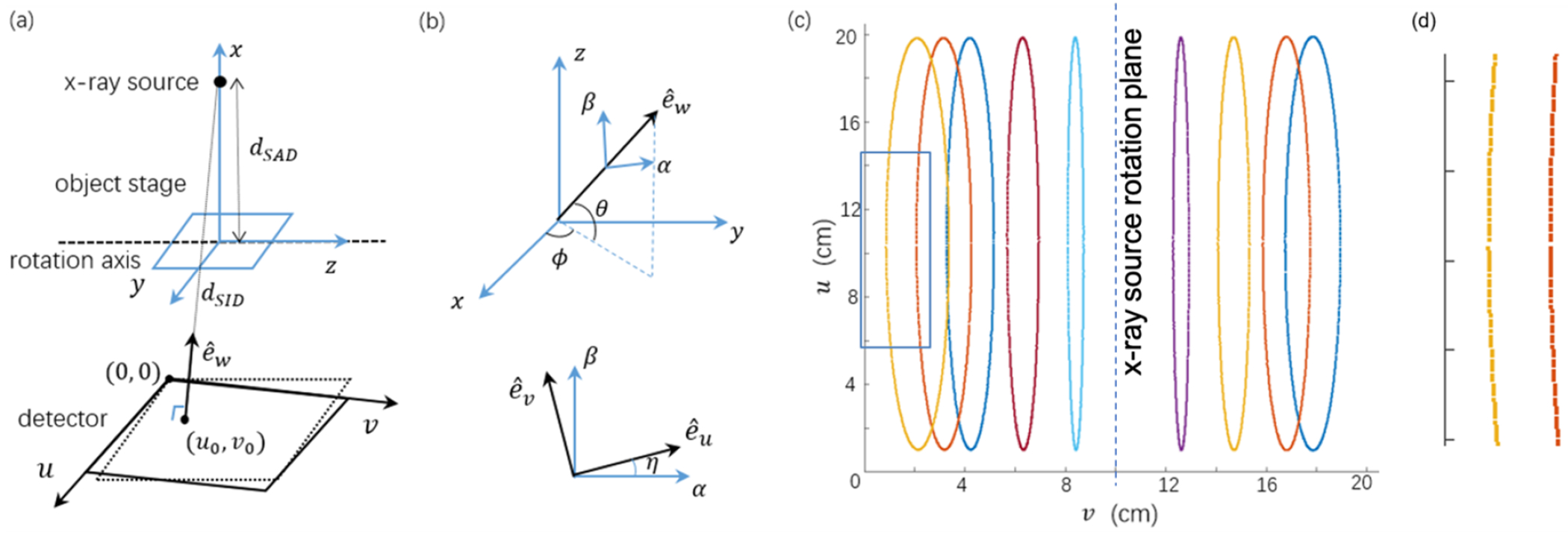
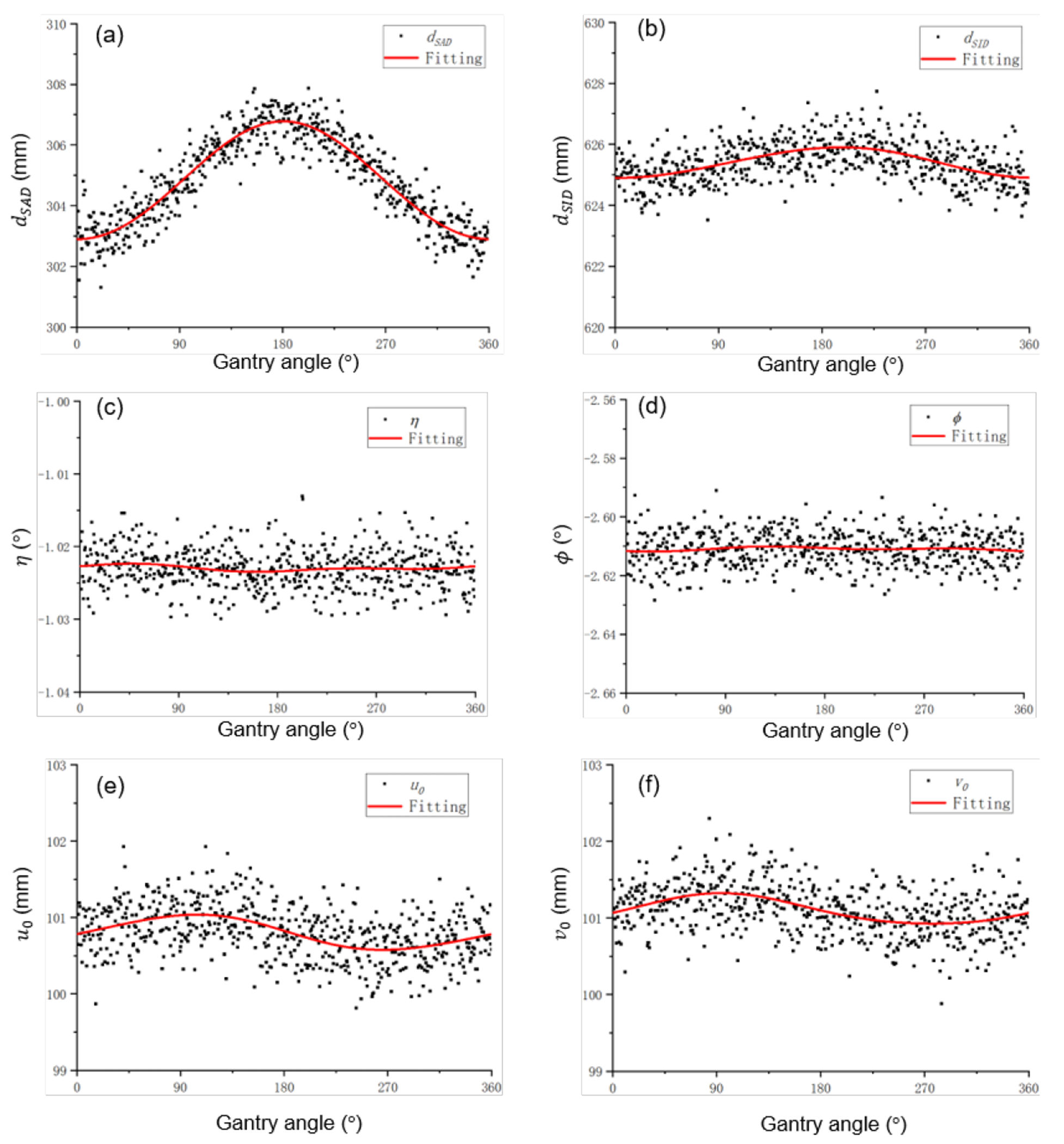
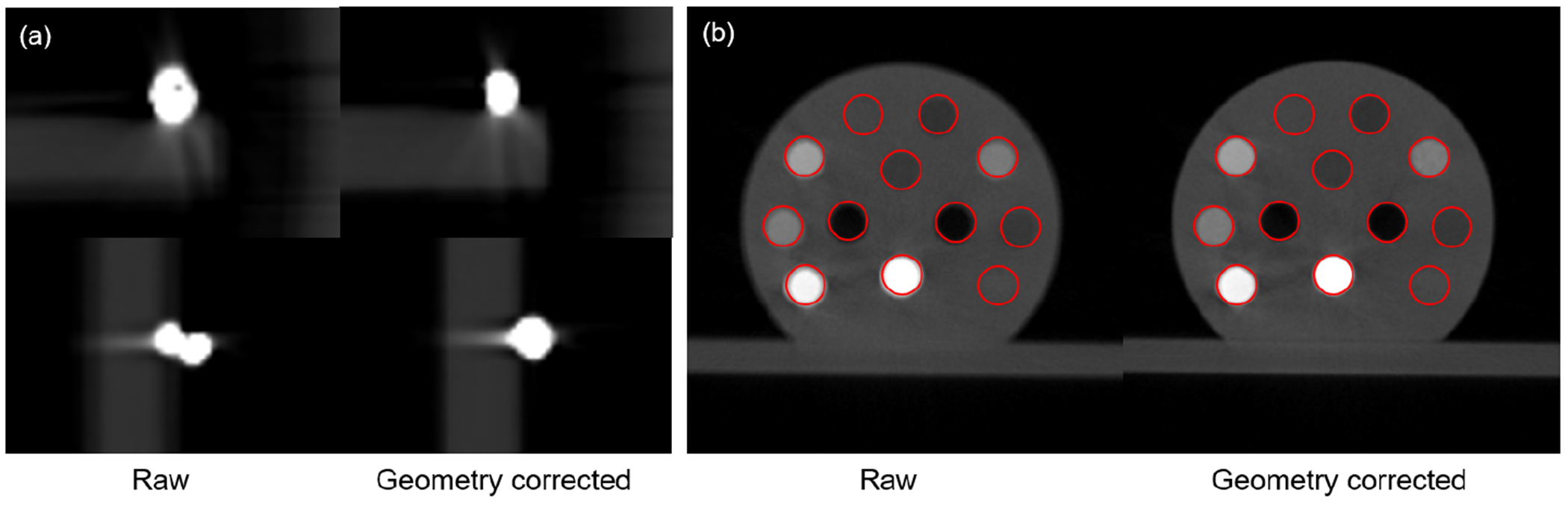
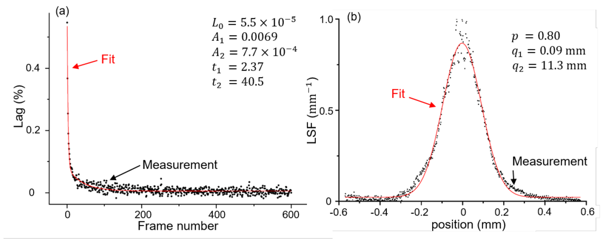
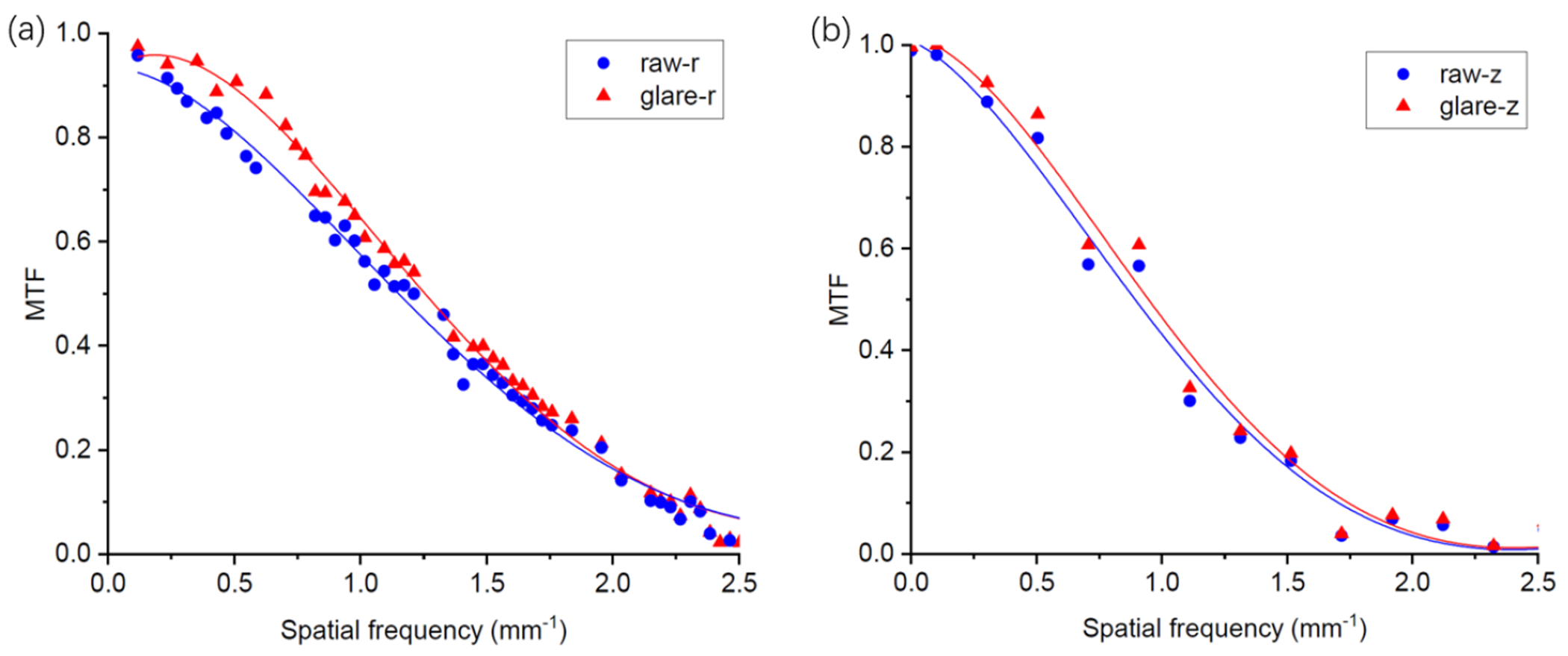
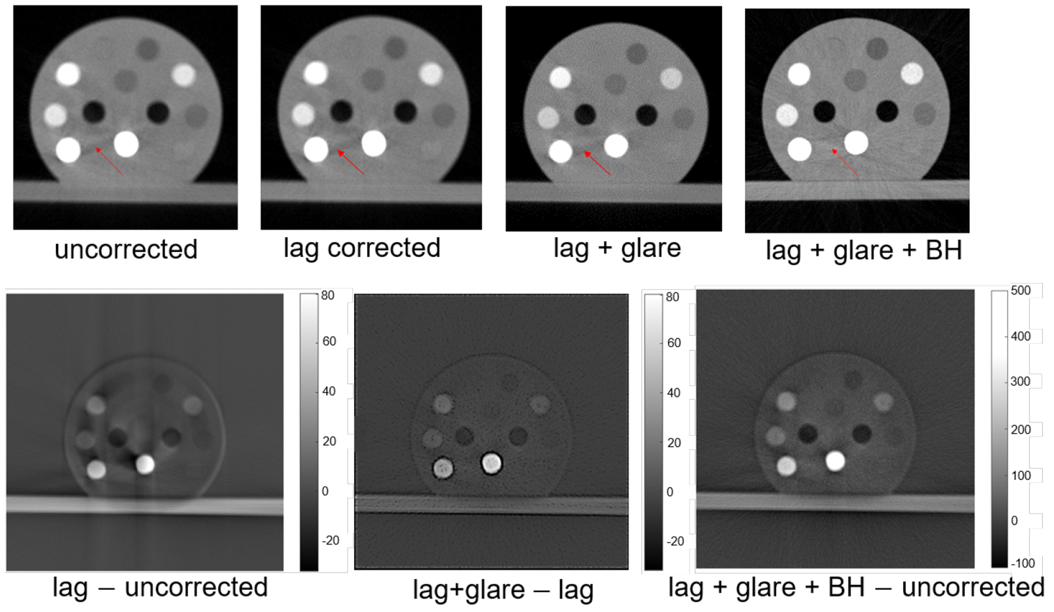
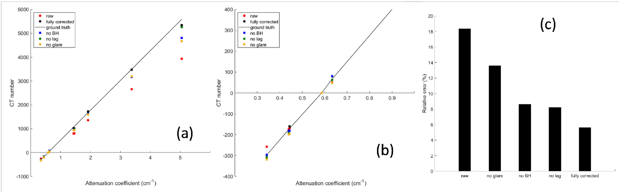
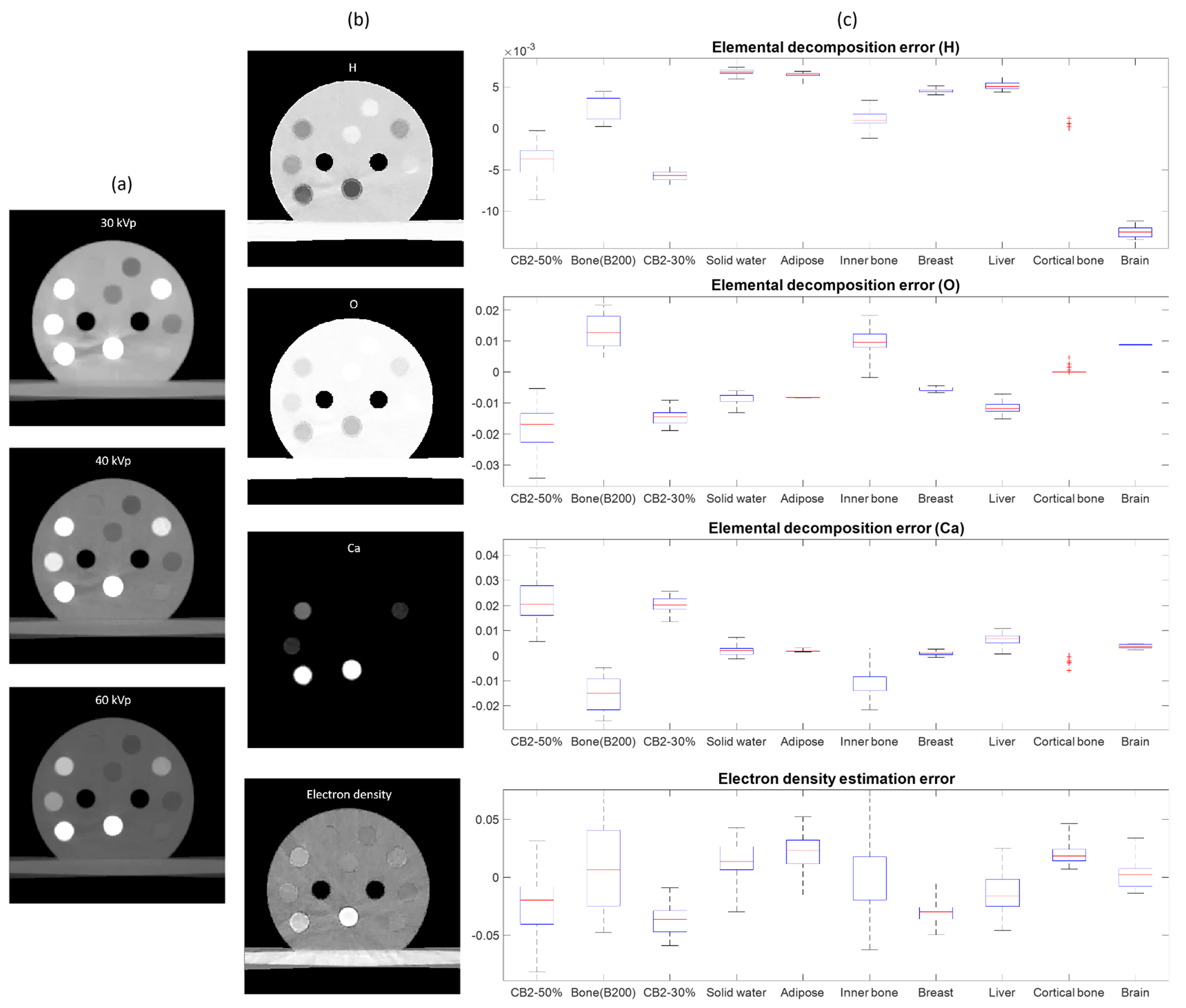
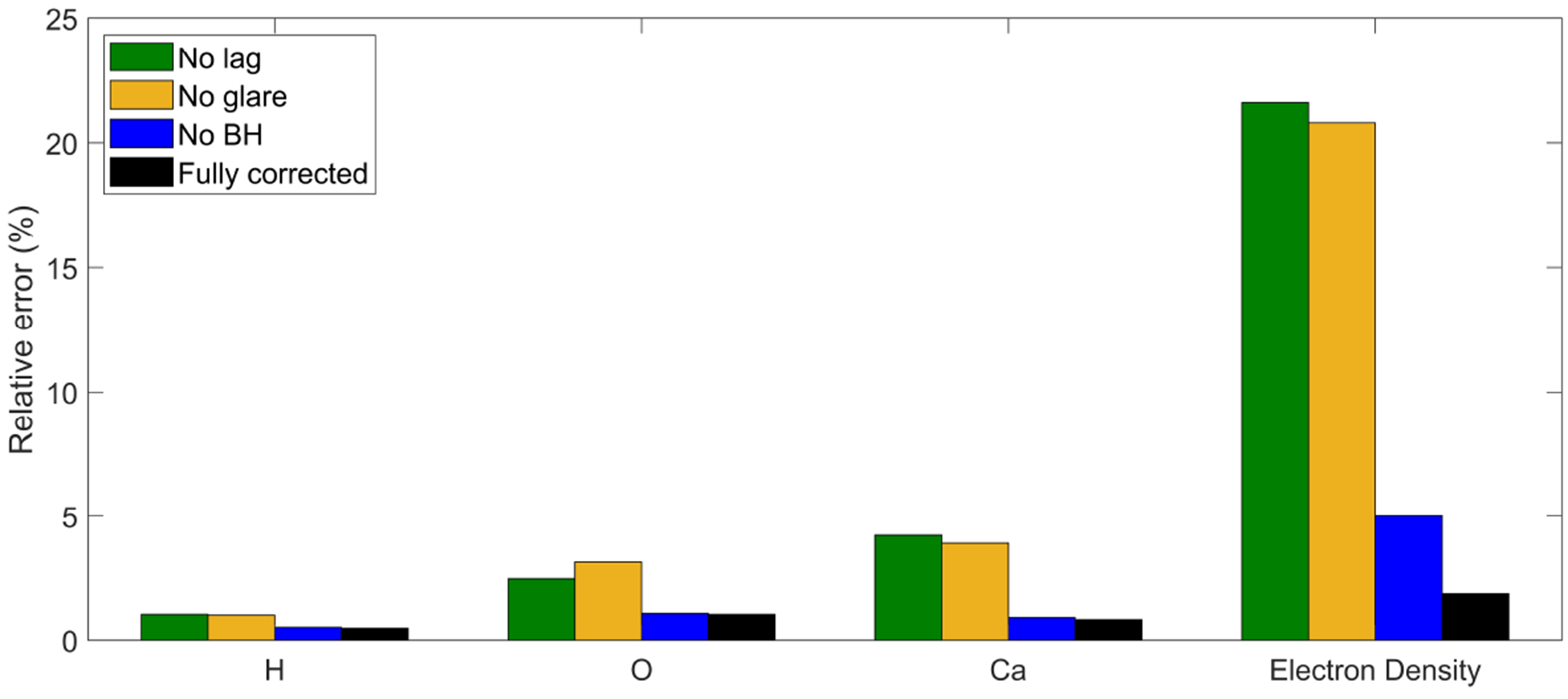
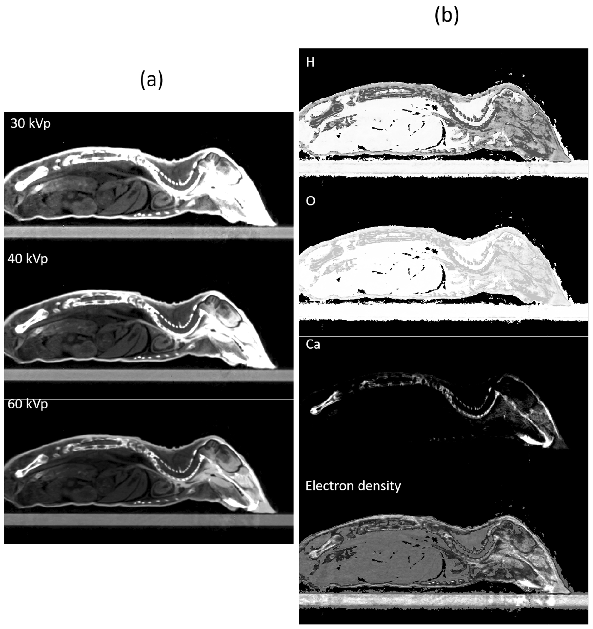
Cone-beam CT (CBCT) in modern pre-clinical small-animal radiation research platforms provides volumetric images for image guidance and experiment planning purposes. In this work, we implemented multi-energy element-resolved (MEER) CBCT using three scans with different kVps on a SmART platform (Precision x-ray Inc.) to determine images of relative electron density (rED) and elemental composition (EC) that are needed for Monte Carlo-based radiation dose calculation.We performed comprehensive calibration tasks to achieve sufficient accuracy for this quantitative imaging purpose. For geometry calibration, we scanned a ball bearing phantom and used an analytical method together with an optimization approach to derive gantry angle specific geometry parameters. Intensity calibration and correction included the corrections for detector lag, glare, and beam hardening. The corrected CBCT projection images acquired at 30, 40, and 60 kVp in multiple scans were used to reconstruct CBCT images using the Feldkamp-Davis-Kress reconstruction algorithm. After that, an optimization problem was solved to determine images of rED and EC. We demonstrated the effectiveness of our CBCT calibration steps by showing improvements in image quality and successful material decomposition in cases with a small animal CT calibration phantom and a plastinated mouse phantom.It was found that artifacts induced by geometry inaccuracy, detector lag, glare, and beam hardening were visually reduced. CT number mean errors were reduced from 19% to 5%. In the CT calibration phantom case, median errors in H, O, and Ca fractions for all the inserts were below 1%, 2%, and 4% respectively, and median error in rED was less than 5%. Compared to the standard approach deriving material type and rED via CT number conversion, our approach improved Monte Carlo simulation-based dose calculation accuracy in bone regions. Mean dose error was reduced from 47.5% to 10.9%.The MEER-CBCT implemented on an existing CBCT system of a small animal irradiation platform achieved accurate material decomposition and significantly improved Monte Carlo dose calculation accuracy.
锥形束 CT(CBCT)在现代临床前小动物放射研究平台中提供容积图像,用于图像引导和实验规划目的。在这项工作中,我们使用 SmART 平台(Precision x-ray Inc.)上的三种不同千伏的扫描实现了多能量元素分辨(MEER)CBCT,以确定用于基于蒙特卡罗的辐射剂量计算所需的相对电子密度(rED)和元素组成(EC)的图像。我们进行了全面的校准任务,以实现这种定量成像目的的足够准确性。对于几何校准,我们扫描了一个球轴承体模,并使用分析方法和优化方法来推导特定于旋转架角度的几何参数。强度校准和校正包括校正探测器滞后、耀斑和束硬化。在多次扫描中,在 30、40 和 60 kVp 下获得的校正后的 CBCT 投影图像使用 Feldkamp-Davis-Kress 重建算法进行重建。之后,通过求解优化问题来确定 rED 和 EC 的图像。我们通过显示在小动物 CT 校准体模和塑化鼠体模情况下图像质量的改善和成功的材料分解,证明了我们的 CBCT 校准步骤的有效性。结果表明,由于几何精度、探测器滞后、耀斑和束硬化引起的伪影得到了明显的减少。CT 数平均误差从 19%降低到 5%。在 CT 校准体模情况下,所有插入物的 H、O 和 Ca 分数的中位数误差分别低于 1%、2%和 4%,rED 的中位数误差小于 5%。与通过 CT 数转换推导材料类型和 rED 的标准方法相比,我们的方法提高了基于蒙特卡罗模拟的骨区域剂量计算精度。平均剂量误差从 47.5%降低到 10.9%。在小型动物照射平台的现有 CBCT 系统上实施的 MEER-CBCT 实现了精确的材料分解,并显著提高了蒙特卡罗剂量计算精度。