• 文献检索
  • 文档翻译
  • 深度研究
  • 学术资讯
  • Suppr Zotero 插件Zotero 插件
  • 邀请有礼
  • 套餐&价格
  • 历史记录
应用&插件
Suppr Zotero 插件Zotero 插件浏览器插件Mac 客户端Windows 客户端微信小程序
定价
高级版会员购买积分包购买API积分包
服务
文献检索文档翻译深度研究API 文档MCP 服务
关于我们
关于 Suppr公司介绍联系我们用户协议隐私条款
关注我们

Suppr 超能文献

核心技术专利:CN118964589B侵权必究
粤ICP备2023148730 号-1Suppr @ 2026

文献检索

告别复杂PubMed语法,用中文像聊天一样搜索,搜遍4000万医学文献。AI智能推荐,让科研检索更轻松。

立即免费搜索

文件翻译

保留排版,准确专业,支持PDF/Word/PPT等文件格式,支持 12+语言互译。

免费翻译文档

深度研究

AI帮你快速写综述,25分钟生成高质量综述,智能提取关键信息,辅助科研写作。

立即免费体验

乳腺癌筛查中放射状硬化病变的恶性升级率。

Malignancy Upgrade Rates of Radial Sclerosing Lesions at Breast Cancer Screening.

机构信息

From the Department of Diagnostic Imaging, Alpert Medical School of Brown University, Rhode Island Hospital, 593 Eddy St, Providence, RI 02903.

出版信息

Radiol Imaging Cancer. 2021 Nov;3(6):e210036. doi: 10.1148/rycan.2021210036.

DOI:10.1148/rycan.2021210036
PMID:34766844
原文链接:https://pmc.ncbi.nlm.nih.gov/articles/PMC8637224/
Abstract

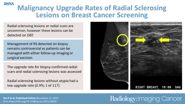
Purpose To determine the upgrade rate for biopsy-proven radial scars and radial sclerosing lesions (RS). Materials and Methods In this retrospective study, radiology and pathology databases from two tertiary breast centers were searched to identify patients with biopsy-confirmed RS between March 1, 2012, and December 31, 2017, during which all mammography was performed with digital breast tomosynthesis (DBT). Adjunct modalities such as MRI or US are performed at our centers to better characterize lesions identified at DBT. Patient demographics, imaging, needle and excisional biopsies, and follow-up data were collected at the patient level. Clopper-Pearson interval estimate for upgrade was calculated for 95% confidence using PropCIs package with R version 4.1.0 (R Foundation for Statistical Computing) (1). Results During the study period, a total of 155 885 DBT examinations were performed. From these examinations, 146 biopsy-proven RS were identified in 142 women (median age, 58 years; age range, 26-87 years). A total of 80.1% (117 of 146) of all RS did not have associated atypia or malignancy, and 19.9% (29 of 146) had associated atypia at initial biopsy. A total of 66.7% (78 of 117) of RS without atypia or malignancy were surgically excised, 25.6% (30 of 117) were followed (median, 3 years; range, 1-7 years) with benign findings on imaging, and 7.7% (nine of 117) were lost to follow-up. The rate of malignancy upgrade was 0.9% (one of 117 [95% CI: 0.02, 4.7]); one RS without concurrent atypia or malignancy demonstrated invasive carcinoma at surgical excision. Conclusion RS without atypia had a low upgrade rate. Mammography, Breast © RSNA, 2021.

摘要

目的

确定经活检证实的放射状瘢痕和放射状硬化性病变(RS)的升级率。

材料与方法

本回顾性研究检索了 2 家三级乳腺中心的放射学和病理学数据库,以确定 2012 年 3 月 1 日至 2017 年 12 月 31 日期间经活检证实为 RS 的患者,在此期间,所有乳腺 X 线摄影均采用数字乳腺断层合成技术(DBT)进行。在我们中心,会采用 MRI 或 US 等辅助手段来更好地对 DBT 发现的病变进行特征描述。以患者为单位收集患者的人口统计学、影像学、针吸活检和切除活检以及随访数据。使用 R 版本 4.1.0(R 基金会用于统计计算)中的 PropCIs 包计算 95%置信区间的 Clopper-Pearson 间隔估计值(1)。

结果

在研究期间,共进行了 155885 次 DBT 检查。在此期间,从这些检查中确定了 142 名女性的 146 例经活检证实的 RS(中位年龄 58 岁;年龄范围 26-87 岁)。所有 RS 中,80.1%(117/146)无相关非典型性或恶性肿瘤,19.9%(29/146)在初次活检时存在相关非典型性。无非典型性或恶性肿瘤的 RS 中,66.7%(117/146)行手术切除,25.6%(30/117)行随访(中位随访时间 3 年;范围 1-7 年),影像学显示良性结果,7.7%(117/117)失访。恶性肿瘤升级率为 0.9%(117 例中的 1 例[95%CI:0.02,4.7]);1 例无伴发非典型性或恶性肿瘤的 RS 在手术切除时显示浸润性癌。

结论

无非典型性的 RS 升级率较低。

乳腺摄影,乳房 ©RSNA,2021。

https://cdn.ncbi.nlm.nih.gov/pmc/blobs/403d/8637224/222d90505aba/rycan.2021210036.VA.jpg

相似文献

1
Malignancy Upgrade Rates of Radial Sclerosing Lesions at Breast Cancer Screening.乳腺癌筛查中放射状硬化病变的恶性升级率。
Radiol Imaging Cancer. 2021 Nov;3(6):e210036. doi: 10.1148/rycan.2021210036.
2
Management of Architectural Distortion on Digital Breast Tomosynthesis With Nonmalignant Pathology at Biopsy.对活检有非恶性病变的数字乳腺断层合成术的结构扭曲进行管理。
AJR Am J Roentgenol. 2022 Jul;219(1):46-54. doi: 10.2214/AJR.21.27161. Epub 2022 Feb 2.
3
Upgrade risk of image-targeted radial scar and complex sclerosing lesions diagnosed at needle-guided biopsy: a retrospective study.在经皮引导活检诊断的影像目标性放射状瘢痕和复杂硬化性病变中升级的风险:一项回顾性研究。
Eur Radiol. 2023 Dec;33(12):8399-8406. doi: 10.1007/s00330-023-09877-6. Epub 2023 Jun 29.
4
Management of radial scars/complex sclerosing lesions of the breast diagnosed on vacuum-assisted large-core biopsy: is surgery always necessary?在真空辅助大芯活检诊断的乳腺放射状瘢痕/复杂硬化性病变的处理:是否总是需要手术?
Histopathology. 2019 Dec;75(6):900-915. doi: 10.1111/his.13950. Epub 2019 Oct 18.
5
Rate of radial scars by core biopsy and upgrading to malignancy or high-risk lesions before and after introduction of digital breast tomosynthesis.在引入数字乳腺断层合成前后,核心活检中放射状瘢痕的发生率以及向恶性肿瘤或高危病变的升级率。
Breast Cancer Res Treat. 2019 Jan;173(1):23-29. doi: 10.1007/s10549-018-4973-x. Epub 2018 Sep 21.
6
Outcome of radial scar/complex sclerosing lesion associated with epithelial proliferations with atypia diagnosed on breast core biopsy: results from a multicentric UK-based study.乳腺核心活检中诊断的伴有非典型上皮增生的放射状瘢痕/复杂硬化性病变的转归:一项基于英国的多中心研究结果。
J Clin Pathol. 2019 Dec;72(12):800-804. doi: 10.1136/jclinpath-2019-205764. Epub 2019 Jul 26.
7
Meta-analysis of upgrade rates in 3163 radial scars excised after needle core biopsy diagnosis.经皮穿刺芯针活检诊断后切除的 3163 例放射状瘢痕升级率的荟萃分析。
Breast Cancer Res Treat. 2019 Feb;174(1):165-177. doi: 10.1007/s10549-018-5040-3. Epub 2018 Nov 20.
8
A Comprehensive Assessment of Radial Scars on Core Needle Biopsy in Patients with or without Breast Cancer: Upgrade Rate and Implications on Management.对有或无乳腺癌患者核心针活检中放射状瘢痕的全面评估:升级率及其对管理的影响。
Clin Breast Cancer. 2024 Jun;24(4):e273-e278. doi: 10.1016/j.clbc.2024.01.020. Epub 2024 Feb 1.
9
High-risk lesions diagnosed at MRI-guided vacuum-assisted breast biopsy: imaging characteristics, outcome of surgical excision or imaging follow-up.MRI引导下真空辅助乳腺活检诊断出的高危病变:影像学特征、手术切除结果或影像学随访
Breast Cancer. 2020 May;27(3):405-414. doi: 10.1007/s12282-019-01032-8. Epub 2019 Dec 14.
10
Radial Scars on Screening Digital Breast Tomosynthesis: Upstaging Rates and Management Strategies.筛查性数字化乳腺断层摄影术的放射状瘢痕:升级率和管理策略。
AJR Am J Roentgenol. 2024 Jun;222(6):e2430845. doi: 10.2214/AJR.24.30845. Epub 2024 Jun 5.

引用本文的文献

1
Navigating the Uncertainty of B3 Breast Lesions: Diagnostic Challenges and Evolving Management Strategies.应对B3级乳腺病变的不确定性:诊断挑战与不断发展的管理策略
J Pers Med. 2025 Jan 18;15(1):36. doi: 10.3390/jpm15010036.
2
Radial sclerosing lesions found on core needle biopsy: excision can be safely avoided.在粗针活检中发现的放射性硬化性病变:可安全避免切除。
Histopathology. 2024 Sep;85(3):397-404. doi: 10.1111/his.15233. Epub 2024 Jun 6.
3
A comparative study on the features of breast sclerosing adenosis and invasive ductal carcinoma via ultrasound and establishment of a predictive nomogram.

本文引用的文献

1
Radial Scars and Complex Sclerosing Lesions of the Breast: Prevalence of Malignancy and Natural History Under Active Surveillance.乳腺放射状瘢痕和复杂硬化性病变:主动监测下的恶性肿瘤患病率和自然病史。
Ann Surg Oncol. 2021 Sep;28(9):5149-5155. doi: 10.1245/s10434-021-09713-5. Epub 2021 Mar 5.
2
Radial scars and complex sclerosing lesions on core needle biopsy of the breast: upgrade rates and long-term outcomes.乳腺芯针活检中的放射状瘢痕和复杂硬化性病变:升级率和长期结果。
Breast Cancer Res Treat. 2020 Oct;183(3):677-682. doi: 10.1007/s10549-020-05806-z. Epub 2020 Jul 21.
3
Digital Mammography Stereotactic Biopsy versus Digital Breast Tomosynthesis-guided Biopsy: Differences in Biopsy Targets, Pathologic Results, and Discordance Rates.
超声对乳腺硬化性腺病与浸润性导管癌特征的对比研究及预测列线图的建立
Front Oncol. 2023 Oct 23;13:1276524. doi: 10.3389/fonc.2023.1276524. eCollection 2023.
4
Third International Consensus Conference on lesions of uncertain malignant potential in the breast (B3 lesions).第三版乳腺交界性病变国际专家共识(B3 病变)
Virchows Arch. 2023 Jul;483(1):5-20. doi: 10.1007/s00428-023-03566-x. Epub 2023 Jun 17.
5
[Interpretation of Image-Guided Biopsy Results and Assessment].[影像引导下活检结果的解读与评估]
J Korean Soc Radiol. 2023 Mar;84(2):361-371. doi: 10.3348/jksr.2022.0164. Epub 2023 Mar 28.
6
An ultrasound-based radiomics model to distinguish between sclerosing adenosis and invasive ductal carcinoma.一种基于超声的影像组学模型,用于鉴别硬化性腺病和浸润性导管癌。
Front Oncol. 2023 Mar 7;13:1090617. doi: 10.3389/fonc.2023.1090617. eCollection 2023.
数字乳腺立体定向活检与数字乳腺断层合成引导活检:活检目标、病理结果和不相符率的差异。
Radiology. 2020 Mar;294(3):518-527. doi: 10.1148/radiol.2019191525. Epub 2020 Jan 21.
4
Reducing indications for radial scar surgical excision in Slovenian breast cancer screening program.减少斯洛文尼亚乳腺癌筛查计划中对放射状瘢痕手术切除的适应证。
Ann Diagn Pathol. 2020 Apr;45:151438. doi: 10.1016/j.anndiagpath.2019.151438. Epub 2019 Dec 13.
5
Outcome of radial scar/complex sclerosing lesion associated with epithelial proliferations with atypia diagnosed on breast core biopsy: results from a multicentric UK-based study.乳腺核心活检中诊断的伴有非典型上皮增生的放射状瘢痕/复杂硬化性病变的转归:一项基于英国的多中心研究结果。
J Clin Pathol. 2019 Dec;72(12):800-804. doi: 10.1136/jclinpath-2019-205764. Epub 2019 Jul 26.
6
Imaging Follow-up Versus Surgical Excision for Radial Scars Identified on Tomosynthesis-Guided Core Needle Biopsy.基于断层合成引导下核心针活检的放射状瘢痕的影像学随访与手术切除的比较。
Acad Radiol. 2020 Mar;27(3):389-394. doi: 10.1016/j.acra.2019.05.012. Epub 2019 Jul 13.
7
Management of radial scars/complex sclerosing lesions of the breast diagnosed on vacuum-assisted large-core biopsy: is surgery always necessary?在真空辅助大芯活检诊断的乳腺放射状瘢痕/复杂硬化性病变的处理:是否总是需要手术?
Histopathology. 2019 Dec;75(6):900-915. doi: 10.1111/his.13950. Epub 2019 Oct 18.
8
Adoption of Digital Breast Tomosynthesis in Clinical Practice.数字乳腺断层合成技术在临床实践中的应用。
JAMA Intern Med. 2019 Sep 1;179(9):1292-1295. doi: 10.1001/jamainternmed.2019.1058.
9
An audit of mammographic screen detected lesions of uncertain malignant potential (B3) diagnosed on initial image guided needle biopsy: how has our practice changed over 10 years?乳腺 X 线摄影筛查检出的初始影像引导下经皮针活检诊断为性质不明的恶性潜能病灶(B3)的审计:我们的实践在 10 年内发生了怎样的变化?
Clin Radiol. 2019 Aug;74(8):653.e19-653.e25. doi: 10.1016/j.crad.2019.04.006. Epub 2019 May 9.
10
Meta-analysis of upgrade rates in 3163 radial scars excised after needle core biopsy diagnosis.经皮穿刺芯针活检诊断后切除的 3163 例放射状瘢痕升级率的荟萃分析。
Breast Cancer Res Treat. 2019 Feb;174(1):165-177. doi: 10.1007/s10549-018-5040-3. Epub 2018 Nov 20.