• 文献检索
  • 文档翻译
  • 深度研究
  • 学术资讯
  • Suppr Zotero 插件Zotero 插件
  • 邀请有礼
  • 套餐&价格
  • 历史记录
应用&插件
Suppr Zotero 插件Zotero 插件浏览器插件Mac 客户端Windows 客户端微信小程序
定价
高级版会员购买积分包购买API积分包
服务
文献检索文档翻译深度研究API 文档MCP 服务
关于我们
关于 Suppr公司介绍联系我们用户协议隐私条款
关注我们

Suppr 超能文献

核心技术专利:CN118964589B侵权必究
粤ICP备2023148730 号-1Suppr @ 2026

文献检索

告别复杂PubMed语法,用中文像聊天一样搜索,搜遍4000万医学文献。AI智能推荐,让科研检索更轻松。

立即免费搜索

文件翻译

保留排版,准确专业,支持PDF/Word/PPT等文件格式,支持 12+语言互译。

免费翻译文档

深度研究

AI帮你快速写综述,25分钟生成高质量综述,智能提取关键信息,辅助科研写作。

立即免费体验

年龄相关性黄斑变性的个性化萎缩风险测绘。

Personalized Atrophy Risk Mapping in Age-Related Macular Degeneration.

机构信息

Department of Ophthalmology, University of Lausanne, Jules-Gonin Eye Hospital, Fondation Asile des Aveugles, Lausanne, Switzerland.

RetinAI Medical AG, Bern, Switzerland.

出版信息

Transl Vis Sci Technol. 2021 Nov 1;10(13):18. doi: 10.1167/tvst.10.13.18.

DOI:10.1167/tvst.10.13.18
PMID:34767623
原文链接:https://pmc.ncbi.nlm.nih.gov/articles/PMC8590159/
Abstract

PURPOSE

To develop and validate an automatic retinal pigment epithelial and outer retinal atrophy (RORA) progression prediction model for nonexudative age-related macular degeneration (AMD) cases in optical coherence tomography (OCT) scans.

METHODS

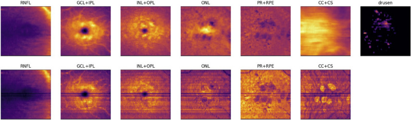
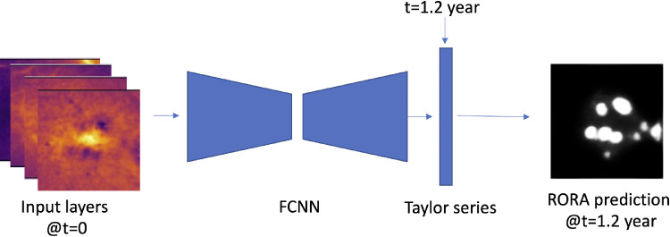
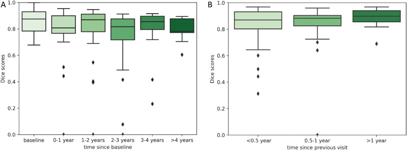
Longitudinal OCT data from 129 eyes/119 patients with RORA was collected and separated into training and testing groups. RORA was automatically segmented in all scans and additionally manually annotated in the test scans. OCT-based features such as layers thicknesses, mean reflectivity, and a drusen height map served as an input to the deep neural network. Based on the baseline OCT scan or the previous visit OCT, en face RORA predictions were calculated for future patient visits. The performance was quantified over time with the means of Dice scores and square root area errors.

RESULTS

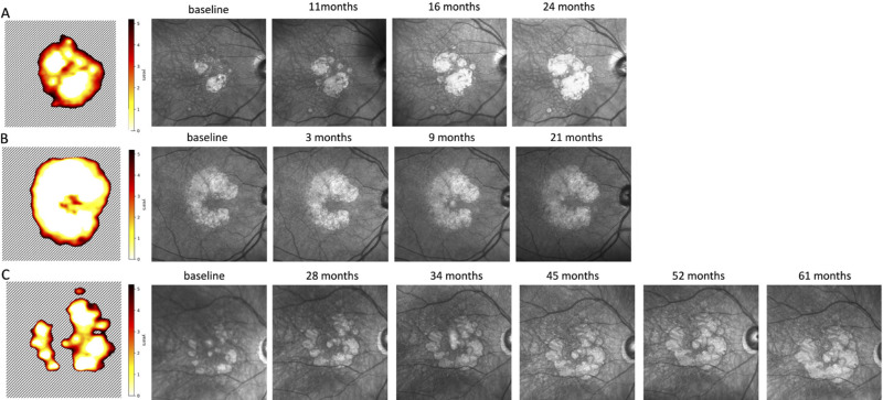
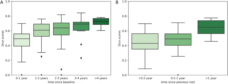
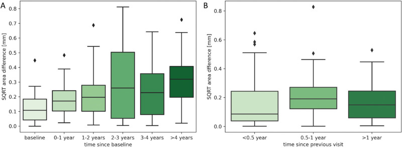
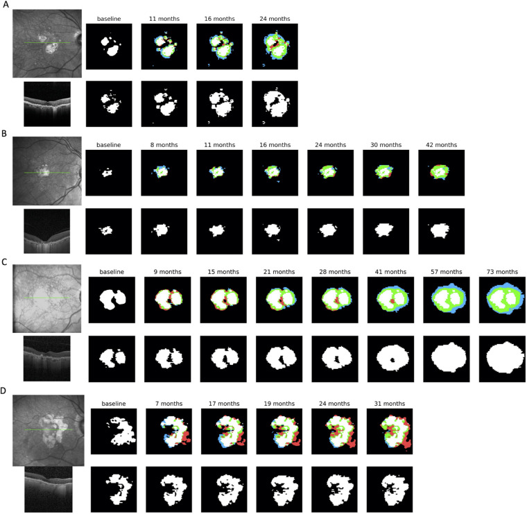
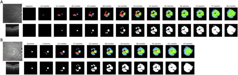
The average Dice score for segmentations at baseline was 0.85. When predicting progression from baseline OCTs, the Dice scores ranged from 0.73 to 0.80 for total RORA area and from 0.46 to 0.72 for RORA growth region. The square root area error ranged from 0.13 mm to 0.33 mm. By providing continuous time output, the model enabled creation of a patient-specific atrophy risk map.

CONCLUSIONS

We developed a machine learning method for RORA progression prediction, which provides continuous-time output. It was used to compute atrophy risk maps, which indicate time-to-RORA-conversion, a novel and clinically relevant way of representing disease progression.

TRANSLATIONAL RELEVANCE

Application of recent advances in artificial intelligence to predict patient-specific progression of atrophic AMD.

摘要

目的

开发和验证一种用于光学相干断层扫描(OCT)扫描中渗出性年龄相关性黄斑变性(AMD)病例的自动视网膜色素上皮和外层视网膜萎缩(RORA)进展预测模型。

方法

收集了 129 只眼/119 例 RORA 的纵向 OCT 数据,并将其分为训练组和测试组。在所有扫描中自动分割 RORA,并在测试扫描中手动注释。OCT 为基础的特征,如层厚度、平均反射率和一个玻璃膜疣高度图,作为深度神经网络的输入。基于基线 OCT 扫描或前一次就诊的 OCT,为未来的患者就诊计算了 RORA 的即时预测。通过 Dice 分数和平方根误差的平均值来定量评估随时间的性能。

结果

基线分割的平均 Dice 分数为 0.85。当从基线 OCT 预测进展时,总 RORA 面积的 Dice 分数范围为 0.73 至 0.80,RORA 增长区域的 Dice 分数范围为 0.46 至 0.72。平方根误差范围为 0.13 毫米至 0.33 毫米。通过提供连续的时间输出,该模型能够创建患者特定的萎缩风险图。

结论

我们开发了一种用于 RORA 进展预测的机器学习方法,它提供了连续时间的输出。它被用来计算萎缩风险图,这是一种表示疾病进展的新的、临床相关的方法,表明 RORA 转化的时间。

翻译是否准确?

https://cdn.ncbi.nlm.nih.gov/pmc/blobs/5d28/8590159/eab0a722d015/tvst-10-13-18-f009.jpg
https://cdn.ncbi.nlm.nih.gov/pmc/blobs/5d28/8590159/a813429ca3f2/tvst-10-13-18-f001.jpg
https://cdn.ncbi.nlm.nih.gov/pmc/blobs/5d28/8590159/7e54d63a6523/tvst-10-13-18-f002.jpg
https://cdn.ncbi.nlm.nih.gov/pmc/blobs/5d28/8590159/dafff38cab0b/tvst-10-13-18-f003.jpg
https://cdn.ncbi.nlm.nih.gov/pmc/blobs/5d28/8590159/54b51e600f19/tvst-10-13-18-f004.jpg
https://cdn.ncbi.nlm.nih.gov/pmc/blobs/5d28/8590159/50728fae37d7/tvst-10-13-18-f005.jpg
https://cdn.ncbi.nlm.nih.gov/pmc/blobs/5d28/8590159/9467d64931ab/tvst-10-13-18-f006.jpg
https://cdn.ncbi.nlm.nih.gov/pmc/blobs/5d28/8590159/a2967517d0ca/tvst-10-13-18-f007.jpg
https://cdn.ncbi.nlm.nih.gov/pmc/blobs/5d28/8590159/b4de33fa05df/tvst-10-13-18-f008.jpg
https://cdn.ncbi.nlm.nih.gov/pmc/blobs/5d28/8590159/eab0a722d015/tvst-10-13-18-f009.jpg

相似文献

1
Personalized Atrophy Risk Mapping in Age-Related Macular Degeneration.年龄相关性黄斑变性的个性化萎缩风险测绘。
Transl Vis Sci Technol. 2021 Nov 1;10(13):18. doi: 10.1167/tvst.10.13.18.
2
Fully-automated atrophy segmentation in dry age-related macular degeneration in optical coherence tomography.光学相干断层扫描在干性年龄相关性黄斑变性中的全自动萎缩分割。
Sci Rep. 2021 Nov 8;11(1):21893. doi: 10.1038/s41598-021-01227-0.
3
Progression Dynamics of Early versus Later Stage Atrophic Lesions in Nonneovascular Age-Related Macular Degeneration Using Quantitative OCT Biomarker Segmentation.采用定量 OCT 生物标志物分割评估非新生血管性年龄相关性黄斑变性早、晚期萎缩病变的进展动态。
Ophthalmol Retina. 2023 Sep;7(9):762-770. doi: 10.1016/j.oret.2023.05.004. Epub 2023 May 9.
4
Prediction of Individual Disease Conversion in Early AMD Using Artificial Intelligence.利用人工智能预测早期 AMD 的个体疾病转化。
Invest Ophthalmol Vis Sci. 2018 Jul 2;59(8):3199-3208. doi: 10.1167/iovs.18-24106.
5
Progression of Photoreceptor Degeneration in Geographic Atrophy Secondary to Age-related Macular Degeneration.年龄相关性黄斑变性继发地图状萎缩中感光细胞变性的进展。
JAMA Ophthalmol. 2020 Oct 1;138(10):1026-1034. doi: 10.1001/jamaophthalmol.2020.2914.
6
Optical Coherence Tomography Reflective Drusen Substructures Predict Progression to Geographic Atrophy in Age-related Macular Degeneration.光学相干断层扫描反射性玻璃膜疣亚结构预测年龄相关性黄斑变性进展为地图状萎缩
Ophthalmology. 2016 Dec;123(12):2554-2570. doi: 10.1016/j.ophtha.2016.08.047. Epub 2016 Oct 25.
7
Validated automatic segmentation of AMD pathology including drusen and geographic atrophy in SD-OCT images.验证了 SD-OCT 图像中 AMD 病变(包括 drusen 和 GA)的自动分割。
Invest Ophthalmol Vis Sci. 2012 Jan 5;53(1):53-61. doi: 10.1167/iovs.11-7640.
8
Detection of Nonexudative Macular Neovascularization on Structural OCT Images Using Vision Transformers.使用视觉Transformer在结构光学相干断层扫描(OCT)图像上检测非渗出性黄斑新生血管形成
Ophthalmol Sci. 2022 Jul 8;2(4):100197. doi: 10.1016/j.xops.2022.100197. eCollection 2022 Dec.
9
Optical coherence tomography-defined changes preceding the development of drusen-associated atrophy in age-related macular degeneration.光学相干断层扫描定义的在年龄相关性黄斑变性相关的 drusen 相关萎缩发展之前的变化。
Ophthalmology. 2014 Dec;121(12):2415-22. doi: 10.1016/j.ophtha.2014.06.034. Epub 2014 Aug 8.
10
Structural Features Associated With the Development and Progression of RORA Secondary to Maternally Inherited Diabetes and Deafness.母系遗传糖尿病和耳聋继发 RORA 结构特征与发育和进展相关。
Am J Ophthalmol. 2020 Oct;218:136-147. doi: 10.1016/j.ajo.2020.05.023. Epub 2020 May 22.

引用本文的文献

1
Modeling pegcetacoplan treatment effect for atrophic age-related macular degeneration with AI-based progression prediction.基于人工智能的进展预测对萎缩性年龄相关性黄斑变性的培西加可普坦治疗效果进行建模。
Int J Retina Vitreous. 2025 Feb 7;11(1):14. doi: 10.1186/s40942-025-00634-z.
2
Deep Learning to Predict the Future Growth of Geographic Atrophy from Fundus Autofluorescence.利用深度学习从眼底自发荧光预测地图样萎缩的未来发展
Ophthalmol Sci. 2024 Oct 23;5(2):100635. doi: 10.1016/j.xops.2024.100635. eCollection 2025 Mar-Apr.
3
Role of Artificial Intelligence in Retinal Diseases.

本文引用的文献

1
Fully-automated atrophy segmentation in dry age-related macular degeneration in optical coherence tomography.光学相干断层扫描在干性年龄相关性黄斑变性中的全自动萎缩分割。
Sci Rep. 2021 Nov 8;11(1):21893. doi: 10.1038/s41598-021-01227-0.
2
Growth Modeling for Quantitative, Spatially Resolved Geographic Atrophy Lesion Kinetics.定量、空间分辨地理萎缩病变动力学的生长建模。
Transl Vis Sci Technol. 2021 Jun 1;10(7):26. doi: 10.1167/tvst.10.7.26.
3
Automated Quantification of Pathological Fluids in Neovascular Age-Related Macular Degeneration, and Its Repeatability Using Deep Learning.
人工智能在视网膜疾病中的作用。
Klin Monbl Augenheilkd. 2024 Sep;241(9):1023-1031. doi: 10.1055/a-2378-6138. Epub 2024 Sep 16.
4
Deep Learning-Based Prediction of Individual Geographic Atrophy Progression from a Single Baseline OCT.基于深度学习从单次基线光学相干断层扫描预测个体地理萎缩进展
Ophthalmol Sci. 2024 Jan 17;4(4):100466. doi: 10.1016/j.xops.2024.100466. eCollection 2024 Jul-Aug.
5
Artificial intelligence in age-related macular degeneration: state of the art and recent updates.人工智能在年龄相关性黄斑变性中的应用:现状与最新进展。
BMC Ophthalmol. 2024 Mar 15;24(1):121. doi: 10.1186/s12886-024-03381-1.
6
The Predictive Capabilities of Artificial Intelligence-Based OCT Analysis for Age-Related Macular Degeneration Progression-A Systematic Review.基于人工智能的光学相干断层扫描分析对年龄相关性黄斑变性进展的预测能力——一项系统评价
Diagnostics (Basel). 2023 Jul 24;13(14):2464. doi: 10.3390/diagnostics13142464.
7
Retinitis Pigmentosa Associated with EYS Gene Mutations: Disease Severity Staging and Central Retina Atrophy.与EYS基因突变相关的色素性视网膜炎:疾病严重程度分期及视网膜中央萎缩
Diagnostics (Basel). 2023 Feb 23;13(5):850. doi: 10.3390/diagnostics13050850.
利用深度学习对新生血管性年龄相关性黄斑变性病理性液体积分的自动量化及其重复性。
Transl Vis Sci Technol. 2021 Apr 1;10(4):17. doi: 10.1167/tvst.10.4.17.
4
Imaging Features Associated with Progression to Geographic Atrophy in Age-Related Macular Degeneration: Classification of Atrophy Meeting Report 5.年龄相关性黄斑变性中与进展为地图样萎缩相关的影像学特征:萎缩会议报告5分类
Ophthalmol Retina. 2021 Sep;5(9):855-867. doi: 10.1016/j.oret.2020.12.009. Epub 2020 Dec 22.
5
An integrated time adaptive geographic atrophy prediction model for SD-OCT images.基于 SD-OCT 图像的时间自适应地理萎缩综合预测模型。
Med Image Anal. 2021 Feb;68:101893. doi: 10.1016/j.media.2020.101893. Epub 2020 Nov 12.
6
Progression of Photoreceptor Degeneration in Geographic Atrophy Secondary to Age-related Macular Degeneration.年龄相关性黄斑变性继发地图状萎缩中感光细胞变性的进展。
JAMA Ophthalmol. 2020 Oct 1;138(10):1026-1034. doi: 10.1001/jamaophthalmol.2020.2914.
7
Growth Rate of Geographic Atrophy Secondary to Age-Related Macular Degeneration: A Meta-Analysis of Natural History Studies and Implications for Designing Future Trials.年龄相关性黄斑变性继发地理萎缩的增长率:自然史研究的荟萃分析及其对未来试验设计的影响。
Ophthalmic Res. 2021;64(2):205-215. doi: 10.1159/000510507. Epub 2020 Jul 28.
8
Automatically Enhanced OCT Scans of the Retina: A proof of concept study.自动增强的视网膜 OCT 扫描:概念验证研究。
Sci Rep. 2020 May 8;10(1):7819. doi: 10.1038/s41598-020-64724-8.
9
Characterization of Drusen and Hyperreflective Foci as Biomarkers for Disease Progression in Age-Related Macular Degeneration Using Artificial Intelligence in Optical Coherence Tomography.利用人工智能在光学相干断层扫描中对年龄相关性黄斑变性的特征进行分析,以评估其作为疾病进展的生物标志物。
JAMA Ophthalmol. 2020 Jul 1;138(7):740-747. doi: 10.1001/jamaophthalmol.2020.1376.
10
Role of Deep Learning-Quantified Hyperreflective Foci for the Prediction of Geographic Atrophy Progression.深度学习量化的高反射焦点在预测地图状萎缩进展中的作用。
Am J Ophthalmol. 2020 Aug;216:257-270. doi: 10.1016/j.ajo.2020.03.042. Epub 2020 Apr 8.