• 文献检索
  • 文档翻译
  • 深度研究
  • 学术资讯
  • Suppr Zotero 插件Zotero 插件
  • 邀请有礼
  • 套餐&价格
  • 历史记录
应用&插件
Suppr Zotero 插件Zotero 插件浏览器插件Mac 客户端Windows 客户端微信小程序
定价
高级版会员购买积分包购买API积分包
服务
文献检索文档翻译深度研究API 文档MCP 服务
关于我们
关于 Suppr公司介绍联系我们用户协议隐私条款
关注我们

Suppr 超能文献

核心技术专利:CN118964589B侵权必究
粤ICP备2023148730 号-1Suppr @ 2026

文献检索

告别复杂PubMed语法,用中文像聊天一样搜索,搜遍4000万医学文献。AI智能推荐,让科研检索更轻松。

立即免费搜索

文件翻译

保留排版,准确专业,支持PDF/Word/PPT等文件格式,支持 12+语言互译。

免费翻译文档

深度研究

AI帮你快速写综述,25分钟生成高质量综述,智能提取关键信息,辅助科研写作。

立即免费体验

基于焦点区域提取的大量微观图像的多焦点图像融合。

Multi-Focus Image Fusion Using Focal Area Extraction in a Large Quantity of Microscopic Images.

机构信息

Department of Biomedical Engineering, College of Software and Digital Healthcare Convergence, Yonsei University, Wonju 26493, Korea.

Department of Mechanical Engineering, Pohang University of Science and Technology (POSTECH), Pohang 37673, Korea.

出版信息

Sensors (Basel). 2021 Nov 5;21(21):7371. doi: 10.3390/s21217371.

DOI:10.3390/s21217371
PMID:34770677
原文链接:https://pmc.ncbi.nlm.nih.gov/articles/PMC8586970/
Abstract

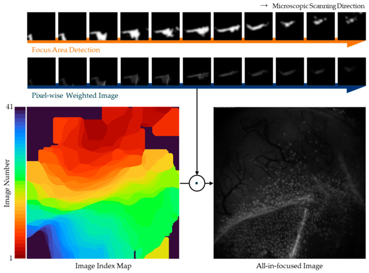
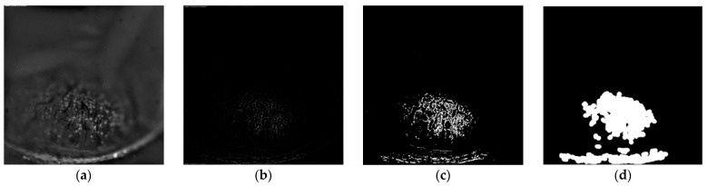
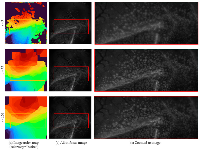
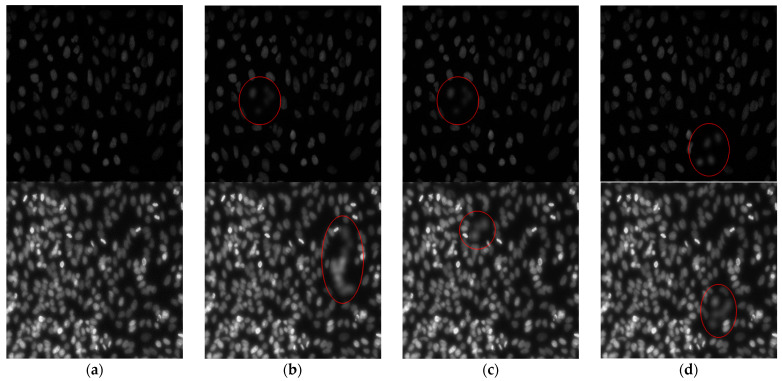
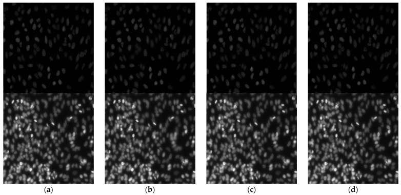
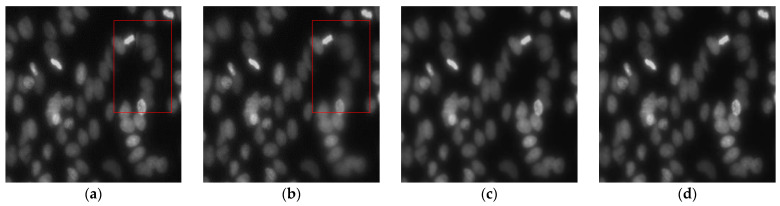
The non-invasive examination of conjunctival goblet cells using a microscope is a novel procedure for the diagnosis of ocular surface diseases. However, it is difficult to generate an all-in-focus image due to the curvature of the eyes and the limited focal depth of the microscope. The microscope acquires multiple images with the axial translation of focus, and the image stack must be processed. Thus, we propose a multi-focus image fusion method to generate an all-in-focus image from multiple microscopic images. First, a bandpass filter is applied to the source images and the focus areas are extracted using Laplacian transformation and thresholding with a morphological operation. Next, a self-adjusting guided filter is applied for the natural connections between local focus images. A window-size-updating method is adopted in the guided filter to reduce the number of parameters. This paper presents a novel algorithm that can operate for a large quantity of images (10 or more) and obtain an all-in-focus image. To quantitatively evaluate the proposed method, two different types of evaluation metrics are used: "full-reference" and "no-reference". The experimental results demonstrate that this algorithm is robust to noise and capable of preserving local focus information through focal area extraction. Additionally, the proposed method outperforms state-of-the-art approaches in terms of both visual effects and image quality assessments.

摘要

使用显微镜对结膜杯状细胞进行非侵入性检查是诊断眼表疾病的一种新方法。然而,由于眼睛的曲率和显微镜的有限景深,很难生成全聚焦图像。显微镜通过轴向焦点平移获取多个图像,并且必须处理图像堆栈。因此,我们提出了一种多聚焦图像融合方法,从多个显微镜图像生成全聚焦图像。首先,对源图像应用带通滤波器,并使用拉普拉斯变换和形态学操作的阈值处理提取焦点区域。接下来,应用自调整导向滤波器进行局部焦点图像之间的自然连接。在导向滤波器中采用窗口大小更新方法来减少参数数量。本文提出了一种可以处理大量图像(10 张或更多)并获得全聚焦图像的新算法。为了定量评估所提出的方法,使用了两种不同类型的评估指标:“全参考”和“无参考”。实验结果表明,该算法对噪声具有鲁棒性,并且能够通过焦点区域提取来保留局部焦点信息。此外,在所提出的方法中,在视觉效果和图像质量评估方面都优于最先进的方法。

https://cdn.ncbi.nlm.nih.gov/pmc/blobs/91c8/8586970/5da54af835d9/sensors-21-07371-g007.jpg
https://cdn.ncbi.nlm.nih.gov/pmc/blobs/91c8/8586970/75886a51a9c9/sensors-21-07371-g001.jpg
https://cdn.ncbi.nlm.nih.gov/pmc/blobs/91c8/8586970/69a7ac341cc3/sensors-21-07371-g002.jpg
https://cdn.ncbi.nlm.nih.gov/pmc/blobs/91c8/8586970/6f5e8a4fd7e7/sensors-21-07371-g003.jpg
https://cdn.ncbi.nlm.nih.gov/pmc/blobs/91c8/8586970/c5606cef5d8f/sensors-21-07371-g004.jpg
https://cdn.ncbi.nlm.nih.gov/pmc/blobs/91c8/8586970/81adf8e809bc/sensors-21-07371-g005.jpg
https://cdn.ncbi.nlm.nih.gov/pmc/blobs/91c8/8586970/1ef2a9251af9/sensors-21-07371-g006.jpg
https://cdn.ncbi.nlm.nih.gov/pmc/blobs/91c8/8586970/5da54af835d9/sensors-21-07371-g007.jpg

相似文献

1
Multi-Focus Image Fusion Using Focal Area Extraction in a Large Quantity of Microscopic Images.基于焦点区域提取的大量微观图像的多焦点图像融合。
Sensors (Basel). 2021 Nov 5;21(21):7371. doi: 10.3390/s21217371.
2
Fast processing of microscopic images using object-based extended depth of field.使用基于对象的扩展景深快速处理显微图像。
BMC Bioinformatics. 2016 Dec 22;17(Suppl 19):516. doi: 10.1186/s12859-016-1373-2.
3
Construction of All-in-Focus Images Assisted by Depth Sensing.基于深度感知的全聚焦图像构建。
Sensors (Basel). 2019 Mar 22;19(6):1409. doi: 10.3390/s19061409.
4
DL-EDOF: Novel Multi-Focus Image Data Set and Deep Learning-Based Approach for More Accurate and Specimen-Free Extended Depth of Focus.DL-EDOF:新型多焦点图像数据集和基于深度学习的方法,实现更准确、无需样本的扩展景深。
J Imaging Inform Med. 2024 Aug;37(4):1991-2013. doi: 10.1007/s10278-024-01076-z. Epub 2024 Mar 25.
5
TransFusion-net for multifocus microscopic biomedical image fusion.TransFusion-net 用于多焦点微观生物医学图像融合。
Comput Methods Programs Biomed. 2023 Oct;240:107688. doi: 10.1016/j.cmpb.2023.107688. Epub 2023 Jun 28.
6
Multi-focus image fusion using a guided-filter-based difference image.基于引导滤波的差分图像的多聚焦图像融合
Appl Opt. 2016 Mar 20;55(9):2230-9. doi: 10.1364/AO.55.002230.
7
Fusion of multi-focus images via a Gaussian curvature filter and synthetic focusing degree criterion.基于高斯曲率滤波器和合成聚焦度准则的多聚焦图像融合
Appl Opt. 2018 Dec 10;57(35):10092-10101. doi: 10.1364/AO.57.010092.
8
Multi-focus image fusion algorithm based on focus detection in spatial and NSCT domain.基于空间域和 NSCT 域中焦点检测的多聚焦图像融合算法。
PLoS One. 2018 Sep 20;13(9):e0204225. doi: 10.1371/journal.pone.0204225. eCollection 2018.
9
Dual-tree complex wavelet transform and image block residual-based multi-focus image fusion in visual sensor networks.视觉传感器网络中基于双树复数小波变换和图像块残差的多聚焦图像融合
Sensors (Basel). 2014 Nov 26;14(12):22408-30. doi: 10.3390/s141222408.
10
Generation of all-in-focus images by noise-robust selective fusion of limited depth-of-field images.通过稳健的抗噪有限景深图像选择性融合生成全聚焦图像。
IEEE Trans Image Process. 2013 Mar;22(3):1242-51. doi: 10.1109/TIP.2012.2231087. Epub 2012 Dec 3.

本文引用的文献

1
Image Fusion Techniques: A Survey.图像融合技术:综述
Arch Comput Methods Eng. 2021;28(7):4425-4447. doi: 10.1007/s11831-021-09540-7. Epub 2021 Jan 24.
2
Moxifloxacin based axially swept wide-field fluorescence microscopy for high-speed imaging of conjunctival goblet cells.基于莫西沙星的轴向扫描宽视野荧光显微镜用于结膜杯状细胞的高速成像
Biomed Opt Express. 2020 Aug 6;11(9):4890-4900. doi: 10.1364/BOE.401896. eCollection 2020 Sep 1.
3
Handheld In Vivo Reflectance Confocal Microscopy for the Diagnosis of Eyelid Margin and Conjunctival Tumors.
手持式体内反射共聚焦显微镜用于睑缘和结膜肿瘤的诊断
JAMA Ophthalmol. 2017 Aug 1;135(8):845-851. doi: 10.1001/jamaophthalmol.2017.2019.
4
Assessment of conjunctival goblet cell density using laser scanning confocal microscopy versus impression cytology.使用激光扫描共聚焦显微镜与印迹细胞学评估结膜杯状细胞密度
Cont Lens Anterior Eye. 2016 Jun;39(3):221-6. doi: 10.1016/j.clae.2016.01.006. Epub 2016 Feb 3.
5
Guided image filtering.引导图像滤波。
IEEE Trans Pattern Anal Mach Intell. 2013 Jun;35(6):1397-409. doi: 10.1109/TPAMI.2012.213.
6
No-reference image quality assessment in the spatial domain.空间域无参考图像质量评估。
IEEE Trans Image Process. 2012 Dec;21(12):4695-708. doi: 10.1109/TIP.2012.2214050. Epub 2012 Aug 17.
7
Image analysis using mathematical morphology.基于数学形态学的图像分析。
IEEE Trans Pattern Anal Mach Intell. 1987 Apr;9(4):532-50. doi: 10.1109/tpami.1987.4767941.
8
Objective Assessment of Multiresolution Image Fusion Algorithms for Context Enhancement in Night Vision: A Comparative Study.客观评估多分辨率图像融合算法在夜视中增强上下文的性能:一项比较研究。
IEEE Trans Pattern Anal Mach Intell. 2012 Jan;34(1):94-109. doi: 10.1109/TPAMI.2011.109. Epub 2011 May 19.
9
Image quality assessment: from error visibility to structural similarity.图像质量评估:从误差可见性到结构相似性。
IEEE Trans Image Process. 2004 Apr;13(4):600-12. doi: 10.1109/tip.2003.819861.
10
Conjunctival goblet cell density in normal subjects and in dry eye syndromes.正常受试者和干眼综合征患者的结膜杯状细胞密度
Invest Ophthalmol. 1975 Apr;14(4):299-302.