• 文献检索
  • 文档翻译
  • 深度研究
  • 学术资讯
  • Suppr Zotero 插件Zotero 插件
  • 邀请有礼
  • 套餐&价格
  • 历史记录
应用&插件
Suppr Zotero 插件Zotero 插件浏览器插件Mac 客户端Windows 客户端微信小程序
定价
高级版会员购买积分包购买API积分包
服务
文献检索文档翻译深度研究API 文档MCP 服务
关于我们
关于 Suppr公司介绍联系我们用户协议隐私条款
关注我们

Suppr 超能文献

核心技术专利:CN118964589B侵权必究
粤ICP备2023148730 号-1Suppr @ 2026

文献检索

告别复杂PubMed语法,用中文像聊天一样搜索,搜遍4000万医学文献。AI智能推荐,让科研检索更轻松。

立即免费搜索

文件翻译

保留排版,准确专业,支持PDF/Word/PPT等文件格式,支持 12+语言互译。

免费翻译文档

深度研究

AI帮你快速写综述,25分钟生成高质量综述,智能提取关键信息,辅助科研写作。

立即免费体验

肝内胆管细胞癌误诊为肝脓肿的影像学特征:8 例分析。

Imaging features of intrahepatic cholangiocarcinoma mimicking a liver abscess: an analysis of 8 cases.

机构信息

Department of Ultrasound, Guangxi Medical University Third Affiliated Hospital, Nanning, 530031, China.

Department of Radiology, Guangxi Medical University Third Affiliated Hospital, Nanning, 530031, China.

出版信息

BMC Gastroenterol. 2021 Nov 12;21(1):427. doi: 10.1186/s12876-021-02002-1.

DOI:10.1186/s12876-021-02002-1
PMID:34772340
原文链接:https://pmc.ncbi.nlm.nih.gov/articles/PMC8588691/
Abstract

BACKGROUND

In rare cases, intrahepatic cholangiocarcinoma can present as a pyogenic liver abscess and are often misdiagnosed. This study aimed to analyze the imaging features of intrahepatic cholangiocarcinoma mimicking a pyogenic liver abscess.

METHODS

The clinical data and imaging results of eight patients with pathologically confirmed intrahepatic cholangiocarcinoma mimicking a liver abscess were retrospectively collected.

RESULTS

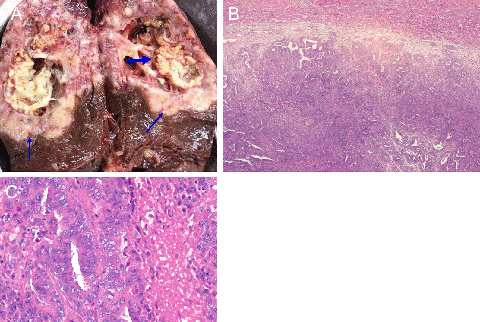
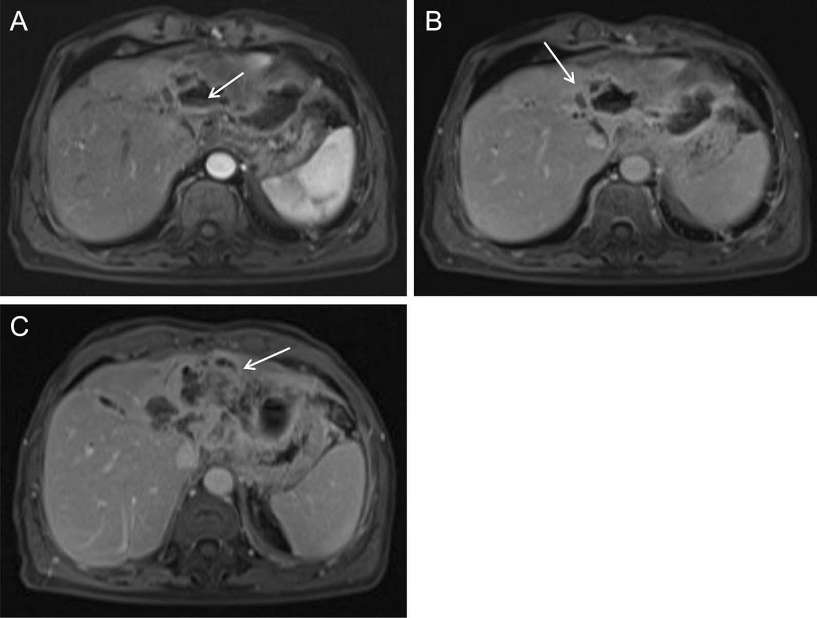
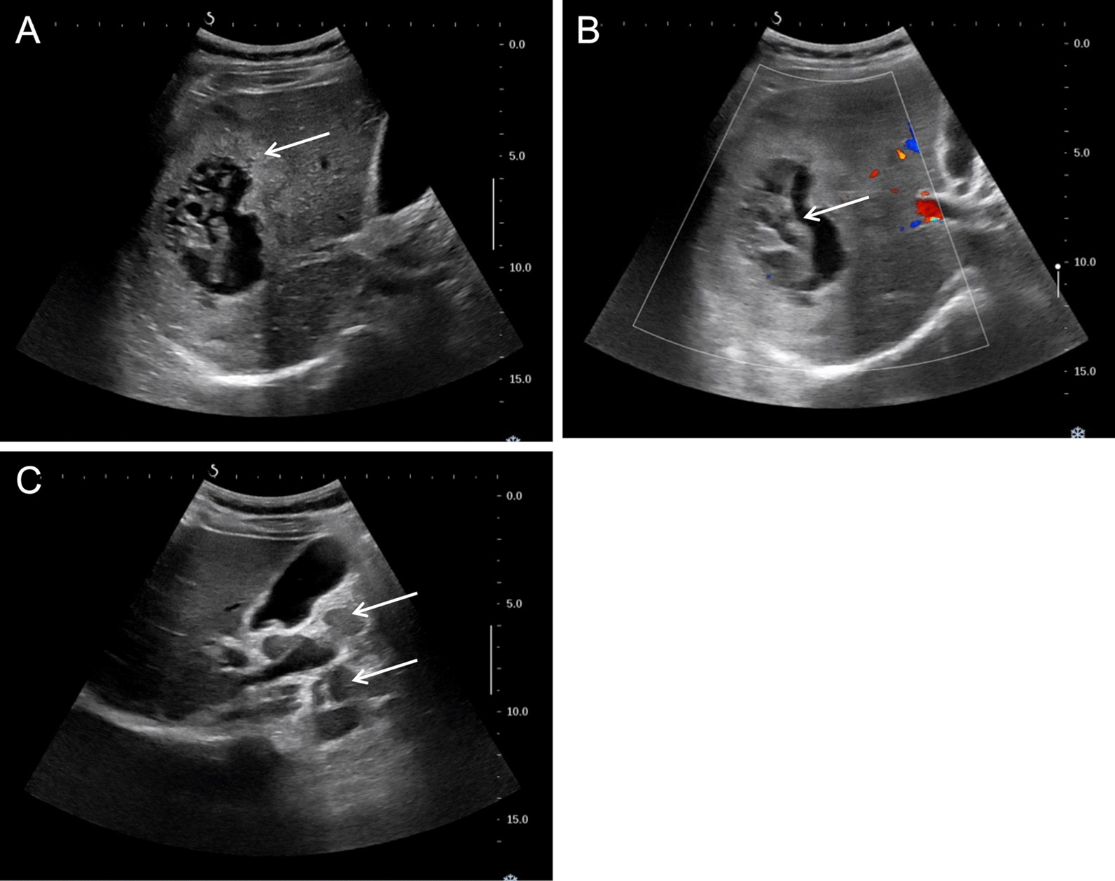
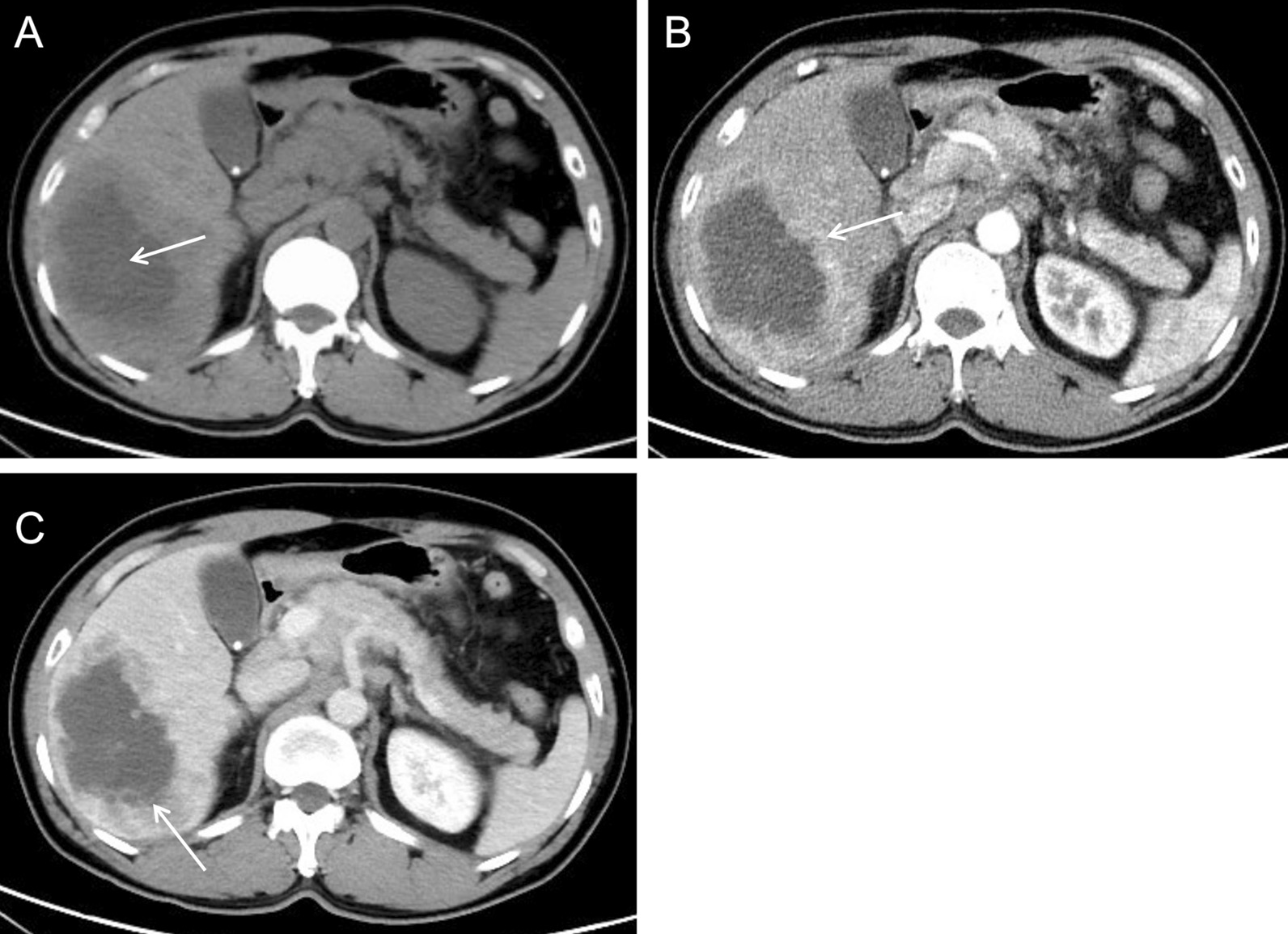
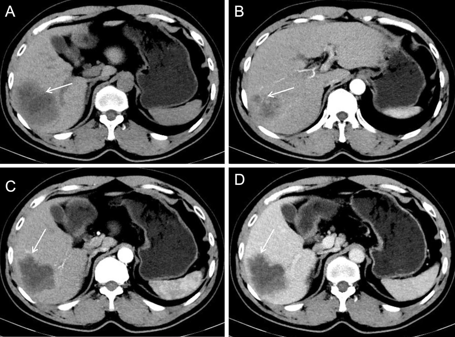
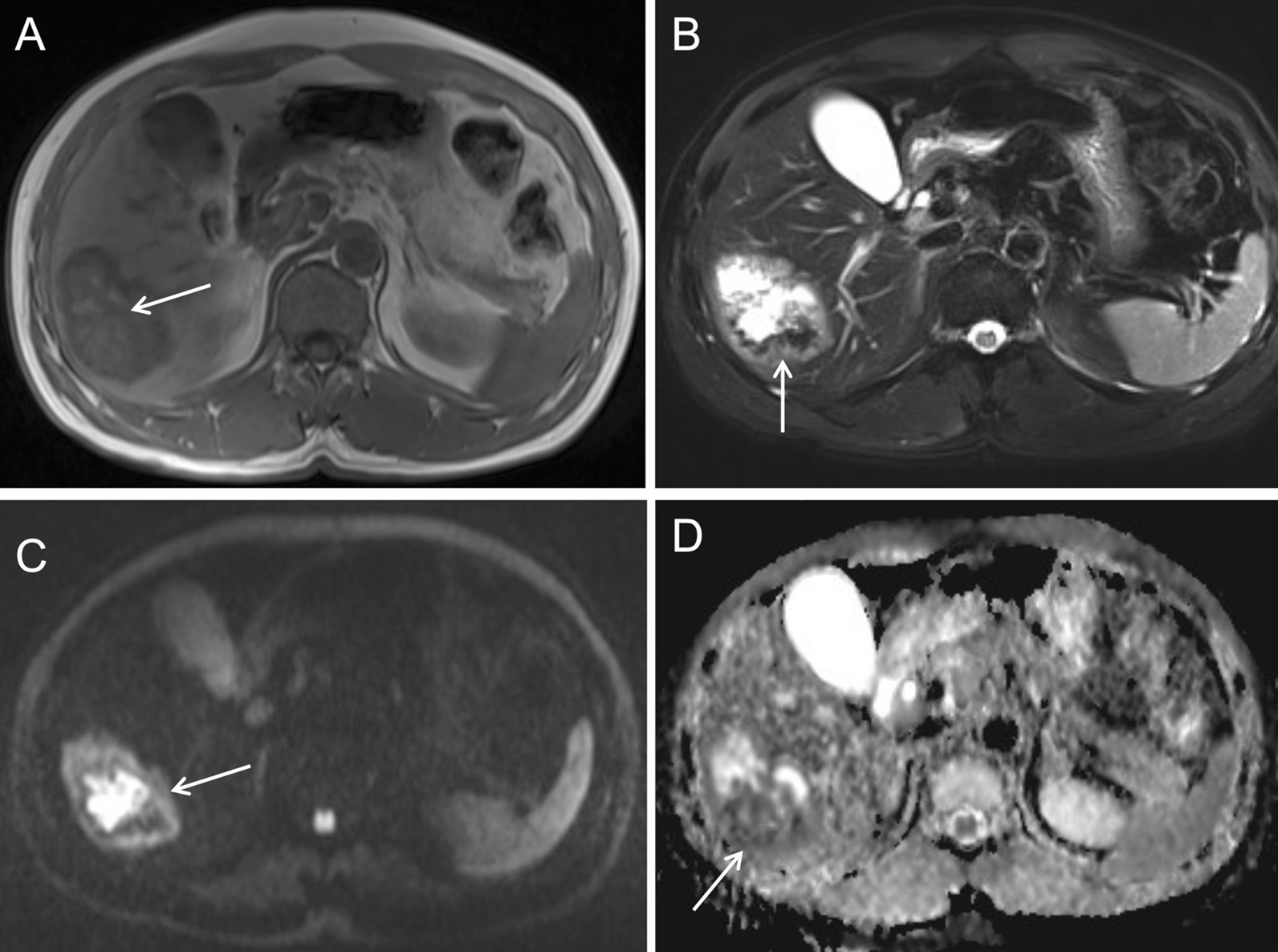
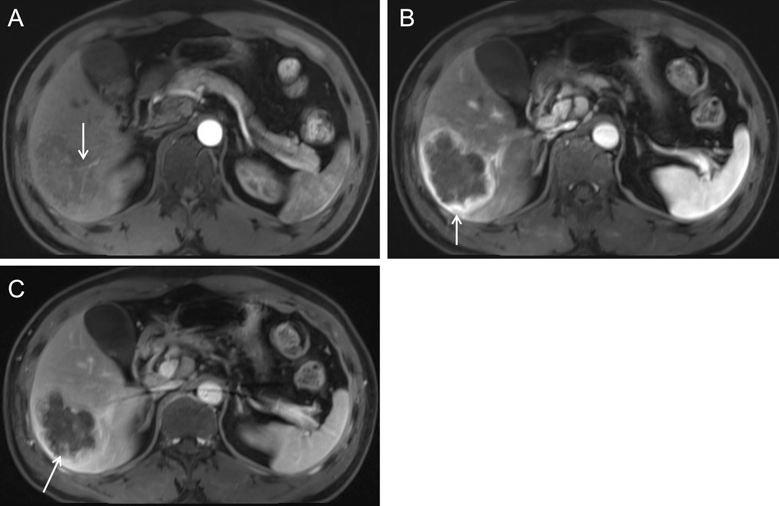
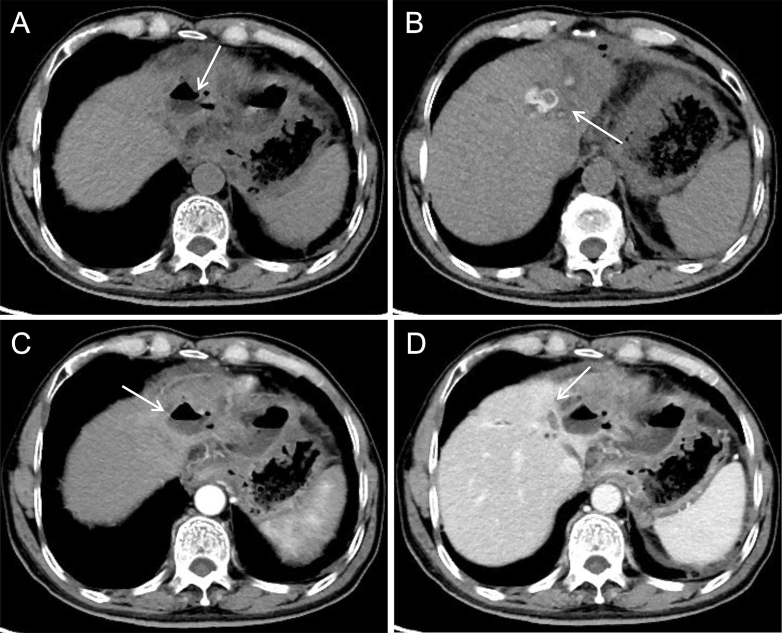
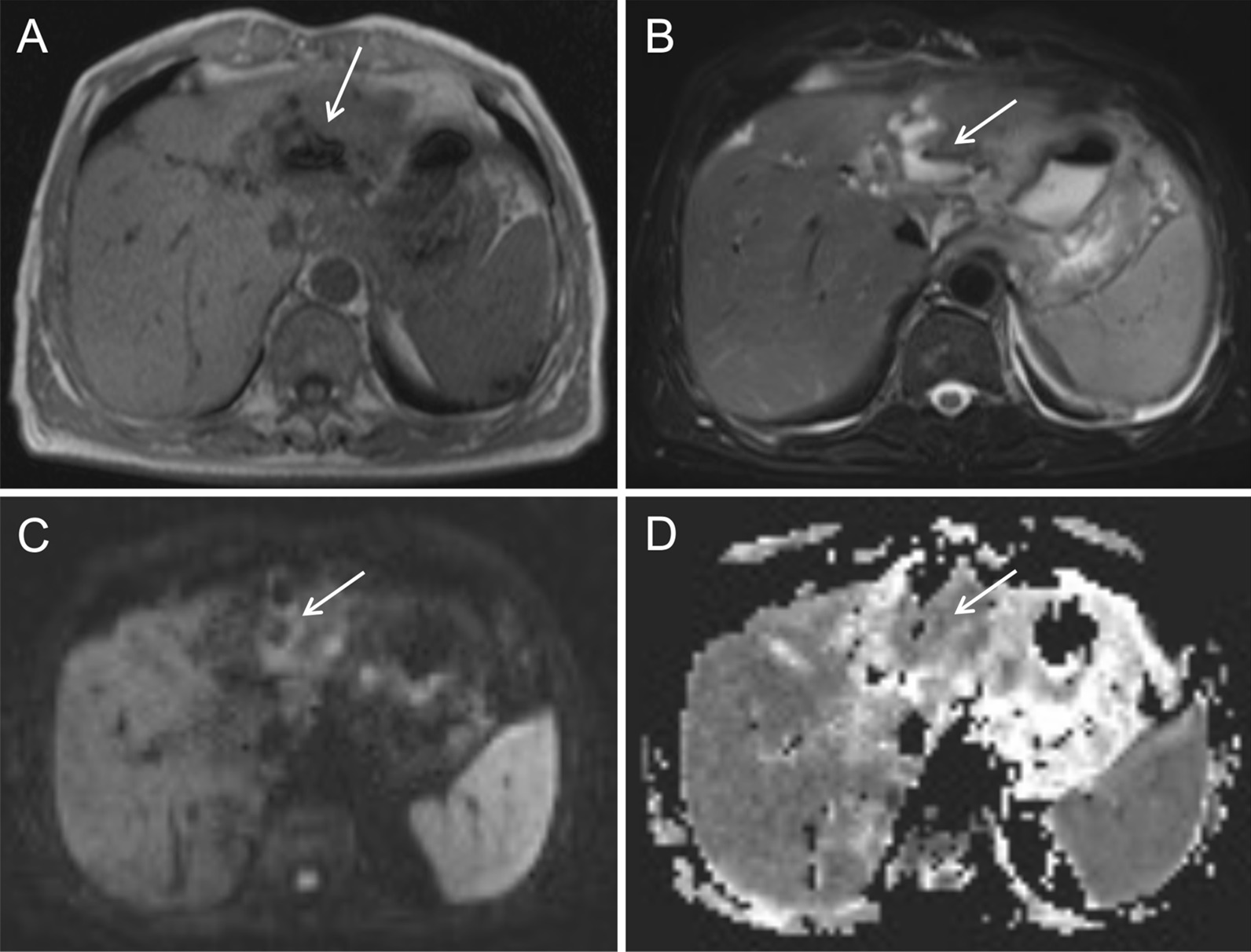
The mean age was 58 years with a range of 46-68 years. Fever and leukocytosis were present in six patients. All the eight lesions were a single mass. Air-liquid levels were present in two patients. Only one patient showed hepatic lobar atrophy and hepatic capsule retraction. The double target sign of liver abscess was not noticed in the CT/MRI images of all eight patients. The inner wall of the lesion was rough and irregular, with multiple dot/patchy and wall nodule enhancements. The abscess wall and the marginal parenchyma were supplied by the hepatic artery in four patients, and the intralesional arteries were rough and disrupted. Bile duct dilatation was seen adjacent to the lesion. In seven patients, diffusion-weighted images showed irregular patchy restricted diffusion in the marginal parenchyma of the necrotic area in addition to the prominent restricted diffusion in the necrotic area. Two patients with cholangiolithiasis showed patchy slight CT hypodensity, slight T1 hypointensity, slight T2 hyperintensity, and patchy delayed enhancement. Multiple lymph nodes enlargement in the hepatic hilar area and the retroperitoneal space were seen in five patients.

CONCLUSION

Intrahepatic cholangiocarcinoma mimicking a pyogenic liver abscess have unique imaging features and require careful image examination to avoid misdiagnosis.

摘要

背景

在罕见情况下,肝内胆管细胞癌可表现为化脓性肝脓肿,且常被误诊。本研究旨在分析肝内胆管细胞癌模拟肝脓肿的影像学特征。

方法

回顾性收集 8 例经病理证实的肝内胆管细胞癌模拟肝脓肿患者的临床资料和影像学结果。

结果

患者的平均年龄为 58 岁(范围 46-68 岁),其中 6 例有发热和白细胞增多。8 个病灶均为单个肿块。2 例有气液平面。仅 1 例出现肝叶萎缩和肝包膜回缩。8 例患者的 CT/MRI 图像均未见肝脓肿的双靶征。病灶的内壁粗糙不规则,呈多个点状/斑片状和壁结节强化。4 例患者的脓肿壁和边缘实质由肝动脉供应,病灶内动脉呈粗糙、中断。病变周围可见胆管扩张。7 例患者弥散加权图像除了坏死区明显弥散受限外,还显示坏死区边缘实质不规则斑片状受限弥散。2 例伴有胆管结石的患者 CT 显示斑片状稍低密度,T1 稍低信号,T2 稍高信号,斑片状延迟强化。5 例患者肝门区和腹膜后可见多个淋巴结肿大。

结论

肝内胆管细胞癌模拟肝脓肿具有独特的影像学特征,需要仔细检查图像以避免误诊。

https://cdn.ncbi.nlm.nih.gov/pmc/blobs/6310/8588691/8b4890d40101/12876_2021_2002_Fig11_HTML.jpg
https://cdn.ncbi.nlm.nih.gov/pmc/blobs/6310/8588691/b66d2d915423/12876_2021_2002_Fig1_HTML.jpg
https://cdn.ncbi.nlm.nih.gov/pmc/blobs/6310/8588691/499cd95d04b3/12876_2021_2002_Fig2_HTML.jpg
https://cdn.ncbi.nlm.nih.gov/pmc/blobs/6310/8588691/311c30d09cf8/12876_2021_2002_Fig3_HTML.jpg
https://cdn.ncbi.nlm.nih.gov/pmc/blobs/6310/8588691/b171b44b8b11/12876_2021_2002_Fig4_HTML.jpg
https://cdn.ncbi.nlm.nih.gov/pmc/blobs/6310/8588691/e06840d0d10d/12876_2021_2002_Fig5_HTML.jpg
https://cdn.ncbi.nlm.nih.gov/pmc/blobs/6310/8588691/bef803d84ab7/12876_2021_2002_Fig6_HTML.jpg
https://cdn.ncbi.nlm.nih.gov/pmc/blobs/6310/8588691/e54c131033cf/12876_2021_2002_Fig7_HTML.jpg
https://cdn.ncbi.nlm.nih.gov/pmc/blobs/6310/8588691/10bbd79ed7a1/12876_2021_2002_Fig8_HTML.jpg
https://cdn.ncbi.nlm.nih.gov/pmc/blobs/6310/8588691/0f67dcf2ac76/12876_2021_2002_Fig9_HTML.jpg
https://cdn.ncbi.nlm.nih.gov/pmc/blobs/6310/8588691/8ca668619107/12876_2021_2002_Fig10_HTML.jpg
https://cdn.ncbi.nlm.nih.gov/pmc/blobs/6310/8588691/8b4890d40101/12876_2021_2002_Fig11_HTML.jpg

相似文献

1
Imaging features of intrahepatic cholangiocarcinoma mimicking a liver abscess: an analysis of 8 cases.肝内胆管细胞癌误诊为肝脓肿的影像学特征:8 例分析。
BMC Gastroenterol. 2021 Nov 12;21(1):427. doi: 10.1186/s12876-021-02002-1.
2
Distinguishing intrahepatic mass-forming biliary carcinomas from hepatocellular carcinoma by computed tomography and magnetic resonance imaging using the Bayesian method: a bi-center study.应用贝叶斯方法通过 CT 和 MRI 鉴别肝内肿块型胆管细胞癌和肝细胞癌:一项双中心研究。
Eur Radiol. 2020 Nov;30(11):5992-6002. doi: 10.1007/s00330-020-06972-w. Epub 2020 Jun 4.
3
Cholangiocarcinoma with a paraneoplastic leukemoid reaction mimicking a pyogenic liver abscess.伴有类白血病反应的胆管癌,酷似肝脓肿。
Korean J Intern Med. 2015 Jan;30(1):110-3. doi: 10.3904/kjim.2015.30.1.110. Epub 2014 Dec 30.
4
A case of intrahepatic mass-forming portal biliopathy mimicking intrahepatic cholangiocarcinoma.肝内肿块型门静脉胆病酷似肝内胆管细胞癌 1 例
Clin J Gastroenterol. 2024 Aug;17(4):671-676. doi: 10.1007/s12328-024-01971-6. Epub 2024 Apr 20.
5
MR imaging of intrahepatic cholangiocarcinoma with pathologic correlation.肝内胆管癌的磁共振成像及其病理对照
AJR Am J Roentgenol. 2001 Jun;176(6):1499-507. doi: 10.2214/ajr.176.6.1761499.
6
[Imaging of peripheral cholangiocarcinoma. Comparison with pathological anatomy].[周围型胆管癌的影像学表现。与病理解剖的比较]
Gastroenterol Clin Biol. 1996 Mar;20(2):139-45.
7
Intrahepatic mass-forming cholangiocarcinoma and solitary hypovascular liver metastases: is the differential diagnosis using diffusion-weighted MRI possible?肝内肿块型胆管癌与孤立性乏血供肝转移瘤:能否通过扩散加权磁共振成像进行鉴别诊断?
Acta Radiol. 2017 Dec;58(12):1417-1426. doi: 10.1177/0284185117695666. Epub 2017 Mar 28.
8
MRI features of combined hepatocellular- cholangiocarcinoma versus mass forming intrahepatic cholangiocarcinoma.MRI 特征在肝细胞癌-胆管细胞癌与肿块型肝内胆管细胞癌的对比。
Cancer Imaging. 2018 Feb 27;18(1):8. doi: 10.1186/s40644-018-0142-z.
9
Mass-forming intrahepatic cholangiocarcinoma: Can diffusion-weighted imaging predict microvascular invasion?巨块型肝内胆管细胞癌:弥散加权成像能否预测微血管侵犯?
J Magn Reson Imaging. 2019 Jul;50(1):315-324. doi: 10.1002/jmri.26566. Epub 2018 Nov 15.
10
The CT and MRI Features of Primary Intrahepatic Lymphoepithelioma-Like Cholangiocarcinoma.原发性肝内淋巴上皮瘤样胆管细胞癌的 CT 和 MRI 特征。
AJR Am J Roentgenol. 2021 Feb;216(2):393-402. doi: 10.2214/AJR.20.22937. Epub 2020 Dec 16.

引用本文的文献

1
Sarcomatoid hepatocellular carcinoma with subdiaphragmatic metastasis misdiagnosed as liver abscess: A case report.伴有膈下转移的肉瘤样肝细胞癌误诊为肝脓肿:一例报告
Medicine (Baltimore). 2024 Dec 6;103(49):e40842. doi: 10.1097/MD.0000000000040842.
2
Values of MRI Imaging Presentations in the Hepatobiliary Phase, DWI and T2WI Sequences in Predicting Pathological Grades of Intrahepatic Mass-Forming Cholangiocarcinoma.磁共振成像在肝胆期、扩散加权成像(DWI)及T2加权成像(T2WI)序列中的表现对肝内肿块型胆管癌病理分级的预测价值
Front Oncol. 2022 Jun 7;12:867702. doi: 10.3389/fonc.2022.867702. eCollection 2022.
3
Imaging Spectrum of Intrahepatic Mass-Forming Cholangiocarcinoma and Its Mimickers: How to Differentiate Them Using MRI.

本文引用的文献

1
Intrahepatic cholangiocarcinoma mimicking a liver abscess.酷似肝脓肿的肝内胆管癌
Clin Case Rep. 2020 Aug 3;8(12):2510-2513. doi: 10.1002/ccr3.3201. eCollection 2020 Dec.
2
Characterization of Indeterminate Liver Lesions on CT and MRI With Contrast-Enhanced Ultrasound: What Is the Evidence?CT 和 MRI 平扫联合超声造影对肝脏局灶性病变的定性诊断:有何证据?
AJR Am J Roentgenol. 2020 Jun;214(6):1295-1304. doi: 10.2214/AJR.19.21498. Epub 2020 Mar 17.
3
Sarcomatoid intrahepatic cholangiocarcinoma mimicking liver abscess: A case report.
肝内肿块型胆管细胞癌及其模拟病变的影像学表现:MRI 如何对其进行鉴别。
Curr Oncol. 2022 Jan 30;29(2):698-723. doi: 10.3390/curroncol29020061.
类肉瘤样肝内胆管癌误诊为肝脓肿:一例报告
World J Clin Cases. 2020 Jan 6;8(1):208-216. doi: 10.12998/wjcc.v8.i1.208.
4
Risk factors for intrahepatic and extrahepatic cholangiocarcinoma: A systematic review and meta-analysis.肝内和肝外胆管癌的危险因素:系统评价和荟萃分析。
J Hepatol. 2020 Jan;72(1):95-103. doi: 10.1016/j.jhep.2019.09.007. Epub 2019 Sep 16.
5
Epidemiology of Hepatocellular Carcinoma and Intrahepatic Cholangiocarcinoma.肝细胞癌和肝内胆管癌的流行病学
Cancer Control. 2017 Jul-Sep;24(3):1073274817729245. doi: 10.1177/1073274817729245.
6
Cholangiocarcinoma: classification, diagnosis, staging, imaging features, and management.胆管癌:分类、诊断、分期、影像学特征和治疗。
Abdom Radiol (NY). 2017 Jun;42(6):1637-1649. doi: 10.1007/s00261-017-1094-7.
7
Intrahepatic cholangiocarcinoma masquerading as liver abscess.伪装成肝脓肿的肝内胆管癌
J Clin Exp Hepatol. 2015 Mar;5(1):89-92. doi: 10.1016/j.jceh.2014.12.006. Epub 2015 Jan 14.
8
MRI of Focal Liver Lesions.局灶性肝病变的磁共振成像
Curr Med Imaging Rev. 2012 May;8(2):107-116. doi: 10.2174/157340512800672216.
9
Primary liver cancer presenting as pyogenic liver abscess: characteristics, diagnosis, and management.原发性肝癌表现为化脓性肝脓肿:特征、诊断和治疗。
J Surg Oncol. 2012 Jun 1;105(7):687-91. doi: 10.1002/jso.22103. Epub 2011 Sep 22.
10
Transarterial hepatic yttrium-90 radioembolization in patients with unresectable intrahepatic cholangiocarcinoma: factors associated with prolonged survival.经动脉肝内钇-90 放射性栓塞治疗不可切除的肝内胆管细胞癌患者:与生存时间延长相关的因素。
Cardiovasc Intervent Radiol. 2012 Feb;35(1):105-16. doi: 10.1007/s00270-011-0142-x. Epub 2011 Mar 24.